
J. Sci. Dev. 2011, 9 (Eng.Iss. 1): 47 - 54 HANOI UNIVERSITY OF AGRICULTURE
INFLUENCE OF TEMPERATURE ON FUSION PROCESS AND MALFORMATION
IN SKELETON OF ZEBRAFISH (
DANIO RERIO
)
Ảnh hưởng của nhiệt độ đến quá trình kết nối các đốt sống và
tạo dị tật xương sống ở cá ngựa vằn (Danio rerio)
Nguyen Thi Hanh Tien1, Ann Huysseune2 and Eckhard Witten2
1Research Institute for Aquaculture No1, Dinh Bang, Tu Son, Bac Ninh, Viet Nam
2Biology Department, Faculty of Science, Ghent University, B-9000 Gent, Belgium
Corresponding author email: hanhtienait8@gmail.com
Received date: 11.03.2011 Accepted date: 18.04.2011
TÓM TẮT
Cá ngựa vằn Danio rerio là loài cá được bán phổ biến trong các cửa hàng cá cảnh và được nhiều
người chơi cá cảnh trên toàn thế giới biết đến. Trong nuôi thủy sản hiện đại, dị tật trên xương sống
làm giảm giá trị của sản phẩm. Người nuôi cá cảnh luôn quan tâm đến điều chỉnh ngoại hình của cá.
Nghiên cứu này thực hiện nhằm tìm hiểu sâu hơn ảnh hưởng của nhiệt độ đến quá trình kết nối các
đốt sống và tạo dị tật xương sống trên cá ngựa vằn nuôi. Tổng số 94 mẫu cá được nghiên cứu. Các
mẫu cá được nhuộm bằng kỹ thuật nhuộm màu cho sụn và cho xương để xác định dị tật. Kết quả của
nghiên cứu cho thấy cá nuôi ở 320C có dị tật ở các đốt sống phần trước và đầu xương cột sống trong
khi cá nuôi ở các nhiệt độ khác không có dị tật này. Tất cả các mẫu cá nghiên cứu đều có sự kết nối
giữa các đốt sống đuôi PU1, U1 và U2. Ngoài ra cá nuôi ở 200C có sự kết nối giữa các đốt sống PU2
với PU3, PU3 với đốt sống đuôi cuối cùng. Nhiệt độ có thể là nguyên nhân ảnh hưởng đến quá trình
nối các đốt sống và tạo dị tật trên cá ngựa vằn. Hiểu biết về ảnh hưởng của nhiệt độ đến quá trình
hình thành dị tật trên cá ngựa vằn nhằm điều chỉnh điều kiện nuôi thích hợp để kiểm soát dị tật và tạo
ra cá cảnh có hình dáng đẹp.
Từ khóa: Cá ngựa vằn, Danio rerio, dị tật, nhiệt độ, xương sống.
SUMMARY
The zebrafish, Danio rerio is an ornamental fish which can be purchased from pet stores and is
very popular amongst hobbyists throughout the world. In commercial aquaculture, the osteological
abnormalities are undesirable because they reduce the value of the product. Ornamental fish
producers are interested in controlling the beauty form of their fish. The present study was
conducted to understand in depth the influence of temperature on vertebrae development in reared
zebrafish. A total of 94 specimens were observed. Juvenile and adult fish were stained with a whole
mount cartilage and bone staining technique to determine vertebral fusion. We present data showing
that larvae reared at 32°C show malformations in precaudal and caudal region while this feature was
not present at other temperatures. The results show that in all fish studied, fusion of caudal vertebrae
occurred between PU1 (preural 1), U1 (ural 1) and U2 (ural 2). Furthermore, fusion of PU2 (preural 2)
with PU3 (preural 3) and PU3 with the last caudal vertebra was seen in fish reared at 20.0°C.
Temperature may affect the fusion process of zebrafish. The understanding about the temperature
effect on fusion process of zebrafish could help to optimize rearing conditions in order to control
malformations and the figure of ornamental fish.
Key words: Danio rerio, fusion, vertebrae, zebrafish.
1. INTRODUCTION
Alterations the rearing temperatures outside the
range of thermal preference of the fish may have an
impact and potentially compromise research in a
number of ways (Westerfield, 2000). The influences
of environment on vertebral body occur during
embryonic development or shortly after hatching
(McDowall, 2003a). Furthermore, the frequency of
abnormal phenotypes can provide a measure of
developmental stability within a population. Skeletal
47

Influence of temperature on fusion process and malformation in skeleton of Zebrafish...
abnormalities are not rare in wild populations and
also occur in laboratory fish. However, problems
because of abnormalities such as vertebral fusions
have often been over looked. Skeletal anomalies in
farmed fish can be caused by genetic and epigenetic
factors such as different sub-optimal environmental
conditions (Lewis et al., 2004). Variations in
temperature and dissolved oxygen can change
morphological characteristics in individuals at
different levels (Leary et al., 1992). The assessment
of malformations could be used as a tool to estimate
the larval quality of reared fish (Ferreri et al., 2000).
This assessment was based on the hypothesis that a
high number of malformations indicate anomalous
developmental conditions (Favaloro and Mazzola,
2003). Therefore, it is necessary to have a better
understanding of the influence of hatchery
conditions on larval development, and in particular,
to characterize the influence of temperature on
vertebral fusion.
In commercial aquaculture, osteological
abnormalities are undesirable because they reduce
the value of the product (Lewisa et al., 2004;
Ørnsrud et al., 2004) and they raise concerns about
animal welfare (Witten et al., 2009). A severe and
recurrent skeletal malformation in farmed fish is
the fusion of two or several vertebral bodies.
Fusion of vertebrae is not always pathological as it
is required for the development of the caudal fin
endoskeleton (Witten et al., 2006). The caudal fin is
supported by a complex of bones which originate
from modified and fused caudal vertebrae. It is well
established that elevated temperatures affect the
number of vertebral bodies in fish. Furthermore, the
elevated temperatures are assumed to cause
pathological vertebral fusion. Under farming
conditions the exact relationship between
temperature and vertebral fusion is difficult to
establish (Witten et al., 2006). This is because often
multiple factors such as imbalance of vitamins,
minerals, bacterial infections, genetic disorders, and
chemical pollution can cause the development of
this pathology (Ørnsrud et al., 2004; Witten et al.,
2005). It is beneficial to study the normal and
abnormal developmental process of the skeletal
system. Thus, we studied the effect of temperature
on the fusion of vertebral bodies, in order to obtain
insights into the basic alterations that cause these
pathological processes.
The zebrafish Danio rerio is one of the most
important vertebrate model organisms for studying
fish biology and human disease (Lamason et al.,
2005). The optimal temperature for rearing zebrafish
is 28.5°C. Different to other farmed fish species such
as salmon or cod, the zebrafish usually shows no
fusion of vertebral bodies under husbandry
conditions. Zebrafish provides the opportunity to
study the effect of temperature on vertebral fusions
that are part of normal development. The effect of
temperature on vertebral fusion in parts of the spine
that usually display well separated vertebral bodies
was studied as well. The present study aims to
investigate how temperature influences the
occurrence of anomalies in the spine and how it
affects the fusion of vertebral bodies. The study will
provide a better understanding of skeletal
abnormalities at different temperatures.
2. MATERIALS AND METHODS
The experiments were carried out at the
laboratory of Vertebrate Morphology &
Developmental Biology, Biology Department,
Faculty of Science of Ghent University, Belgium.
The experiment was carried out at different
temperatures including 20.0, 22.0, 26.0, 28.5 and
32.0°C to determine the role of temperature in a
very common type of malformation and to
investigate how temperature influences early and
late fusion of vertebral bodies.
2.1. Zebrafish maintenance
Adult zebrafish (Danio rerio) were maintained
at standard temperature (28.5°C) in aquaria with a
14 hour light regime. The aquaria were covered
with black plastic sheets to minimize exposure to
outside light. The fish were fed daily with a variety
of foods including brine shrimp larvae, TetraMin
and granular commercial feed containing 52-60%
protein in order to fulfil the requirement of n-6
polyunsaturated fatty acids for growth and
fertilization (Siccardi et al., 2009). A layer of
plastic marbles on the bottom of the aquaria was
used to protect eggs from being eaten by the adults
(Ferreri et al., 2000). Fish were allowed to natural
spawning and embryos were then pipetted in a
plastic container containing a solution of 1‰
Methylene Blue in embryo medium to prevent
fungal infection.
Depending on the purpose of the experiments,
embryos were placed in plastic containers. These
containers were incubated at different temperatures
in covered mini glass aquarium with a 12 hours
light/darkness cycle. Water baths were cleaned
every day and the temperature was regulated within
the aquaria themselves. Larvae from 5 dpf onward
were fed with commercial feed (52-60% protein
with the size of 30-500 µ). Feeds with increasing
48

Nguyen Thi Hanh Tien, Ann Huysseune and Eckhard Witten
particle size were used starting from ZM-000, ZM
100, ZM 200, ZM 300 and Artemia nauplii.
2.3.2. A two-color acid-free cartilage and bone stain
A two color acid-free cartilage and bone stain
method for zebrafish larvae (Walker and Kimmel,
2007) was used to stain cartilage and bones. The
staining procedure includes five steps: Tissue
fixation, staining in acid-free double stain solution,
bleaching, clearing, and storage. After staining, the
number of vertebrates was counted and
photographs were taken with a digital camera
attached to the binocular microscope.
2.2. Sample collection
Kimmel et al. (1995) suggested that when
comparing the development of embryos, the
developmental stage of a particular rearing
temperature should be converted to the "standard
developmental time" in order to bring embryos from
different stages at different temperatures to an
equivalent developmental time. Therefore, the larval
developmental stages at different temperatures were
converted to standard hours (h) post-fertilization at
28.5°C by using the following equation:
2.4. Visualizing, counting vertebrae and measurement
Vertebrae were counted based on the number
of vertebral bodies. Vertebrae were counted as two
if partially fused and counted as one when
completely fused (Morin-Kensicki et al., 2002).
Vertebral counts exclude the compound of the
hypural centrum (McDowall, 2003a). The
malformation of vertebrates was photographed with
a digital camera attached to the binocular
microscope. Standard length (SL) of specimens
with flexed notochords was measured from the
anterior end of the upper jaw to the posterior end of
the hypurals (Bird and Mabee, 2003) and the total
length (TL) of the fish was also measured.
Sampling time = (developmental time at
28.5°C × incubation temperature) / 28.5°C
A total of 94 specimens were s collected. Fish
were anaesthetized by MS 222 and fixed in 4%
buffered paraformaldehyde (PFA) (Table 1).
2.3. Staining procedures
2.3.1. Alizarin red staining
Whole mounts of adult zebrafish were stained
with Alizarin Red to visualize vertebrae. Fish were
anesthetized by an overdose of MS 222, fixed in
PFA for 48 hours and transferred to an ethanol
series (70%, 50%, 20% ethanol in phosphate
buffered saline (PBS)) and to PBS. Specimens were
stained by 0.1% Alizarin red solution in 1%
potassium hydroxide (KOH) overnight at room
temperature until bones were distinctly red. Fish
were then bleached by 1% hydrogen peroxide
(H2O2) in 1% KOH for 4 hours. After removing the
scales with forceps, the fish were washed two times
in PBS before being transferred to 20% glycerol in
2% KOH in a rocker overnight at room temperature
to clear the muscle. Finally, the fish were
transferred to 50% glycerol in 1% KOH for
visualizing and storage. At the end of this
procedure, the vertebrae were clearly visible
(adapted from Wassersug, 1976).
2.5. Data analysis
Frequencies (%) of abnormal individuals were
evaluated as the number of zebrafish showing a
particular type of anomaly out of the total number
of individuals per group of fish.
Microsoft Excel was used to calculate mean
values and standard deviation (SD). Statistical
software of Statistical Package for the Social
Sciences (SPSS) 16.0 was used. Because
assumptions of normality and equal variances were
not fulfilled, all data were subjected to non-
parametric Kruskal-Wallis-test to test the
significance between somite number and number of
vertebrates at different temperatures. Then, a
Mann-Whitney test was used to compare the means
of two independent samples. Differences were
considered to be significant if P-value ≤ 0.05.
Table 1. Sampling summary
Temperature Sampling time Number of specimens Staining method Analytical procedure
Juvenile (42 days) 18 Double staining Vertebrae number (VN)
and fusion
32.0°C
Adult ( > 90 days) 2 Alizarin Red VN and fusion
Juvenile (65 days) 22 Double staining VN and fusion
28.5°C Adults ( > 90 days) 2 Alizarin red VN and fusion
Juvenile (60 days) 20 Double staining VN and fusion
26.0 °C Adult ( > 90 days) 6 Alizarin red VN and fusion
22.0 °C Adult ( > 90 days) 4 Alizarin red VN and fusion
20.0 °C Juvenile (45 days) 20 Double staining VN and fusion
49
Influence of temperature on fusion process and malformation in skeleton of Zebrafish...
3. RESULTS
3.1. Temperature and fusion process
The vertebrae were examined to assess
malformations and fusion processes. The rate at
which the zebrafish larvae developed was quite
variable, so the time at which vertebral
development was completed also varied
considerably. The observations of fusions were
subdivided anatomically into different features
(Figure 1). One fish may show more than one
fusion.
3.2. Malformations in the precaudal vertebrae
Experimental results showed that 16.6% of the
larvae reared at 32.0°C bear malformations in
precaudal vertebrae (Figure 3). Fish reared at other
temperatures did not show this feature...................
B
A
C D
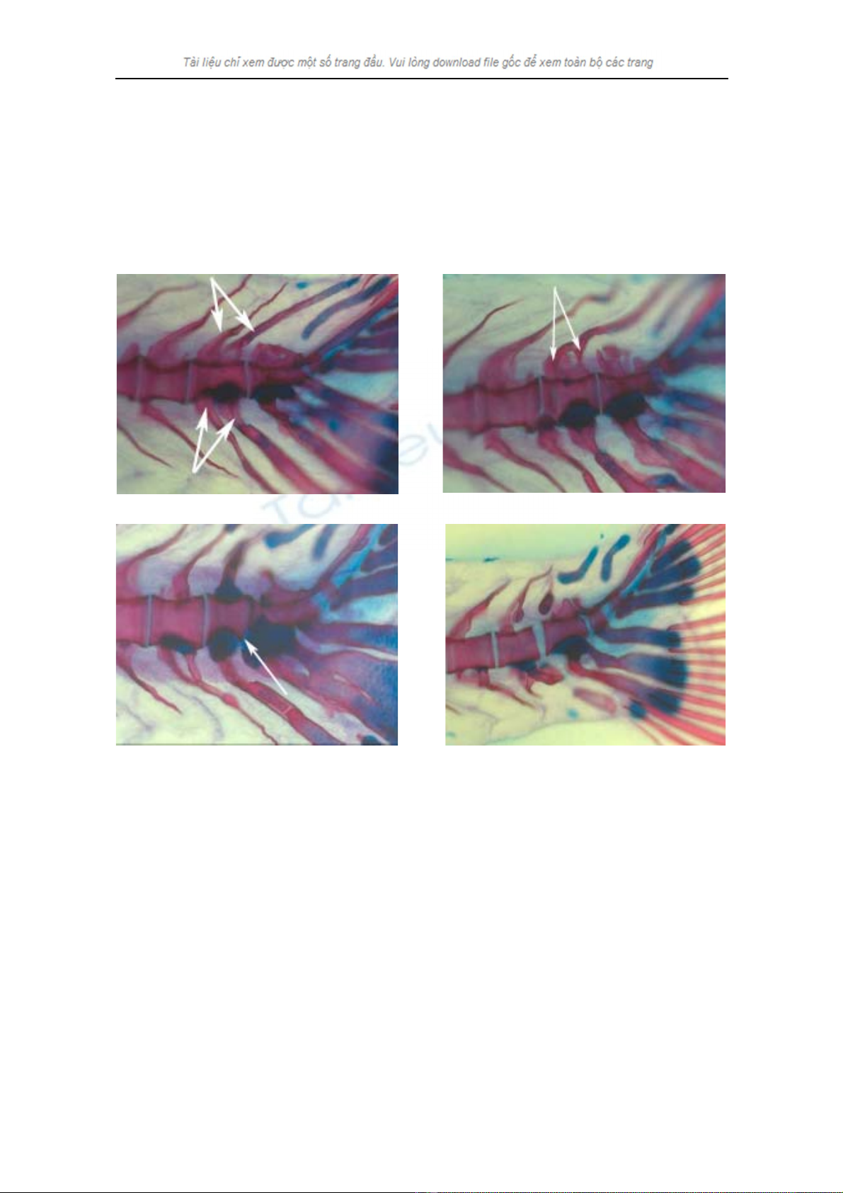
Figure 1. Fusion in caudal vertebrae
(Anterior to the left, Posterior to the right, Dorsal is to the top)
(A) Arrows indicate double Neural Spine (NS) and Haemal Spine (HS) in PU2: 5.5% of fish reared at
32.0°C showed this feature. In addition, 5.5% of fish reared at 32.0 °C and 5% of fish reared at 20.0°C
showed double NS but single HS in PU2;
(B) Arrows indicate fusion of two NS in PU2: 5.5% of fish reared at 32.0°C and 10% of fish reared at
20.0°C showed this feature;
(C) Arrow indicates a fusion of PU2 with [PU1+U1] - 5.5% of the fish reared at 32.0°C and 10% of fish
reared at 20.0°C showed this feature;
(D) No fusion between PU1, U1, U2 - 11.2% of fish reared at 32.0°C showed this feature.
50
Nguyen Thi Hanh Tien, Ann Huysseune and Eckhard Witten
Error!
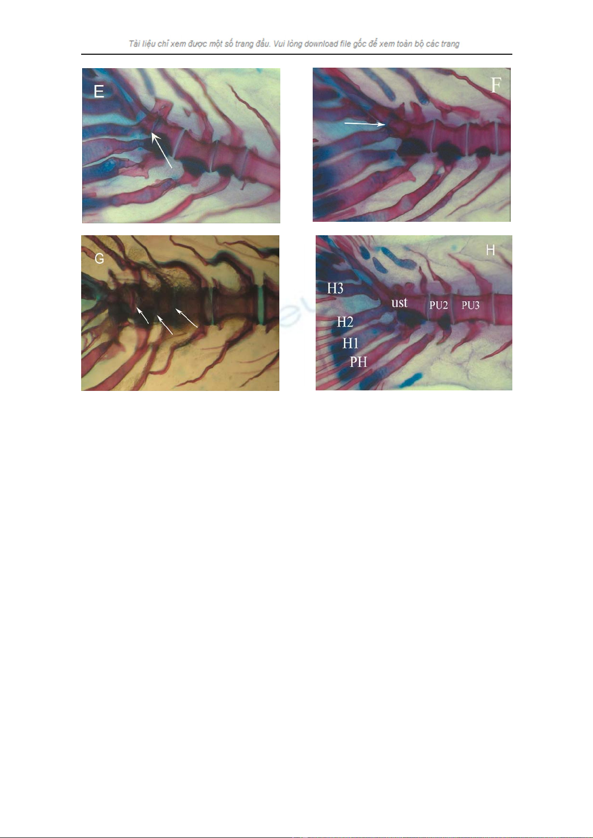
Figure 2. Fusion in caudal and caudal fin vertebrae
(Anterior to the right, Posterior to the left, Dorsal is to the top, PH: Parhypural, H1-3: Hypural 1 to 3)
(E) Arrow indicates a fusion of PU1 and U1: 72.2% of the fish reared at 32.0°C and 35% of the fish
reared at 20.0°C showed this feature;
(F) Arrow indicates incomplete fusion of U2 with [PU1 + U1]: 16.7% of the fish reared at 32.0°C showed
this feature;
(G) Arrows indicate fusion of urostyle with PU2 (10% of the fish reared at 20.0°C), PU2 with PU3 (15%
of the fish reared at 20.0°C) and 20% of the fish reared at 20.0°C showed the fusion of PU3 with last
caudal vertebrate;
(H) Caudal fin vertebrae end with urostyle (ust) (fusion of U2, U1 and PU1): 16.6% of the fish reared at
32.0°C, 65% of the fish reared at 20.0°C and 100% of the fish reared at 26.0 and 28.5°C showed this
feature.
51


%20--%3e%3cdefs%3e%3cstyle%3e%20.st0%20{%20fill:%20%23fff;%20}%20.st1%20{%20fill:%20%237800fa;%20}%20%3c/style%3e%3c/defs%3e%3cpath%20class='st1'%20d='M117.78,12.18H43.11c2.9,3.47,4.65,7.94,4.65,12.82,0,5.6-2.3,10.66-6.01,14.29h76.02l7.22-13.56-7.22-13.56Z'/%3e%3cg%3e%3cpath%20class='st0'%20d='M53.58,26.17h-.59v-1.46h.59v-4.96h2.83c1.78,0,2.67.94,2.67,2.82v5.76c0,1.87-.89,2.81-2.67,2.81h-2.83v-4.96ZM55.36,21.37v3.34h1.1v1.46h-1.1v3.34h1.01c.61,0,.91-.37.91-1.1v-5.93c0-.74-.3-1.1-.91-1.1h-1.01Z'/%3e%3cpath%20class='st0'%20d='M65.99,31.14h-1.8l-.31-2.07h-2.19l-.31,2.07h-1.64l1.82-11.39h2.62l1.82,11.39ZM65.28,18.04c-.25.46-.51.77-.75.94-.21.15-.47.22-.79.22-.26,0-.57-.07-.92-.22l-.38-.15c-.14-.05-.26-.07-.37-.07-.3,0-.53.18-.71.54l-.91-.68c.25-.46.51-.77.75-.94.21-.14.48-.21.79-.21.26,0,.57.07.92.21l.38.15c.14.05.26.07.37.07.3,0,.53-.18.71-.54l.91.68ZM61.91,27.52h1.73l-.87-5.76-.87,5.76Z'/%3e%3cpath%20class='st0'%20d='M74.53,26.89v1.52c0,1.91-.89,2.86-2.67,2.86s-2.67-.95-2.67-2.86v-5.93c0-1.91.89-2.86,2.67-2.86s2.67.95,2.67,2.86v1.11h-1.69v-1.22c0-.75-.31-1.12-.93-1.12s-.93.37-.93,1.12v6.15c0,.74.31,1.11.93,1.11s.93-.37.93-1.11v-1.63h1.69Z'/%3e%3cpath%20class='st0'%20d='M81.4,31.14h-1.8l-.31-2.07h-2.19l-.31,2.07h-1.64l1.82-11.39h2.62l1.82,11.39ZM75.9,19.2l1.52-1.91h1.71l1.51,1.91h-1.61l-.76-.95-.75.95h-1.61ZM77.32,27.52h1.73l-.87-5.76-.87,5.76ZM83.1,15.99l-1.76,1.91h-1.26l1.17-1.91h1.86Z'/%3e%3cpath%20class='st0'%20d='M84.86,19.75c1.78,0,2.67.94,2.67,2.82v1.48c0,1.87-.89,2.81-2.67,2.81h-.85v4.28h-1.79v-11.39h2.64ZM84.01,21.37v3.86h.85c.58,0,.87-.36.87-1.08v-1.71c0-.71-.29-1.07-.87-1.07h-.85Z'/%3e%3cpath%20class='st0'%20d='M93.51,19.75c1.78,0,2.67.94,2.67,2.82v1.48c0,1.87-.89,2.81-2.67,2.81h-.85v4.28h-1.79v-11.39h2.64ZM92.66,21.37v3.86h.85c.58,0,.87-.36.87-1.08v-1.71c0-.71-.29-1.07-.87-1.07h-.85Z'/%3e%3cpath%20class='st0'%20d='M98.8,31.14h-1.79v-11.39h1.79v4.88h2.03v-4.88h1.83v11.39h-1.83v-4.88h-2.03v4.88Z'/%3e%3cpath%20class='st0'%20d='M105.36,24.55h2.46v1.62h-2.46v3.34h3.09v1.63h-4.88v-11.39h4.88v1.63h-3.09v3.18ZM108.17,17.29l-1.76,1.91h-1.26l1.17-1.91h1.86Z'/%3e%3cpath%20class='st0'%20d='M112.2,19.75c1.78,0,2.67.94,2.67,2.82v1.48c0,1.87-.89,2.81-2.67,2.81h-.85v4.28h-1.79v-11.39h2.64ZM111.35,21.37v3.86h.85c.58,0,.87-.36.87-1.08v-1.71c0-.71-.29-1.07-.87-1.07h-.85Z'/%3e%3c/g%3e%3ccircle%20class='st1'%20cx='25'%20cy='25'%20r='20'/%3e%3cpath%20class='st0'%20d='M32.78,19.27c2.92,0,4.43,2.55,5.28,5.33l.71,2.17c.14.38-.33.75-.71.75h-5.61c.19-.33.24-.71.09-1.08l-.75-2.45c-.43-1.32-.99-2.64-1.79-3.77.75-.57,1.65-.94,2.78-.94h0ZM25,18.38c3.25,0,4.9,2.78,5.89,5.89l.76,2.45c.14.42-.33.8-.8.8h-11.69c-.42,0-.94-.38-.8-.8l.75-2.45c.99-3.11,2.64-5.89,5.89-5.89h0ZM25,11.35c1.74,0,3.11,1.37,3.11,3.11s-1.37,3.11-3.11,3.11-3.11-1.41-3.11-3.11,1.41-3.11,3.11-3.11h0ZM17.27,19.27c1.08,0,1.98.38,2.73.94-.8,1.13-1.37,2.45-1.74,3.77l-.8,2.45c-.14.38-.05.75.09,1.08h-5.56c-.42,0-.9-.38-.75-.75l.71-2.17c.9-2.78,2.41-5.33,5.33-5.33h0ZM17.27,12.91c1.51,0,2.78,1.27,2.78,2.83s-1.27,2.83-2.78,2.83-2.83-1.27-2.83-2.83,1.27-2.83,2.83-2.83h0ZM32.78,12.91c1.56,0,2.78,1.27,2.78,2.83s-1.23,2.83-2.78,2.83-2.83-1.27-2.83-2.83,1.27-2.83,2.83-2.83h0ZM27.07,28.56v.09c0,.57-.24,1.08-.61,1.46h0v.05c-.38.33-.9.57-1.46.57s-1.08-.24-1.46-.61h0c-.38-.38-.61-.9-.61-1.46v-.09h1.41v.09c0,.19.05.38.19.47v.05c.09.09.28.19.47.19s.38-.09.47-.19v-.05c.14-.09.24-.28.24-.47t-.05-.09h1.41ZM30.99,28.56v.09c0,1.65-.66,3.16-1.74,4.24-1.08,1.08-2.59,1.79-4.24,1.79s-3.16-.71-4.24-1.79l-.05-.05c-1.04-1.08-1.7-2.55-1.7-4.2v-.09h1.41v.09c0,1.27.47,2.4,1.27,3.25h.05c.85.85,1.98,1.37,3.25,1.37s2.4-.52,3.25-1.37c.85-.8,1.37-1.98,1.37-3.25v-.09h1.37ZM34.99,28.56v.09c0,2.78-1.13,5.28-2.92,7.07-1.79,1.79-4.29,2.92-7.07,2.92s-5.23-1.13-7.07-2.92c-1.79-1.79-2.92-4.29-2.92-7.07v-.09h1.41v.09c0,2.4.94,4.53,2.5,6.08,1.56,1.56,3.72,2.5,6.08,2.5s4.52-.94,6.08-2.5c1.56-1.56,2.5-3.68,2.5-6.08v-.09h1.41Z'/%3e%3c/svg%3e)