
SHORT REPOR T Open Access
Identification of host proteins associated with
HIV-1 preintegration complexes isolated from
infected CD4
+
cells
Nidhanapati K Raghavendra
1
, Nikolozi Shkriabai
2
, Robert LJ Graham
3
, Sonja Hess
3
, Mamuka Kvaratskhelia
2
,LiWu
1*
Abstract
An integrated HIV-1 genomic DNA leads to an infected cell becoming either an active or a latent virus-producing
cell. Upon appropriate activation, a latently infected cell can result in production of progeny viruses that spread the
infection to uninfected cells. The host proteins influence several steps of HIV-1 infection including formation of the
preintegration complex (PIC), a key nucleoprotein intermediate essential for integration of reverse transcribed viral
DNA into the chromosome. Much effort has gone into the identification of host proteins contributing to the
assembly of functional PICs. Experimental approaches included the use of yeast two-hybrid system, co-immunopre-
cipitation, affinity tagged HIV-1 viral proteins and in vitro reconstitution of salt-stripped PIC activity. Several host
proteins identified using these approaches have been shown to affect HIV-1 replication in cells and influence cata-
lytic activities of recombinant IN in vitro. However, the comprehensive identification and characterization of host
proteins associated with HIV-1 PICs of infected cells have been hindered in part by the technical limitation in
acquiring sufficient amount of catalytically active PICs. To efficiently identify additional host factors associated with
PICs in infected cells, we have developed the following novel approach. The catalytically active PICs from HIV-1-
infected CD4
+
cells were isolated using biotinylated target DNA, and the proteins selectively co-purifying with PICs
have been analyzed by mass spectrometry. This technology enabled us to reveal at least 19 host proteins that are
associated with HIV-1 PICs, of which 18 proteins have not been described previously with respect to HIV-1 integra-
tion. Physiological functions of the identified proteins range from chromatin organization to protein transport. A
detailed characterization of these host proteins could provide new insights into the mechanism of HIV-1 integra-
tion and uncover new antiviral targets to block HIV-1 integration.
Findings
Human immunodeficiency virus type 1 (HIV-1) inte-
grase (IN) is a 288 amino-acid protein with three func-
tional domains: N-terminal domain (NTD), catalytic
core domain (CCD) and C-terminal domain (CTD). The
NTD contains a zinc binding motif, the CCD has three
acidic residues, D64, D116 and E152, which co-ordinate
the catalytic divalent metal ions; and the CTD is sug-
gested to nonspecifically bind the DNA substrate [1]. IN
catalyzes two endonucleolytic reactions - 3′processing:
the removal of two deoxynucleotides from viral DNA
ends; and DNA strand transfer: the covalent ligation of
viral DNA 3′ends to host chromosomal DNA. While a
recombinant IN can catalyze 3′processing and strand
transfer reactions [2], the activity of HIV-1 integrase in
the context of preintegration complex (PIC) is assisted
and modulated by several host factors during proviral
DNA formation. The PIC is thought to be derived from
the reverse transcription complex and consists of the
full length viral DNA and both viral and host proteins
that participate in generation of the proviral DNA [3,4].
The PIC formed following reverse transcription is in
limiting amounts to permit biochemical purification of
the pure complexes and identification of constituent
proteins [5]. Previous studies to identify IN-interacting
host proteins have primarily used yeast two-hybrid sys-
tem and co-immunoprecipitations involving ectopically
expressed viral and host proteins (Table 1). Another
approach has been the in vitro reconstitution of salt-
stripped PIC activity (PICs treated with high salt result
* Correspondence: wu.840@osu.edu
1
Center for Retrovirus Research, Department of Veterinary Biosciences, The
Ohio State University, Columbus, Ohio 43210, USA
Full list of author information is available at the end of the article
Raghavendra et al.Retrovirology 2010, 7:66
http://www.retrovirology.com/content/7/1/66
© 2010 Raghavendra et al; licensee BioMed Central Ltd. This is an Open Access article distributed under the terms of the Creative
Commons Attribution License (http://creativecommons.org/licenses/by/2.0), which permits unrestricted use, distribution, and
reproduction in any medium, provided the original work is properly cited.
in integration-defective complexes) using purified or
recombinant host proteins. These approaches have
helped to identify host proteins that physically interact
with HIV-1 IN or stimulate HIV-1 IN catalytic activity.
A recent study involving use of a biotinylated IN as a
tool to detect interacting host proteins concluded that
activity of the modified IN was adversely affected [6].
In the current study, a novel approach to identify the
host proteins associated with PIC is presented. The pro-
tocol involves using a biotinylated target DNA in the
standard in vitro PIC reaction assay, and the isolation of
the protein complex covalently attached to target DNA
using streptavidin beads (Figure 1). As a stable complex
that is imported into the nucleus for integration into
host chromosome, it is possible that the proteins
associated with the HIV-1 DNA remain bound even
after catalysis of the integration into a biotinylated target
DNA. This assumption is the basis of the approach
described here. A well-established protocol has been
used to isolate the cytoplasmicPICs(whichisacyto-
plasmic extract of HIV-1-infected cells) and perform an
in vitro integration assay [6]. The H9/HTLVIIIB cell line
is a chronically HIV-1 infected H9-derived CD4
+
cell
line that releases infectious HIV-1 into the culture
supernatant [7]. Stimulation of the H9/HTLVIIIB cell
line with phorbol 12-myristate 13-acetate (PMA)
increases viral production several fold and also increases
the cell-to-cell transmission of the virus in co-culture
experiments [6]. The parental H9 cells that do not pro-
duce HIV-1 were used as a negative control. Co-culture
of HIV-1 producing H9/HTLVIIIB cells with HIV-1 sus-
ceptible cells such as CD4
+
SupT1 cells typically leads
to a high proportion of infected cells. SupT1 cells (2.5 ×
10
9
) were co-cultured for 6 hours with PMA-treated
H9/HTLVIIIB or H9 cells (2.5 × 10
8
) in the supernatant
(600 ml) obtained from a 24 hour culture of H9/
HTLVIIIB or H9 cells, respectively. (All cells were
grown to a density of 1-1.5 × 10
6
per ml prior to co-cul-
ture). The PICs isolated from such co-cultured cells
were demonstrated to exhibit high integration activity
into naked plasmid DNA [8]. The cytoplasmic PICs gen-
erated here (isolated in 50 ml of digitonin-containing
lysis buffer) have been used for in vitro integration into
a ~1.5 kb biotinylated target DNA (100 μg, prepared by
PCR amplification of non-viral DNA in pNL4-3 plasmid
using the following primer pair: Biotin - 5′CAA AGT
GCT GGG ACA ACC GGG 3′and 5′GCG CTC GGC
CCT TCC GGC TGG C 3′). At the end of the integra-
tion assay, the biotinylated target DNA-PIC complex
was bound to streptavidin magnetic beads (MyOne
Streptavidin T1 beads, Invitrogen) at room temperature
for 30 minutes (Figure 2A). To efficiently remove the
majority of non-specific proteins bound to the streptavi-
din beads, 10 washes of 15 ml each were performed
using the assay buffer [5]. The use of a biotinylated tar-
get DNA to isolate the PIC precludes the requirement
of a tagged HIV-1 protein, and the potential alteration
of protein-protein interactions or activity caused by a
tagged-protein. The isolation of the DNA-protein com-
plex based on the catalytic activity of PIC makes it pos-
sible to identify physiologically relevant viral-host
protein interactions.
The integration activity of the isolated complex was
confirmed by real-time PCR analysis using primers spe-
cific to the target and viral DNA [5]. As expected, no
activity was detected either in the absence of a target
DNA or with the cytoplasmic extract from the SupT1-
H9 co-culture as compared to the activity of PICs iso-
lated from the SupT1-H9/HTLVIIIB co-culture (set to
Table 1 Summary of previously characterized host
proteins interacting with HIV-1 IN
Host proteins Methods References
BAF SS [11]
Gemin2 IP [14]
HAT p300 IP [26]
HMGA1 SS [8]
HSP 60 PD [27]
Human EED protein THS; PD [28]
Importin 7 IP [13]
Integrase interactor 1 THS [29]
LEDGF/p75 IP [30]
UNG2 PD [31]
A list of host proteins interacting with HIV-1 integrase and the experimental
procedure used to identify the protein-protein interaction. THS: two hybrid
system; PD: Pull down; SS: reconstitution of salt stripped PIC activity; IP:
immunoprecipitation.
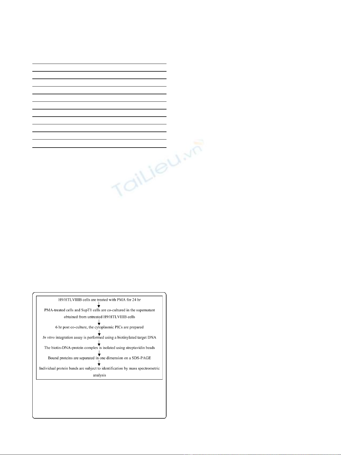
Figure 1 Experimental design for the identification of host
proteins associated with HIV-1 PIC. The PICs were generated
following a protocol described previously [5]. PICs covalently bound
to biotinylated DNA were isolated using streptavidin beads. The
proteins in the isolated complex were identified by mass
spectrometric analysis. PMA: phorbol 12-myristate 13-acetate.
Raghavendra et al.Retrovirology 2010, 7:66
http://www.retrovirology.com/content/7/1/66
Page 2 of 7
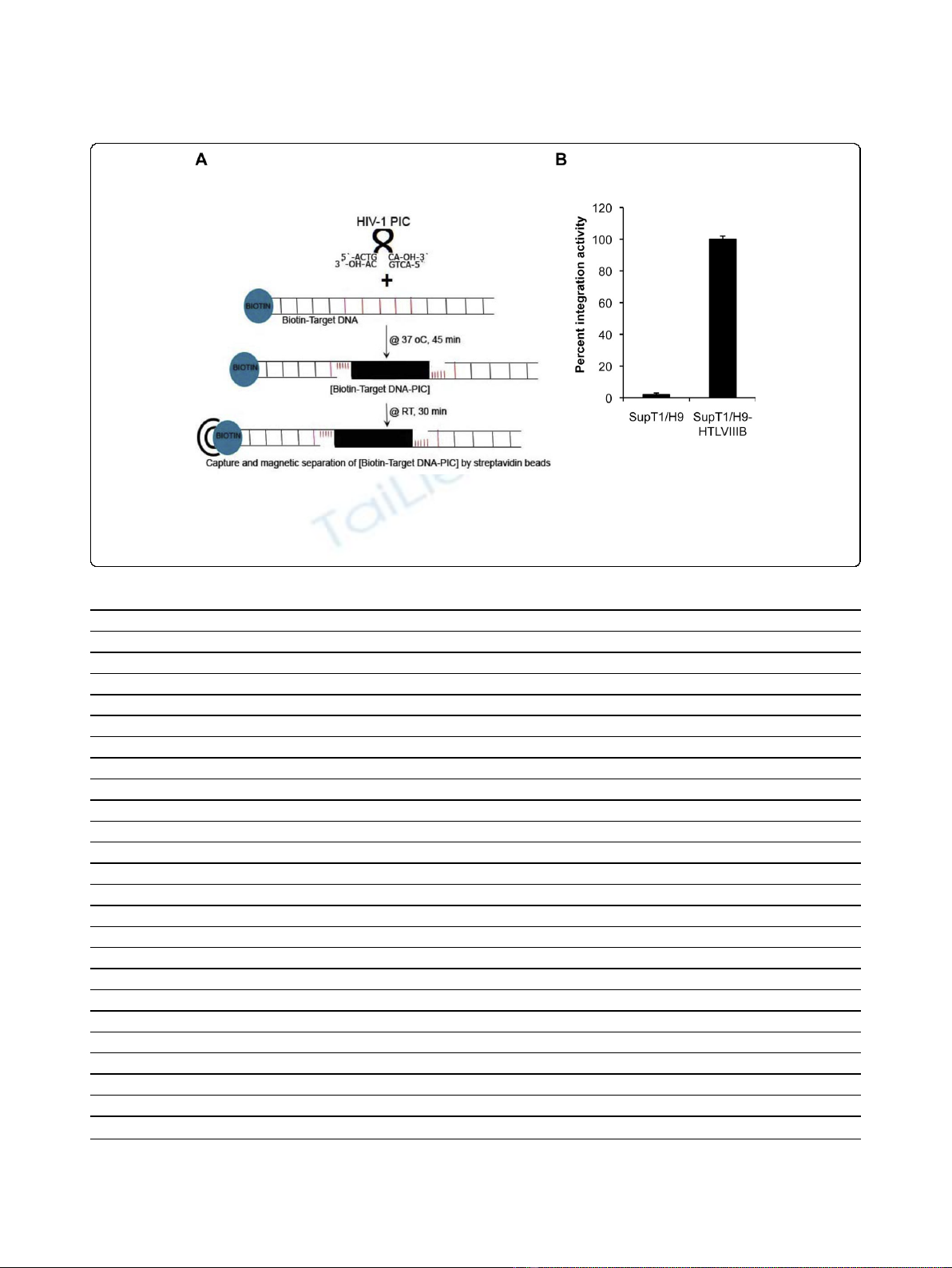
Figure 2 Isolation of HIV-1 PICs and their activity. (A) Magnetic separation of functional HIV-1 PICs. The PIC integrates HIV-1 DNA into the ~1.5
kb biotinylated non-viral DNA from pNL4-3 plasmid that serves as a target DNA in the in vitro assay. The biotin-target DNA-protein complex is then
isolated using streptavidin magnetic beads after incubation at room temperature (RT) for 30 minutes. (B) Integration activity of HIV-1 PICs. The
integration activity of the PICs isolated from SupT1/H9-HTLVIIIB cell co-cultures (HIV-1-infected, set to 100 percent) and control cytoplasmic extract
from SupT1/H9 cell co-cultures (control cells), using biotin-target DNA, is shown. The analysis was performed as described previously [5].
Table 2 Host proteins selectively co-purifying with HIV-1 PICs
Host Proteins Accession numbers Molecular Weight No. of peptides
Chromatin organization
Barrier-to-autointegration factor baf_bovin 10 kDa 3
Nucleosome assembly protein 1-like 1 np1l1_bovin 45 kDa 5
Histone-binding protein RBBP4 rbbp4_bovin 48 kDa 3
Transcription regulation
Acidic leucine-rich nuclear phosphoprotein 32 family member A an32a_bovin 29 kDa 4
Acidic leucine-rich nuclear phosphoprotein 32 family member E an32e_human 31 kDa 3
Calreticulin calr_cerae 48 kDa 5
NF-kappa-B essential modulator nemo_bovin 49 kDa 7
Non-POU domain-containing octamer-binding protein nono_human 54 kDa 8
RNA polymerase-associated protein LEO1 leo1_human 75 kDa 6
RNA processing/localization
ATP-dependent RNA helicase DDX19A dd19a_bovin 54 kDa 4
Double-stranded RNA-binding protein Staufen homolog 1 stau1_human 63 kDa 2
Heterogeneous nuclear ribonucleoprotein H1 hnrh1_human 49 kDa 2
Heterogeneous nuclear ribonucleoprotein H3 hnrh3_human 37 kDa 3
Plasminogen activator inhibitor 1 RNA-binding protein pairb_human 45 kDa 7
Splicing factor 3B subunit 2 sf3b2_human 98 kDa 19
Splicing factor, arginine/serine-rich 3 sfrs3_bovin 19 kDa 3
U4/U6.U5 tri-snRNP-associated protein 1 snut1_human 90 kDa 17
Translation
Eukaryotic translation initiation factor 4 gamma 1 if4g1_human 176 kDa 3
Cytoplasmic trafficking
Dynactin subunit 2 dctn2_human 44 kDa 3
The host proteins have been broadly categorized based on the known physiological function. The number of peptides identified by mass spectroscopic analysis
and assigned to the specific protein in the database is shown.
Raghavendra et al.Retrovirology 2010, 7:66
http://www.retrovirology.com/content/7/1/66
Page 3 of 7
100%) (Figure 2B). Proteins from complex mixtures such
as cell lysates have been identified successfully by using
mass spectrometric (MS) techniques [9]. The proteins
from the complexes bound to streptavidin beads were
eluted by boiling the beads in 30 μloftheSDS-PAGE
running buffer at 95°C for 5 minutes. The eluted-boiled
proteins were loaded into a single well of a 4-15%
gradient SDS-PAGE gel (Bio-Rad), and the proteins
were separated in one dimension. Differences between
the SupT1-H9/HTLVIIIB and SupT1-H9 co-culture
samples could not be readily delineated from visual
inspection of Coomassie Blue stained gels. This is not
surprising considering the minute amounts of PIC pro-
teins in an infected cell and the several cellular proteins
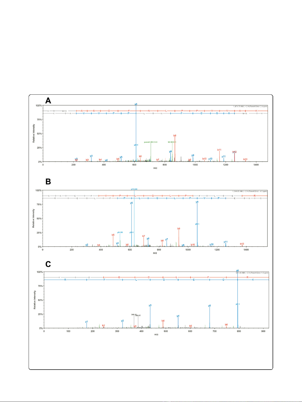
Figure 3 Representative MS/MS data for HIV-1 and host proteins associated with PICs. (A) HIV-1 integrase peptide AMASDFNLPPVVAK. (B)
HIV-1 Rev peptide SAEPVPLQLPPLER. (C) Cellular barrier-to-autointegration factor (BAF) peptide KDEDLFR. The ‘b”and “y”ion series derived from
the amide bond cleavage during collision induced dissociation of the peptide provide amino acid sequence information. The b-ion series
(shown in red) is read from the N-terminus to C-terminus, while the y-ion series (shown in blue) is read from the C-terminus to N-terminus,
providing thus complementary sequence information [25]. Other minor fragments resulted from peptide fragmentations at other sites are shown
in green.
Raghavendra et al.Retrovirology 2010, 7:66
http://www.retrovirology.com/content/7/1/66
Page 4 of 7

that can bind non-specifically to DNA or biotin or
streptavidin [10]. To identify the proteins associated
specifically with PIC, the protein bands ranging in size
from 10 kDa to 250 kDa were sliced into ~ 25 indivi-
dual gel pieces and subjected to semi-quantitative MS
analysis. The false discovery rate as determined using
Peptide and Protein Prophet methods was less than
0.6% for all proteins identified. Given the reduced sam-
ple complexity, undersampling was not observed.
The output from the MS analysis of two independent
experiments of SupT1-H9/HTLVIIIB co-culture infec-
tions was compared against that of the SupT1-H9 co-cul-
ture control. The list of proteins present in SupT1-H9
co-culture experiment serves to eliminate the non-specifi-
cally binding cytoplasmic proteins from those identified
in the SupT1-H9/HTLVIIIB co-culture samples. For a
more sensitive and accurate analysis of the proteins asso-
ciated with HIV-1 PICs, the following criteria have been
employed: (a) identification of at least two peptides from
each protein, and (b) identification of the protein in two
independent SupT1-H9/HTLVIIIB co-culture samples. A
total of 19 host proteins (~ 6% of the total proteins
revealed by the MS analysis) were identified to be specifi-
cally associated with the HIV-1 PICs (Table 2). While
barrier-to-autointegration factor (BAF) is the only host
protein that was characterized previously [11,12], the
identification of 18 new host proteins associated with
HIV-1 PICs reflects the uniqueness of our approach.
Two previously characterized proteins, Importin 7 [13]
and Gemin2 [14] were detected in one of the two
SupT1-H9/HTLVIIIB co-culture samples, cautioning that
some of the characterized and uncharacterized proteins
associated with PICs might not have been identified due
to detection limits of MS. Lamina-associated polypeptide
2 isoform alpha (LAP2a) protein [15] was identified in
the SupT1-H9/HTLVIIIB co-culture samples; however,
its presence in SupT1-H9 samples suggests a non-specific
interaction with biotinylated DNA. The integrase inter-
acting protein, LEDGF/p75 (lens epithelium-derived
growth factor), was not identified in the cytoplasmic
PICs. The current analysis is limited to the identification
of proteins associated with PIC assembling in the cyto-
plasm. A similar analysis of nuclear PICs is expected to
reveal proteins such as LEDGF/p75 that function at the
site of integration in the nucleus [16]. Importantly, the
peptides corresponding to two HIV-1 proteins, IN and
Rev were identified in SupT1-H9/HTLVIIIB co-culture
samples (Figure 3). Recently, Rev has been suggested to
regulate HIV-1 integration in infected cells based on its
ability to interact with both IN and LEDGF/p75 [17]. Rev
could therefore potentially contribute to the formation of
PICs. Figure 4 shows a representative immunoblotting of
SupT1-H9 and SupT1-H9/HTLVIIIB samples confirming
the host proteins that specifically associated with HIV-1
PICs.
Of the host proteins identified here to be specifically
associated with HIV-1 PICs, histone-binding protein
RBBP4 is known to influences transcription activation
by facilitating histone acetylation [18], and non-POU
domain-containing octamer-binding protein is charac-
terized to function with respect to double strand DNA
break repair [19]. Nucleosome assembly protein 1-like 1
protein has been shown to interact with HIV-1 Tat and
promote viral transcription [20], while splicing factor 3B
subunit 2 protein interacts with HIV-1 Vpr and activates
G2 checkpoint activation [21]. Moreover, the double-
stranded RNA-binding protein Staufen homolog 1 is
incorporated in HIV-1 and plays a role in viral genomic
RNA encapsidation and viral particle assembly [22-24].
It is tempting to speculate that such factors might play
a role in the assembly of PICs and assist the formation
of proviral DNA similar to that of BAF or LEDGF/p75
andfulfilltherolesnotattributedtothepreviously
characterized host factors. A detailed characterization of
the host proteins identified here is essential to elucidate
their role in HIV integration and to verify their potential
Figure 4 Immunoblotting for host proteins that are specifically
associated with HIV-1 PIC. The proteins bound to the streptavidin
magnetic beads after integration assay were probed with specific
antibodies. SupT1-H9/HTLVIIIB represents HIV-1 infected cell samples,
and SupT1-H9 represents non-infected control samples. The host
proteins are indicated by accession names on the left. The ‘NONO’is
Non-POU domain-containing octamer-binding protein, ‘NP1L1’is
Nucleosome assembly protein 1-like 1 protein and ‘CALR’is
Calreticulin for which 8, 5 and 5 peptides were identified by MS
analysis respectively. Beta-actin found in both samples is also shown.
Raghavendra et al.Retrovirology 2010, 7:66
http://www.retrovirology.com/content/7/1/66
Page 5 of 7


%20--%3e%3cdefs%3e%3cstyle%3e%20.st0%20{%20fill:%20%23fff;%20}%20.st1%20{%20fill:%20%237800fa;%20}%20%3c/style%3e%3c/defs%3e%3cpath%20class='st1'%20d='M117.78,12.18H43.11c2.9,3.47,4.65,7.94,4.65,12.82,0,5.6-2.3,10.66-6.01,14.29h76.02l7.22-13.56-7.22-13.56Z'/%3e%3cg%3e%3cpath%20class='st0'%20d='M53.58,26.17h-.59v-1.46h.59v-4.96h2.83c1.78,0,2.67.94,2.67,2.82v5.76c0,1.87-.89,2.81-2.67,2.81h-2.83v-4.96ZM55.36,21.37v3.34h1.1v1.46h-1.1v3.34h1.01c.61,0,.91-.37.91-1.1v-5.93c0-.74-.3-1.1-.91-1.1h-1.01Z'/%3e%3cpath%20class='st0'%20d='M65.99,31.14h-1.8l-.31-2.07h-2.19l-.31,2.07h-1.64l1.82-11.39h2.62l1.82,11.39ZM65.28,18.04c-.25.46-.51.77-.75.94-.21.15-.47.22-.79.22-.26,0-.57-.07-.92-.22l-.38-.15c-.14-.05-.26-.07-.37-.07-.3,0-.53.18-.71.54l-.91-.68c.25-.46.51-.77.75-.94.21-.14.48-.21.79-.21.26,0,.57.07.92.21l.38.15c.14.05.26.07.37.07.3,0,.53-.18.71-.54l.91.68ZM61.91,27.52h1.73l-.87-5.76-.87,5.76Z'/%3e%3cpath%20class='st0'%20d='M74.53,26.89v1.52c0,1.91-.89,2.86-2.67,2.86s-2.67-.95-2.67-2.86v-5.93c0-1.91.89-2.86,2.67-2.86s2.67.95,2.67,2.86v1.11h-1.69v-1.22c0-.75-.31-1.12-.93-1.12s-.93.37-.93,1.12v6.15c0,.74.31,1.11.93,1.11s.93-.37.93-1.11v-1.63h1.69Z'/%3e%3cpath%20class='st0'%20d='M81.4,31.14h-1.8l-.31-2.07h-2.19l-.31,2.07h-1.64l1.82-11.39h2.62l1.82,11.39ZM75.9,19.2l1.52-1.91h1.71l1.51,1.91h-1.61l-.76-.95-.75.95h-1.61ZM77.32,27.52h1.73l-.87-5.76-.87,5.76ZM83.1,15.99l-1.76,1.91h-1.26l1.17-1.91h1.86Z'/%3e%3cpath%20class='st0'%20d='M84.86,19.75c1.78,0,2.67.94,2.67,2.82v1.48c0,1.87-.89,2.81-2.67,2.81h-.85v4.28h-1.79v-11.39h2.64ZM84.01,21.37v3.86h.85c.58,0,.87-.36.87-1.08v-1.71c0-.71-.29-1.07-.87-1.07h-.85Z'/%3e%3cpath%20class='st0'%20d='M93.51,19.75c1.78,0,2.67.94,2.67,2.82v1.48c0,1.87-.89,2.81-2.67,2.81h-.85v4.28h-1.79v-11.39h2.64ZM92.66,21.37v3.86h.85c.58,0,.87-.36.87-1.08v-1.71c0-.71-.29-1.07-.87-1.07h-.85Z'/%3e%3cpath%20class='st0'%20d='M98.8,31.14h-1.79v-11.39h1.79v4.88h2.03v-4.88h1.83v11.39h-1.83v-4.88h-2.03v4.88Z'/%3e%3cpath%20class='st0'%20d='M105.36,24.55h2.46v1.62h-2.46v3.34h3.09v1.63h-4.88v-11.39h4.88v1.63h-3.09v3.18ZM108.17,17.29l-1.76,1.91h-1.26l1.17-1.91h1.86Z'/%3e%3cpath%20class='st0'%20d='M112.2,19.75c1.78,0,2.67.94,2.67,2.82v1.48c0,1.87-.89,2.81-2.67,2.81h-.85v4.28h-1.79v-11.39h2.64ZM111.35,21.37v3.86h.85c.58,0,.87-.36.87-1.08v-1.71c0-.71-.29-1.07-.87-1.07h-.85Z'/%3e%3c/g%3e%3ccircle%20class='st1'%20cx='25'%20cy='25'%20r='20'/%3e%3cpath%20class='st0'%20d='M32.78,19.27c2.92,0,4.43,2.55,5.28,5.33l.71,2.17c.14.38-.33.75-.71.75h-5.61c.19-.33.24-.71.09-1.08l-.75-2.45c-.43-1.32-.99-2.64-1.79-3.77.75-.57,1.65-.94,2.78-.94h0ZM25,18.38c3.25,0,4.9,2.78,5.89,5.89l.76,2.45c.14.42-.33.8-.8.8h-11.69c-.42,0-.94-.38-.8-.8l.75-2.45c.99-3.11,2.64-5.89,5.89-5.89h0ZM25,11.35c1.74,0,3.11,1.37,3.11,3.11s-1.37,3.11-3.11,3.11-3.11-1.41-3.11-3.11,1.41-3.11,3.11-3.11h0ZM17.27,19.27c1.08,0,1.98.38,2.73.94-.8,1.13-1.37,2.45-1.74,3.77l-.8,2.45c-.14.38-.05.75.09,1.08h-5.56c-.42,0-.9-.38-.75-.75l.71-2.17c.9-2.78,2.41-5.33,5.33-5.33h0ZM17.27,12.91c1.51,0,2.78,1.27,2.78,2.83s-1.27,2.83-2.78,2.83-2.83-1.27-2.83-2.83,1.27-2.83,2.83-2.83h0ZM32.78,12.91c1.56,0,2.78,1.27,2.78,2.83s-1.23,2.83-2.78,2.83-2.83-1.27-2.83-2.83,1.27-2.83,2.83-2.83h0ZM27.07,28.56v.09c0,.57-.24,1.08-.61,1.46h0v.05c-.38.33-.9.57-1.46.57s-1.08-.24-1.46-.61h0c-.38-.38-.61-.9-.61-1.46v-.09h1.41v.09c0,.19.05.38.19.47v.05c.09.09.28.19.47.19s.38-.09.47-.19v-.05c.14-.09.24-.28.24-.47t-.05-.09h1.41ZM30.99,28.56v.09c0,1.65-.66,3.16-1.74,4.24-1.08,1.08-2.59,1.79-4.24,1.79s-3.16-.71-4.24-1.79l-.05-.05c-1.04-1.08-1.7-2.55-1.7-4.2v-.09h1.41v.09c0,1.27.47,2.4,1.27,3.25h.05c.85.85,1.98,1.37,3.25,1.37s2.4-.52,3.25-1.37c.85-.8,1.37-1.98,1.37-3.25v-.09h1.37ZM34.99,28.56v.09c0,2.78-1.13,5.28-2.92,7.07-1.79,1.79-4.29,2.92-7.07,2.92s-5.23-1.13-7.07-2.92c-1.79-1.79-2.92-4.29-2.92-7.07v-.09h1.41v.09c0,2.4.94,4.53,2.5,6.08,1.56,1.56,3.72,2.5,6.08,2.5s4.52-.94,6.08-2.5c1.56-1.56,2.5-3.68,2.5-6.08v-.09h1.41Z'/%3e%3c/svg%3e)