
BioMed Central
Page 1 of 6
(page number not for citation purposes)
Journal of Immune Based Therapies
and Vaccines
Open Access
Original research
CTLA-4 blockade during dendritic cell based booster vaccination
influences dendritic cell survival and CTL expansion
Anders E Pedersen*1 and Franca Ronchese2
Address: 1Department of International Health, Immunology and Microbiology, The Panum Institute, University of Copenhagen, Denmark and
2Malaghan Institute of Medical Research, Wellington, New Zealand
Email: Anders E Pedersen* - elmpedersen@hotmail.com; Franca Ronchese - fronchese@malaghan.org.nz
* Corresponding author
Abstract
Dendritic cells (DCs) are potent antigen-presenting cells and critical for the priming of CD8+ T
cells. Therefore the use of these cells as adjuvant cells has been tested in a large number of
experimental and clinical vaccination studies, in particular cancer vaccine studies. A number of
protocols are emerging that combine vaccination with CTL expanding strategies, such as e.g.
blockade of CTLA-4 signalling. On the other hand, the lifespan and in vivo survival of therapeutic
DCs have only been addressed in a few studies, although this is of importance for the kinetics of
CTL induction during vaccination. We have previously reported that DCs loaded with specific
antigens are eliminated by antigen specific CTLs in vivo and that this elimination affects the potential
for in vivo CTL generation. We now show that CTLA-4 blockade increases the number of DC
vaccine induced LCMV gp33 specific CTLs and the lysis of relevant in vivo targets. However, the
CTLA-4 blockage dependent expansion of CTLs also affect DC survival during booster DC
injections and our data suggest that during a booster DC vaccine, the largest increase in CTL levels
is already obtained during the first vaccination.
Background
Dendritic cells are sentinel cells in the peripheral tissues.
After exposure to inflammatory cytokines together with
pathogen associated molecular patterns they undergo
maturation, migrate to the regional lymph nodes and ini-
tiate CD4+ and CD8+ T cells responses [1-3]. In particular
the potent priming of CD8+ T cells into CTLs with the
capacity for recognition and killing of target cells has
attracted much attention in cancer vaccination protocols
[4,5].
A number of strategies have been identified for the expan-
sion of CTL's such as PD-1 ligand blockade [6], agonistic
4-1BB monoclonal antibody [7] and CTLA-4 blockade.
CTLA-4 normally competes with CD28 for CD80 and
CD86 binding and thereby acts as a negative regulator of
T cell activation [8]. In addition CTLA-4 is expressed by
CD4+CD25+ natural occurring regulatory T cells which in
this way inhibit DC function and bystander T cells [9-11].
CTLA-4 blockade is therefore a potent strategy for the
amplification of immune responses against weak anti-
gens, e.g. tumour antigens, during vaccination [12-14]
and is currently being tested in clinical cancer trials
[15,16].
The survival of injected DC is of critically importance for
the in vivo induction of CTLs during DC based vaccina-
tion. We have previously shown that in antigen primed
mice, injected DCs are eliminated before they reach the
draining lymph node (DLN) and their interaction with
Published: 29 July 2007
Journal of Immune Based Therapies and Vaccines 2007, 5:9 doi:10.1186/1476-8518-5-9
Received: 10 May 2007
Accepted: 29 July 2007
This article is available from: http://www.jibtherapies.com/content/5/1/9
© 2007 Pedersen and Ronchese; licensee BioMed Central Ltd.
This is an Open Access article distributed under the terms of the Creative Commons Attribution License (http://creativecommons.org/licenses/by/2.0),
which permits unrestricted use, distribution, and reproduction in any medium, provided the original work is properly cited.

Journal of Immune Based Therapies and Vaccines 2007, 5:9 http://www.jibtherapies.com/content/5/1/9
Page 2 of 6
(page number not for citation purposes)
memory or naïve T cells is therefore limited at this site.
This elimination is performed by activated CD8+ T cells
and is dependent on perforin secreted from these cells
[17,18]. Under normal physiological conditions this
probably acts as a feedback mechanism to prevent exag-
gerated expansion of CD8+ T cells during a viral infection
[19,20], but the phenomenon might at the same time
limit the potential of DC based vaccines in therapeutic set-
tings [18,21].
We now show that CTLA-4 blockade increases the number
of DC vaccine induced CTLs and the lysis of in vivo target
cells. However, antigen-loaded DCs are eliminated after
repeated injection in primed animals and the CTLA-4
blockage dependent expansion of CTLs leads to a decrease
in surviving DC reaching the lymph node after a second
DC injection. Our data suggest that repeated DC vaccine
combined with e.g. CTLA-4 blockade does not increase
the CTL expansion over time due to elimination of
injected DCs in a primed host whereas CTLA-4 blockade
provide a potent increase in CTL numbers when delivered
together with the primary DC vaccination.
Methods
Mice
Conventional 6–8 week old female C57Bl/6 mice were
purchased from Taconic Europe (Ry, Denmark) and kept
under controlled microbial conditions at the local animal
facility.
Generation of BM-DC
DCs were generated from BM cells derived from C57Bl/6
mice. BM-cells from femurs and tibias were washed and
cultured overnight in 6-well plates (TPP, Trasadingen,
Schwitzerland) at 2 × 10⁶ cells/ml in 3 ml culture
medium/well. Culture medium (CM) was RPMI-1640
with Glutamax supplemented with 10% FCS (Harlan
Sera-Lab Ltd, Hillcrest, England) and antibiotics. The next
day, non-adherent cells were harvested and resuspended
in CM containing 10 ng/ml GM-CSF plus 20 ng/ml IL-4
(both from Peprotech, Rocky Hill, NJ, USA) and cultured
at 1 × 10⁶ cells/ml in 3 ml CM/well. Fresh cytokines and
medium were added on day 3. Day 6 DCs were harvested
as non-adherent and loosely adherent cells. These cells
have previously been described to be 60–90 % CD11c
positive cells with DC characteristics [22].
Immunization with BM-DC and CTLA-4 blockade
Day 6 DC were harvested and incubated with 40 µM of
the H-2 Dbbinding 33–41 fragment of LCMV glycoprotein
gp33 KAVYNFATM peptide (from Schäfer-N, Copenha-
gen, Denmark) for 2 hours at 37°C and then adminis-
tered by subcutaneous injection as 1*106 cells/mouse. The
hybridoma 9H10 which produces monoclonal hamster
anti mouse CTLA-4 antibody was kindly provided by Dr.
Rienk Offringa and has been described previously [14].
The 9H10 antibody was administered as 100 µg/mouse
i.p. on the first day together with DC vaccination and 50
µg/mouse i.p. on the third and fifth day. We and others
have previously demonstrated that control hamster anti-
body is without effect in similar experiments (data not
shown) [12-14].
ELISPOT assay
For the ELISPOT assay splenocytes (5 × 10⁶/well in 2 mL/
well in 24-well plates (Invitrogen)) were cultured for 8
days with 10 µM peptide (KAVYNFATM) with addition of
100 IU/mL recombinant human IL-2 (Proleukin, Chiron)
at day 1 and then used in the ELISPOT assay. 96-well
nitrocellulose plates (Millititer, Millipore, Bedford, MA)
were coated with anti-mouse IFN-γ (551216 from BD-
Pharmingen) in PBS overnight at room temperature.
Then, wells were washed with PBS and blocked with
Ultraculture medium (BioWhittaker (BE12-725F), Berk-
shire, England) for 2 h at 37°C. Titrated numbers of the ex
vivo restimulated cells, with or without the addition of 10
µM peptide, were incubated for 20 h in the antibody-
coated plates at 37°C and 5% CO2. Plates were then
developed with biotinylated anti-mouse IFN-γ (554410
from BD-Pharmingen) and streptavidin-conjugated per-
oxidase (Dako, Copenhagen, Denmark) followed by 200
µl of substrate [including 1 tablet 4-chloro-1-naphthol 30
mg (057h8927, Sigma) and 5 µl H2O2 30% (H1009,
Sigma)].
VITAL assay in vivo
In vivo cytotoxicity was assessed on fluorescence labelled
syngeneic spleen cell populations administered i.v. into
mice in equal proportions. Labelling was performed as
described previously [23]. The peptide Ag- targets were
labeled with CMTMR (orange fluorescent dye chlorome-
thyl-benzoyl-aminotetramethyl-rhodamine, Molecular
Probes), and the peptide Ag+ populations were labeled
with CFSE (fluorescent dye carboxyfluorescein succinimi-
dyl ester, Molecular Probes, Eugene, OR), thereby provid-
ing discreet populations discernible by FACS. The mixed
target cell preparation was injected as 4*106 cells i.v. into
different groups of mice including naïve hosts to assess for
skewing of population size at the outset of the experi-
ment. Specific lysis of the Ag+ populations was assessed at
24 h after target cell administration by FACS analysis of
blood taken from the lateral tail vein. Ag-CMTMR labelled
cells were detected in FL-2 and Ag+CFSE in FL-1 channel.
The percentage of surviving Ag+spleen cells in immunized
mice could then be calculated on the basis of Ag- cells
which were not deleted in immunized mice compared to
naïve mice and cytotoxicity was calculated as specific lysis
according to the following formula:
%specific lysis = 100 - %adjusted survival

Journal of Immune Based Therapies and Vaccines 2007, 5:9 http://www.jibtherapies.com/content/5/1/9
Page 3 of 6
(page number not for citation purposes)
where
adjusted%survival = 100 × (%survival of Ag+ cells/(aver-
age % survival of Ag+ cells in naïve mice in the absence of
effector cells))
DC labelling, in vivo transfer and recovery
DC were labeled with CFSE by incubation at 5 × 10⁶ cells/
ml in PBS containing 1 µM CFSE for 10 min at 37°C, fol-
lowed by one wash in 5 vol of ice-cold PBS and two
washes in IMDM and loaded with KAVYNFATM peptide.
Another fraction of DC were labeled with CMTMR by
incubation at 5 × 10⁶ cells/ml in pre-warmed CM supple-
mented with 10 µM CMTMR at 37°C for 15 min, followed
by incubation in CM alone for a further 20 min as pub-
lished previously [17].
Mice received 1 × 10⁶ CFSE-labeled DC loaded with pep-
tide and 1 × 10⁶ CMTMR-labeled antigen unloaded DC in
a total volume of 50 µl IMDM by subcutaneous (s.c.)
injections into the distal forelimb (volar aspect). The pres-
ence of fluorescent cells in the draining axillary and bra-
chial lymph nodes was then determined after 48 hours.
DLN were removed and digested in 2.4 mg/ml colla-
genase type II (Gibco-Life Technologies) and 1 mg/ml
DNAse I (Sigma) for 90 min at 37°C. Lymph node cell
suspensions were analyzed using a FACSort (Becton-Dick-
inson, Mountain View, CA) and CellQuest software (Bec-
ton-Dickinson). The region containing DC was identified
on the basis of FSC-SSC profile. Data are expressed as the
mean percentage of fluorescent cells found within this
gate for each experimental group. CTL-mediated elimina-
tion of antigen-loaded DC is expressed as a ratio of DC
loaded with antigen over DC without antigen. No differ-
ence in propidium iodide uptake was observed in har-
vested DCs from immunized or naïve mice.
Statistics
Significant differences between sample means were deter-
mined with the one-tailed Student's t test for independent
samples, and results were considered significant when p <
0.05. Only results presented in the last figure were also sig-
nificant with a two-tailed Student's t test for independent
samples.
Results
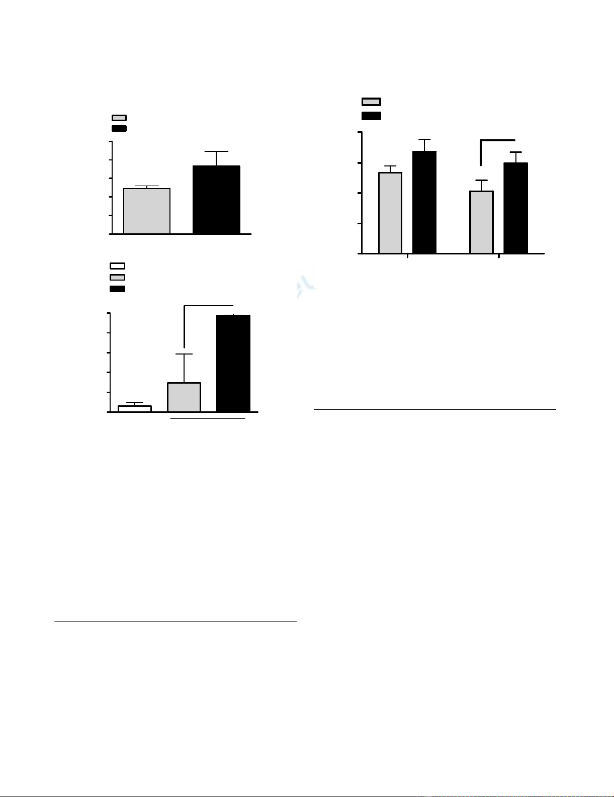
CTLA-4 blockade increases CTL number and in vivo lysis of
target cells during DC vaccination
DC based vaccination is effective for in vivo generation of
CTL's specific for H-2 Db binding LCMV gp33 derived
KAVYNFATM peptide [24] and in vivo treatment with anti-
CTLA-4 mAb augments the accumulation and activation
of adoptively transferred gp33 33–41 specific transgenic T
cells [25]. We tested the ability of CTLA-4 blockade to
expand the number of wildtype CTLs during a single DC
vaccination with LCMV gp3333–41. As shown in fig 1A, the
number of specific CTLs identified in an ELISPOT assay of
spleen cells tend to increase, although this was not signif-
icant (p = 0.11). We then assessed whether this CTL
expansion lead to an increased lysis of target cells in vivo.
We and others have previously shown that specific killing
of fluorescence-labeled peptide loaded syngeneic spleno-
cytes can be used to assess T-cell-mediated cytotoxic activ-
ity in vivo [23]. Using this assay, cytotoxic capacity of the
induced CTLs was assessed 10 days after DC vaccination
with LCMV gp3333–41 as the % specific lysis of i.v. admin-
istered LCMV gp3333–41 loaded syngeneic splenocytes.
Specific lysis was observed only in the immunized ani-
mals, and was significantly increased (p = 0.04) in ani-
mals co-treated with CTLA-4 blocking antibody (Fig 1B).
CTL numbers during repetitive DC vaccination and CTLA-
4 blockade
Repetitive vaccination is a common strategy for boosting
of immune responses, by e.g. increasing specific CTL lev-
els. However, the strategy might have potential flaws and
limits during DC vaccination. We tested the number of
LCMV gp3333–41 specific CTLs induced after 1 and 2 vacci-
nations with LCMV gp3333–41 loaded DCs in an IFN-γ
ELISPOT assay (Fig 2). To our surprise the number of CTLs
was not increased after the second vaccination, but rather
exhibited a small non-significant decrease instead. Like-
wise, two vaccinations combined with CTLA-4 blockade
did also not improve CTL expansion (Fig 2) compared to
treatment with a single vaccination + CTLA-4 blockade.
However, CTLA-4 blockade at the second vaccination sig-
nificantly increased the number of specific CTLs at the sec-
ond vaccination (p < 0.05)
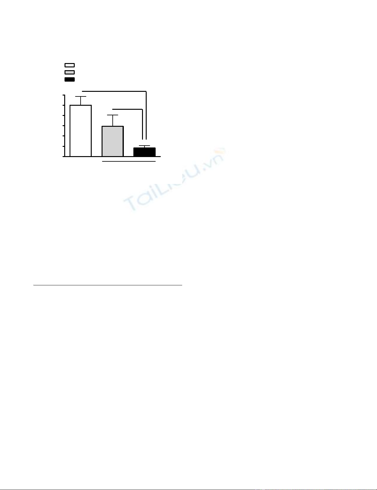
CTLA-4 blockade increase DC elimination during
repetitive DC vaccination
We next tested the effect of CTLA-4 blockade on DC elim-
ination during a second vaccination. Using a method to
directly compare the proportion of antigen-loaded to
non-antigen-loaded DC within the same inoculum of
cells and in the same host [17] we have shown in previous
experiments that in the course of DC based vaccination,
DC appearance in the draining lymph node of immu-
nized mice is decreased. A CFSE+ labeled DC population
was loaded with LCMV gp3333–41 peptide prior to injec-
tion, while the non-antigen-loaded CMTMR+ labeled DC
population served as a control. The two populations of
DCs were then mixed together in equal numbers before
injection in vivo, so that the numbers of antigen-loaded
DCs and non-antigen-loaded DCs could be evaluated
within the same recipient lymph node. DCs were then
harvested from DLN 48 h later, a time point where DC
elimination has previously been shown to be suboptimal
[17]. When DCs were administered to animals that were
immunized with LCMV gp3333–41 loaded DC, only 58 %
Journal of Immune Based Therapies and Vaccines 2007, 5:9 http://www.jibtherapies.com/content/5/1/9
Page 4 of 6
(page number not for citation purposes)
of the antigen-loaded DC had survived and reached the
DLN 48 h later. None of the unloaded DCs were elimi-
nated and no elimination of antigen-loaded DCs was
observed in naïve control mice. However, in mice co-
treated with CTLA-4 blocking antibody only 17 % of the
DC had survived and reached the DLN 48 h post injection
(Fig 3). This survival was significantly decreased com-
pared to survival in immunized mice with no CTLA-4
blockade and in naïve mice. We did not identify any dif-
ference in surface expression of costimulatory molecules
such as CD80 and CD86 on injected DCs from mice
treated with CTLA-4 blockade compared to untreated
mice (data not shown).
Discussion
The present study demonstrates that CTLA-4 blockade
increases the number of DC vaccine induced LCMV
gp3333–41 specific CTLs and the lysis of relevant in vivo tar-
gets. General vaccination approaches take advantage of
repetitive vaccinations as a mean to boost the immune
response and expand the number of specific CTLs. How-
ever, the expansion of CTLs mediated by CTLA-4 blockade
also affects DC elimination during repetitive DC injec-
tion. Our data suggest that repetitive DC vaccination with
or without CTL expanding strategies, e.g. CTLA-4 block-
ade does not increase CTL expansion compared to the lev-
els obtained after the primary vaccination and that this is
due to elimination of injected DCs in a primed host.
Previous reports have documented that CTLA-4 blockade
is a feasible strategy for potent in vivo expansion of antigen
specific T cells, in particular in the context of cancer vacci-
nation [14,15]. Even unspecific expansion elicited by anti-
CTLA-4 mAb can be useful both in experimental models
LCMV gp3333–41 specific CTL levels are stable during repeti-tive DC vaccinationFigure 2
LCMV gp3333–41 specific CTL levels are stable during repeti-
tive DC vaccination. C57/Bl6 mice were immunized with
peptide LCMV gp3333–41 loaded DCs day -14 and -7 in combi-
nation with i.p. injection of anti-CTLA-4 mAb. Spleen cells
from individual mice were isolated day 0, cocultured with
LCMV gp3333–41 peptide for 7–10 days and tested for reactiv-
ity against the peptide in an IFN-γ ELISPOT assay. Results are
shown as mean ± SD of eight mice from two separate exper-
iments. (* p < 0.05)
1 vaccination 2 vaccinations
0
50
100
150
200
- CTLA-4 blockade
+ CTLA-4 blockade
*
SFC/25.000 spleen cells
CTLA-4 blockade increases the induction of LCMV gp3333–41
specific CTLs and in vivo lysis of target cellsFigure 1
CTLA-4 blockade increases the induction of LCMV gp3333–41
specific CTLs and in vivo lysis of target cells. (A) C57/Bl6 mice
were immunized with peptide LCMV gp3333–41 loaded DCs in
combination with i.p. injection of anti-CTLA-4 mAb. Spleen
cells were isolated 7–10 days after the primary immunization
and cocultured with LCMV gp3333–41 peptide + IL-2 and then
tested for reactivity against the peptide in an IFN-γ ELISPOT
assay. (B) Alternatively, peptide LCMV gp3333–41 loaded CFSE
labeled and peptide unloaded CMTMR labeled spleen cells
were injected i.v. in immunized mice and naïve mice and tar-
get cell lysis was analyzed after 24 hours by the in vivo VITAL
assay. Results are shown as mean ± SD of three mice in 1
representative experiment out of 2. (* p < 0.05)
0
50
100
150
200
250
- CTLA-4 blockade
+ CTLA-4 blockade
SFC/25.000 spleen cells
0
10
20
30
40
50
naïve mice
- CTLA-4 blockade
+ CTLA-4 blockade
LCMV
33-41
immunized mice
*
% specific lysis
A
B
Journal of Immune Based Therapies and Vaccines 2007, 5:9 http://www.jibtherapies.com/content/5/1/9
Page 5 of 6
(page number not for citation purposes)
and clinical settings [13,15]. Similar, we observed an
increase in LCMV gp3333–41 specific CTLs and an increased
in vivo lysis of target cells after LCMV gp3333–41 targeting
DC based vaccine combined with CTLA-4 blockade. How-
ever, since LCMV gp3333–41 is already a strong immuno-
dominant epitope, this relative increase is probably
smaller compared to relative increases observed for CTLs
specific for weaker antigens, such as tumour antigens [14].
This might explain, why in vivo tumour prophylactic
experiment with DC based vaccination against gp33 posi-
tive tumour cells did not clearly show an increased effect
of CTLA-4 blockade despite increased CTL levels (data not
shown). Also, the level of specific CTLs shown was low as
we tested the effect of CTLA-4 blockade after the primary
vaccination.
We have previously shown that DC elimination during
DC based vaccination is due to the presence of primed
antigen specific CTLs and is dependent on perforin expres-
sion [17,18]. This phenomenon is likely to limit the
potential of DC based booster vaccines in therapeutic set-
tings [18,21]. Indeed, in a number of DC based vaccina-
tion studies, in particular in cancer patients, CTL
responses are either observed in a low fraction of patients
or with great fluctuation and even a decrease in CTL
number during vaccination has been reported [5,26,27].
In these early studies, repetitive vaccination with imma-
ture or intermediate mature DCs unexposed to potent
maturation reagents was used for booster vaccination
with the same antigen. Thus, the low fraction of CTLs
induced in these studies might be a result of time depend-
ent elimination of injected DCs at booster vaccinations.
Unfortunately CTL responses were most often measured
after several vaccinations and make it difficult to compare
the CTL levels with the levels after first vaccination. In
contrast, at least in in vitro studies, DC elimination is min-
imal when LPS matured DCs are applied due to expres-
sion of the serpin serine protease inhibitor 6 [28]. In this
study, we demonstrate that also the application of CTL
expanding strategies such as CTLA-4 blockade lead to a
massive loss of surviving DCs during booster vaccination.
Since our tumour challenge experiments with addition of
CTLA-4 blockade didn't correlate well with CTL levels in
an experimental LCMV tumour model, it is unknown if
this DC depletion will influence the outcome of a tumour
vaccine. Indeed, CTLs might be reactivated during the kill-
ing of DCs, and the remaining DC's might be particular
potent CTL activators. However, previous studies from
our laboratory suggest that the induction of tumour
immunity is limited by DC elimination [21]. Therefore,
DC elimination, in addition to TH1/TH2 promoting
capacities and migration of the DCs to DLN, is an impor-
tant issue, when designing maturation regimens for DCs
used in vaccination studies, in particular in human studies
where toll-like receptor ligands such as LPS are not
approved for clinical trials. Also, recent research has estab-
lished that mature DCs are more potent than immature
DCs in DC based vaccination studies [3,31] and elimina-
tion of immature DCs during vaccination might be one of
the reasons.
In conclusion, CTLA-4 blockade dependent expansion of
CTLs increases DC elimination during repetitive DC injec-
tion and suggests that alternative strategies, such as prime-
boost strategies with exclusion of DCs at booster vaccina-
tions [29] or heterologous booster vaccinations [30]
designed with alternate epitope loading of DCs during
vaccination, should be applied when DC are used for
repetitive vaccination with or without inclusion of CTL
expanding strategies, such as CTLA-4 blockade.
Authors' contributions
AEP conceived the study, carried out the in vivo experi-
ments and flowcytometry, performed the statistical analy-
sis and drafted the manuscript. FR participated in the
design and coordination of the study and drafted the
Enhanced DC elimination during DC vaccination combined with CTLA-4 blockadeFigure 3
Enhanced DC elimination during DC vaccination combined
with CTLA-4 blockade. C57/Bl6 mice were immunized with
peptide LCMV gp3333–41 loaded DCs with or without i.p.
injection of anti-CTLA-4 mAb. After 7–10 days, an inoculum
consisting of peptide LCMV 33–41 loaded CFSE labeled
together with peptide unloaded CMTMR labeled DCs was
injected subcutaneously into the distal forelimb of naïve mice
(control), immunized mice or immunized mice cotreated
with anti-CTLA-4 mAb. DCs were recovered from the
draining lymph node for FACS analysis and determination of
% surviving DCs. Results are shown as mean ± SD of nine
mice from 3 separate experiments. (* p < 0.05; ***p <
0.0001)
0
20
40
60
80
100
120
Naïve mice
- CTLA-4 blockade
+ CTLA-4 blockade
***
*
LCMV
33-41
immunized mice
% adjusted survival


![Vaccine và ứng dụng: Bài tiểu luận [chuẩn SEO]](https://cdn.tailieu.vn/images/document/thumbnail/2016/20160519/3008140018/135x160/652005293.jpg)


%20--%3e%3cdefs%3e%3cstyle%3e%20.st0%20{%20fill:%20%23fff;%20}%20.st1%20{%20fill:%20%237800fa;%20}%20%3c/style%3e%3c/defs%3e%3cpath%20class='st1'%20d='M117.78,12.18H43.11c2.9,3.47,4.65,7.94,4.65,12.82,0,5.6-2.3,10.66-6.01,14.29h76.02l7.22-13.56-7.22-13.56Z'/%3e%3cg%3e%3cpath%20class='st0'%20d='M53.58,26.17h-.59v-1.46h.59v-4.96h2.83c1.78,0,2.67.94,2.67,2.82v5.76c0,1.87-.89,2.81-2.67,2.81h-2.83v-4.96ZM55.36,21.37v3.34h1.1v1.46h-1.1v3.34h1.01c.61,0,.91-.37.91-1.1v-5.93c0-.74-.3-1.1-.91-1.1h-1.01Z'/%3e%3cpath%20class='st0'%20d='M65.99,31.14h-1.8l-.31-2.07h-2.19l-.31,2.07h-1.64l1.82-11.39h2.62l1.82,11.39ZM65.28,18.04c-.25.46-.51.77-.75.94-.21.15-.47.22-.79.22-.26,0-.57-.07-.92-.22l-.38-.15c-.14-.05-.26-.07-.37-.07-.3,0-.53.18-.71.54l-.91-.68c.25-.46.51-.77.75-.94.21-.14.48-.21.79-.21.26,0,.57.07.92.21l.38.15c.14.05.26.07.37.07.3,0,.53-.18.71-.54l.91.68ZM61.91,27.52h1.73l-.87-5.76-.87,5.76Z'/%3e%3cpath%20class='st0'%20d='M74.53,26.89v1.52c0,1.91-.89,2.86-2.67,2.86s-2.67-.95-2.67-2.86v-5.93c0-1.91.89-2.86,2.67-2.86s2.67.95,2.67,2.86v1.11h-1.69v-1.22c0-.75-.31-1.12-.93-1.12s-.93.37-.93,1.12v6.15c0,.74.31,1.11.93,1.11s.93-.37.93-1.11v-1.63h1.69Z'/%3e%3cpath%20class='st0'%20d='M81.4,31.14h-1.8l-.31-2.07h-2.19l-.31,2.07h-1.64l1.82-11.39h2.62l1.82,11.39ZM75.9,19.2l1.52-1.91h1.71l1.51,1.91h-1.61l-.76-.95-.75.95h-1.61ZM77.32,27.52h1.73l-.87-5.76-.87,5.76ZM83.1,15.99l-1.76,1.91h-1.26l1.17-1.91h1.86Z'/%3e%3cpath%20class='st0'%20d='M84.86,19.75c1.78,0,2.67.94,2.67,2.82v1.48c0,1.87-.89,2.81-2.67,2.81h-.85v4.28h-1.79v-11.39h2.64ZM84.01,21.37v3.86h.85c.58,0,.87-.36.87-1.08v-1.71c0-.71-.29-1.07-.87-1.07h-.85Z'/%3e%3cpath%20class='st0'%20d='M93.51,19.75c1.78,0,2.67.94,2.67,2.82v1.48c0,1.87-.89,2.81-2.67,2.81h-.85v4.28h-1.79v-11.39h2.64ZM92.66,21.37v3.86h.85c.58,0,.87-.36.87-1.08v-1.71c0-.71-.29-1.07-.87-1.07h-.85Z'/%3e%3cpath%20class='st0'%20d='M98.8,31.14h-1.79v-11.39h1.79v4.88h2.03v-4.88h1.83v11.39h-1.83v-4.88h-2.03v4.88Z'/%3e%3cpath%20class='st0'%20d='M105.36,24.55h2.46v1.62h-2.46v3.34h3.09v1.63h-4.88v-11.39h4.88v1.63h-3.09v3.18ZM108.17,17.29l-1.76,1.91h-1.26l1.17-1.91h1.86Z'/%3e%3cpath%20class='st0'%20d='M112.2,19.75c1.78,0,2.67.94,2.67,2.82v1.48c0,1.87-.89,2.81-2.67,2.81h-.85v4.28h-1.79v-11.39h2.64ZM111.35,21.37v3.86h.85c.58,0,.87-.36.87-1.08v-1.71c0-.71-.29-1.07-.87-1.07h-.85Z'/%3e%3c/g%3e%3ccircle%20class='st1'%20cx='25'%20cy='25'%20r='20'/%3e%3cpath%20class='st0'%20d='M32.78,19.27c2.92,0,4.43,2.55,5.28,5.33l.71,2.17c.14.38-.33.75-.71.75h-5.61c.19-.33.24-.71.09-1.08l-.75-2.45c-.43-1.32-.99-2.64-1.79-3.77.75-.57,1.65-.94,2.78-.94h0ZM25,18.38c3.25,0,4.9,2.78,5.89,5.89l.76,2.45c.14.42-.33.8-.8.8h-11.69c-.42,0-.94-.38-.8-.8l.75-2.45c.99-3.11,2.64-5.89,5.89-5.89h0ZM25,11.35c1.74,0,3.11,1.37,3.11,3.11s-1.37,3.11-3.11,3.11-3.11-1.41-3.11-3.11,1.41-3.11,3.11-3.11h0ZM17.27,19.27c1.08,0,1.98.38,2.73.94-.8,1.13-1.37,2.45-1.74,3.77l-.8,2.45c-.14.38-.05.75.09,1.08h-5.56c-.42,0-.9-.38-.75-.75l.71-2.17c.9-2.78,2.41-5.33,5.33-5.33h0ZM17.27,12.91c1.51,0,2.78,1.27,2.78,2.83s-1.27,2.83-2.78,2.83-2.83-1.27-2.83-2.83,1.27-2.83,2.83-2.83h0ZM32.78,12.91c1.56,0,2.78,1.27,2.78,2.83s-1.23,2.83-2.78,2.83-2.83-1.27-2.83-2.83,1.27-2.83,2.83-2.83h0ZM27.07,28.56v.09c0,.57-.24,1.08-.61,1.46h0v.05c-.38.33-.9.57-1.46.57s-1.08-.24-1.46-.61h0c-.38-.38-.61-.9-.61-1.46v-.09h1.41v.09c0,.19.05.38.19.47v.05c.09.09.28.19.47.19s.38-.09.47-.19v-.05c.14-.09.24-.28.24-.47t-.05-.09h1.41ZM30.99,28.56v.09c0,1.65-.66,3.16-1.74,4.24-1.08,1.08-2.59,1.79-4.24,1.79s-3.16-.71-4.24-1.79l-.05-.05c-1.04-1.08-1.7-2.55-1.7-4.2v-.09h1.41v.09c0,1.27.47,2.4,1.27,3.25h.05c.85.85,1.98,1.37,3.25,1.37s2.4-.52,3.25-1.37c.85-.8,1.37-1.98,1.37-3.25v-.09h1.37ZM34.99,28.56v.09c0,2.78-1.13,5.28-2.92,7.07-1.79,1.79-4.29,2.92-7.07,2.92s-5.23-1.13-7.07-2.92c-1.79-1.79-2.92-4.29-2.92-7.07v-.09h1.41v.09c0,2.4.94,4.53,2.5,6.08,1.56,1.56,3.72,2.5,6.08,2.5s4.52-.94,6.08-2.5c1.56-1.56,2.5-3.68,2.5-6.08v-.09h1.41Z'/%3e%3c/svg%3e)