TẠP CHÍ KHOA HỌC - ĐẠI HỌC ĐỒNG NAI, SỐ 30 – 2024 ISSN 2354-1482
DEPSIDONES WITH ALPHA-GLUCOSIDASE INHIBITION FROM THE LICHEN USNEA CERATINA
Huynh Bui Linh Chi Dong Nai University Email: huynhbuilinhchi@gmail.com (Received: 21/3/2024, Revised: 25/3/2024, Accepted for publication: 27/3/2024)
ABSTRACT
From lichen Usnea ceratina, eight (1), 8’-hydroxycryptostictinolide (2),
γ-lactonic depsidones, 3’- the demethylcryptostictinolide 8’- ethoxycryptostictinolide (3), vesuvianic acid (4), 8’-O-methylstictic acid (5), stictic acid (6), norstictic acid (7) and bailesidone (8) were identified by HR-ESI-MS, and NMR analyses. For the first time, compounds (2, 4-7) were detected from the Usnea genus, whereas compounds 1 & 3 were previously reported from this species. Their α-glucosidase inhibitory properties were evaluated. All purified depsidones except 1 & 7 possessed better α-glucosidase inhibitory activity (IC50 values ranged from 38.05 to 143.94 μM) than the standard drug acarbose (IC50 value of 214.50 μM). Keywords: Usnea ceratina, lichen, depsidone, γ-lactonic depsidone, α-
glucosidase inhibitory activity. 1. Introduction
fruticose gray-green 2016). eight depsidones from U. ceratina collected from the bark trees at Paksong town, Paksong district, Champasack province, Laos. 2. Materials and methods 2.1 Materials of Usnea depsides collected town, Paksong
the Department inhibitors
this isolation and
The thalli of the lichen Usnea at ceratina Arch were Paksong district, Champasack province, Laos in April 2015. The scientific name of the lichen was recognized by Dr. Harrie J. M. Sipman, Freie Universitaet, Berlin, Germany. A voucher specimen (US- B030) was deposited in the herbarium of of Organic Chemistry, University of Science, National University – Ho Chi Minh City, Vietnam. 2.2 Methods 2.2.1. General experimental procedures structural for identification The Usnea genus comprising of more than 360 species belongs the Parmeliaceae family. It is one of mostly lichens pale Phytochemical (Prateeksha, species investigations and phenolics, declared depsidones were determined as the main compounds (Prateeksha, 2016). Further, benzofurans, terpenoids and (Prateeksha, steroids were notified 2016). In our earlier papers, depsidones (Bui, 2020), dibenzofuran and phenolic acid (Bui, 2021a) were purified from U. ceratina. As part of our favored studies on of α-glucosidase (Nguyen, 2015; Vietnamese plants Nguyen, 2016a; Nguyen 2016b) as well as bioactive constituents of lichens (Huynh, 2020; Huynh, 2021a; Huynh, the study disclosed 2021b), separation, structural identification, and α-glucosidase inhibitory property of
103
TẠP CHÍ KHOA HỌC - ĐẠI HỌC ĐỒNG NAI, SỐ 30 – 2024 ISSN 2354-1482
silica on
The HR-ESI-MS were recorded on an Exactive mass spectrometer (Thermo Fisher Scientific). The NMR spectra were measured on a Bruker Avance III spectrometer (500 MHz for 1H NMR and 125 MHz for 13C NMR). TLC was carried out on silica gel 60 F254 or silica gel 60 RP-18 F254S (Merck) and spots were visualized by spraying with a solution of 5% vanillin in ethanol, followed by heating at 100oC. Column chromatography was performed with silica gel 60 (0.040 – 0.063 mm, Merck). 2.2.2. Extraction and isolation
The ethyl acetate extract (12.0 g) was separated on silica gel CC using chloroform–methanol mixture with increasing methanol to yield seven sub- fractions (EA1 EA7). The sub-fraction EA4 (1.5 g) was rechromatographed chloroform–methanol eluting with (98:2, v/v) to deliver 1 (5.0 mg), and 3 (6.5 mg). The sub-fraction EA5 (2.05 g) gel purified was using chromatographic column chloroform–methanol–acetic acid (95:5:1, v/v/v) to give 2 (7.0 mg). The sub-fraction EA7 (1.8 g) was applied to silica gel CC, eluted with chloroform– methanol (9:1, v/v) to afford 1 (4.5 mg) and 7 (5.0 mg).
The fresh lichen thalli (1.60 kg) were cleaned under running tap water and air-dried. The ground powder (1.15 kg) was extracted with acetone at room temperature. After filtration, the solvent was removed under reduced pressure to yield the crude acetone residue (163.0 g). This residue was then subjected to silica gel solid phase extraction and further eluted with chloroform, ethyl acetate to deliver chloroform extract (60.0 g) and ethyl acetate (12.0 g) extract.
(calcd
3’-Demethylcryptostictinolide (1): [M-H]- HR-ESI-MS m/z 357.0577 (calcd for C18H13O8, 357.0611); 1H & 13C-NMR data (DMSO-d6) see Table 1. (2): 8’-Hydroxycryptostictinolide HR-ESI-MS m/z 389.0882 [M+H]+ (calcd for C19H17O9, 389.0873); 1H & 13C-NMR data (DMSO-d6) see Table 1. (3): 8’-Ethoxycryptostictinolide [M-H]- HR-ESI-MS m/z 415.0987 (calcd for C21H19O9, 415.1029); 1H & 13C-NMR data (DMSO-d6) see Table 1. Vesuvianic acid (4): HR-ESI-MS [M+Na]+ m/z 437.0826 for C21H18O9Na, 437.0849); 1H & 13C- NMR data (DMSO-d6) see Table 1. (5.05
8’-O-Methylstictic acid (5): HR- ESI-MS m/z 423.0651 [M+Na]+ (calcd for C20H16O9Na, 423.0692). 1H & 13C- NMR data (DMSO-d6) see Table 1.
1H &
solvents eluted
The chloroform extract (60.0 g) was fractionated using silica gel column chromatography (CC) with the solvent systems of n-hexanechloroform to afford nine sub-fractions (C1C9). The sub-fraction C5 g) was rechromatographed on silica gel CC with solvents n-hexane–chloroform (8:2, v/v) to give 5 (6.0 mg), 6 (5.0 mg) and 8 (6.0 mg). The sub-fraction C6 (4.83 g) was applied to silica gel CC n-hexane– using chloroform (7:3) to get 4 (6.5 mg). Stictic acid (6): HR-ESI-MS m/z 385.0557 [M-H]- (calcd for C19H13O9, 13C-NMR data 385.0560); (DMSO-d6) see Table 1.
104
TẠP CHÍ KHOA HỌC - ĐẠI HỌC ĐỒNG NAI, SỐ 30 – 2024 ISSN 2354-1482
(calcd
1H &
Norstictic acid (7): HR-ESI-MS [M-H]- m/z 371.0461 for C18H11O9, 371.0403); 1H & 13C-NMR data (DMSO-d6) see Table 1.
measured the optical density at 401 nm. The IC50 values were calculated as the concentration of α-glucosidase inhibitor that inhibited 50% of α-glucosidase activity. Acarbose was used as the positive control. 3. Results and discussion Bailesidone (8): HR-ESI-MS m/z 413.0854 [M-H]- (calcd for C21H17O9, 13C-NMR data 413.0854); (acetone-d6) see Table 1.
powder. amorphous
6.37 (1H, s, H-3’),
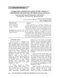
Figure 1: Chemical structures of compounds 1-9.
2.2.3. α-Glucosidase inhibitory activity assay
The
inhibitory α-glucosidase activity was determined according to those presented in our previous paper (Nguyen, 2016a; 2015; Nguyen, Nguyen 2016b). 25 L of p- nitrophenyl-α-D-glucopyranoside (3 mM), 25 L of α-glucosidase enzyme 0.2 U/mL in 0.01 M phosphate buffer solution (pH = 7) were added to 625 L of the sample solution (compounds 1- 8). Each reaction was carried out at 37°C for 30 minutes, and stopped by adding 375 L of Na2CO3 (0.1 M), Compound 1 was afforded as a white The molecular formula was confirmed as C18H14O8 by HR-ESI-MS data ([M-H]- m/z 357.0577, calcd. 357.0611). The 1H-NMR data of 1 (Table 1) showed two hydroxyl protons at δH 11.20 (1H, s, OH-2’), 5.02 (1H, s, OH-8), two aromatic protons at δH 6.96 (1H, s, H- 5), two oxymethylene groups at δH 4.58 (2H, s, H-8), 5.64 (2H, s, H-8’), one oxymethyl group at δH 3.87 (3H, s, H-9), and three methyl protons at δH 2.43 (3H, s, H-10). The 13C-NMR data of 1 (Table 1) displayed eighteen carbons, including two carbonyl carbons at 167.2 (C-7), 172.0 (C-7’), one methyl carbon at δC 21.0 (C-10), one methoxy carbon at δC 56.3 (C-9), two oxymethylene carbons at δC 51.0 (C-8), 66.1 (C-8’), and twelve aromatic carbons with five of those were oxygenated, two of them were methine aromatic carbons. On other (H-8’) hands, protons at δH 5.64 correlated with carbons at δC 107.7 (C- 1’), 140.9 (C-6’), and 172.0 (C-7’) in HMBC, were signified γ-lactone ring. Those data of 1 were suggested a depsidone with γ-lactone moeity similar cryptostictinolide (9) lacked one methyl carbon at C-3’. The HMBC spectrum of 1 (Figure 2) exhibited correlations between proton at δH 6.96 (H-5) and
105
TẠP CHÍ KHOA HỌC - ĐẠI HỌC ĐỒNG NAI, SỐ 30 – 2024 ISSN 2354-1482
the arrangement of
the in carbon C-8’, were evenced that an ethoxy group was linked to be C-8’ of skeleton. Therefore, the structure of 3 verified as 8’-ethoxycryptostictinolide (Bui, 2021b) was elucidated for the first time from this species.
as powder. amorphous
carbon at δC 21.0 (C-10), protons at δH 4.58 (H-8), 3.87 (H-9) and carbon at δC 161.5 (C-4), proton at δH 6.37 (H-3’) and carbon at δC 154.9 (C-2’), were these indicated substituents depsidone framework. Hence, the structure of 1 identified 3’- demethylcryptostictinolide (Dévéhat, 2007) was determined for the first time from this species.
389.0882,
Compound 4 was delivered as a white The molecular formula was illustrated as by HR-ESI-MS data C21H18O9 ([M+Na]+ m/z 437.0826, calcd. 437.0849). The NMR spectral data of 4 (Table 1) were similar to those of 3, except for the arriving of one formyl function at C-3 at δC 186.9 (C-8)/δH 10.53 (1H, s, H-8) in 4, instead of carbinol group at δC 51.1 (C-8) in 3. Furthermore, this proton at δH 10.53 (H- 10) correlated with carbon at δC 115.0 (C-3) in HMBC (Figure 2), and besides upfiled shift of this carbon were assigned that a formyl moiety was connected to be C-3 of depsidone. Thus, the structure of 4 was testified as vesuvianic acid (Huneck, 1987).
Compound 2 was given as a white amorphous powder. The molecular formula was affirmed as C19H16O9 ([M+H]+ m/z calcd. 389.0873). The 13C & 1H-NMR spectra of 2 were similar to those of 9, but missed one oxymethylene carbon C-8’, and reavealed one acetal carbon at δC 95.4 (C-8’)/δH 6.96 (1H, d, 7.5, H-8’) in 2, further the upfield shift of carbon C- 7’ at δC 166.5, which were evenced that a hydroxyl group was attached to be C- 8’ of a depsidone skeleton. Thence, the structure of 2 was elucidated as 8’- (Ismed, hydroxycryptostictinolide 2017). powder. amorphous
powder. amorphous
Compound 5 was yielded as a white The molecular formula was evidenced as by HR-ESI-MS data C20H16O9 ([M+Na]+ m/z 423.0651, calcd. 423.0692). The 13C & 1H-NMR data of 5 (Table 1) were similar to those of 4, except for the disappearing of one ethoxy group in 4, instead of methoxy moiety at δC 57.5 (C-10’)/δH 3.63 (3H, s, H-10’) in 5. Moreover, the correlation between protons at δH 3.63 (H-10’) and carbon at δC 102.9 (C-8’) observed in HMBC (Figure 2) was verfied that a methoxy group was linked to be C-8’ of depsidone. Consequently, the structure Compound 3 was yielded as a The white molecular formula was evinced as C21H20O9 ([M-H]- m/z 415.0987, calcd. 415.1029). The 13C & 1H-NMR spectra of 3 exposed signals of a depsidone frame were similar to that of 2, except for the presence of an ethoxy function at δC 64.6 (C-10’)/3.85 (2H, m, H-10’), 15.1 (C-11’)/1.25 (3H, t, 7.0, H-11’), and further downfield shift of carbon C- 8’ at δC 99.3 in 3. The HMBC spectrum of 3 (Figure 2) proved correlations between protons at δH 3.85 (H-10’) and
106
TẠP CHÍ KHOA HỌC - ĐẠI HỌC ĐỒNG NAI, SỐ 30 – 2024 ISSN 2354-1482
of 5 testified as 8’-O-methylstictic acid (Shimada, 1980) was reported form this species (Bui, 2020).
molecular formula was proved as C18H12O9 by HR-ESI-MS data ([M-H]- m/z 371.0461, calcd. 371.0403). The 13C & 1H-NMR data of 7 (Table 1) were similar to those of 6, but missed methoxy carbon at C-4 at δC 56.9 (C- 9)/δH 3.91 (1H, s, H-9) in 6, and downfield shift of carbon C-4 at δC 164.2 in 7, were testified a hydroxyl was attached to be C-4. Thus, the structure of 7 was determined as norstictic acid (Shimada, 1980).
(1H, s, H-8)
([M-H]-
to those of 1, but
Compound 6 was got as a white amorphous powder. The molecular formula was determined as C19H14O9 by HR-ESI-MS data ([M-H]- m/z 385.0557, calcd. 385.0560). The 13C & 1H-NMR data of 7 (Table 1) were similar to those of 2, except disappeared carbinol function in 2, and replaced formyl group at C-3 at δC 186.8 (C- in 6. 8)/δH 10.46 Additionly, the correlation between this proton at δH 10.46 (H-8) and carbon at δC 114.5 (C-3) in HMBC (Figure 2) was clarified that hydroxyl was substitued to be C-3. Accordingly, the structure of 6 designated as stictic acid (Shimada, 1980) was isolated form this species (Bui, 2020).
(C-10’), 9.0
Figure 2: Key HMBC correlations of compounds 1, 3-6, 8.
Compound 7 was afforded as a The amorphous powder. white Compound 8 was gave as a white amorphous powder. The molecular formula was proved as C21H18O9 by m/z HR-ESI-MS data 413.0854, calcd. 413.0854). The 13C & 1H-NMR data of 8 (Table 1) were lacked similar oxymethylene carbon at C-3 at δC 51.0 (C-8)/δH 4.58 (2H, s, H-8) in 1, and downfield shift of carbon C-3 at δC 133.3 in 8, were testified a hydroxyl was attached to be C-3. Moreover, 13C & 1H-NMR data of 8 were further possessed an acetomethyl moiety, following one carbonyl, one methylene, and one methyl carbons at δC 202.6 (C- (C-12’), 11’), 46.6 respectively, which were correlated with protons at δH 3.09 (1H, dd, 17.5 & 9.0, H-10’a), 3.71 (1H, dd, 17.5 & 2.5, H-10’b), and 2.23 (3H, s, H-12’), respectively. Additionaly, protons at δH 3.09 (H-10’a), 3.71 (H-10’b) correlated with carbon at δC 77.2 (C-8’) in HMBC (Figure 2), were identified that an acetomethyl function was linked to be C-8’. Thus, the structure of 8 signed as bailesidone was identified form this species (Bui, 2020).
107
TẠP CHÍ KHOA HỌC - ĐẠI HỌC ĐỒNG NAI, SỐ 30 – 2024 ISSN 2354-1482
Table 2: α-Glucosidase inhibition of compounds 1-8
Inhibition (%) Samples IC50 (µM)
- Inhibition < 1%
100 7.30 ± 1.41 79.52 ± 1.14 46.42 0.30 99.41 ± 0.57 46.79 ± 0.75 37.05 ± 0.76 17.72 ± 2.13 53.02 ± 0.42 50 1.01 ± 0.59 24.95 ± 1.93 35.40 0.61 68.52 ± 1.53 19.35 ± 0.69 21.12 ± 1.10 6.94 ± 1.12 29.61 ± 0.32 25 – 9.62 ± 1.70 23.84 0.41 30.01 ± 0.75 2.43 ± 1.02 6.61 ± 1.03 1.35 ± 0.97 18.44 ± 0.21 10 – 2.17 ± 0.70 - 7.47 ± 0.63 – – – 14.81 ± 0.33 1 2 3 4 5 6 7 8 Acarbose >250 73.13 126.62 38.05 110.11 143.94 >250 97.12 214.50
the
All separated depsidones except 1 & 7 expressed potential inhibition against α-glucosidase enzyme (IC50 values ranged from 38.05 to 143.94 μM). Among them, 4 displayed the strongest effect on α-glucosidase inhibition (IC50 value of 38.05 μM) comparing with the acarbose drug (IC50 value of 214.50 μM). 4. Conclusion
acid (5), stictic acid (6), norstictic acid (8) were (7), bailesidone and elucidated from lichen Usnea ceratina using HR-ESI-MS, and NMR spectrocopic data. All γ-lactone-type depsidones except 1 & 3 were the genus Usnea, informed from while, compounds 1 & 3 was announced from this species for the first time. All isolated depsidones except 1 & 7 exhibited better α- glucosidase inhibition (IC50 values ranged from 38.05 to 143.94 μM) than the acarbose drug (IC50 value of 214.50 μM).
Eight depsidones with γ-lactone 3’- moiety, including (1), demethylcryptostictinolide (2), 8’-hydroxycryptostictinolide 8’-ethoxycryptostictinolide (3), vesuvianic acid (4), 8’-O-methylstictic
REFERENCES
J. Nat. Prod. 70 activities, (7)
Dévéhat F. L. L., Tomasi S., Elix J. A., Bernard A., Rouaud I., Uriac P. and Boustie J. (2007). Stictic acid derivatives from the lichen Usnea articulata and their (2007) 1218-1220. antioxidant https://doi.org/10.1021/np070145k.
Bui V. M., Duong T. H., Chavasiri W., Nguyen K. P. P. and Huynh, B. L. C. (2020) A new depsidone from the lichen Usnea ceratina Arch, Nat. Prod. Res. (2020). https://doi.org/10.1080/14786419.2020.1828405.
Bui V. M., Huynh B. L. C., Pham N. K. T., Nguyen T. A. T., Nguyen T. T. T., Nguyen K. P. P. and Nguyen T. P. (2021a). Usneaceratins A and B, two new secondary metabolites from the lichen Usnea ceratina, Nat. Prod. Res. (2021). https://doi.org/10.1080/14786419.2021.1901288.
109
TẠP CHÍ KHOA HỌC - ĐẠI HỌC ĐỒNG NAI, SỐ 30 – 2024 ISSN 2354-1482
Bui V. M., Duong T. H., Nguyen T. A. M., Nguyen T.N.V, Nguyen N. H., Nguyen H. H., Chavasiri W., Nguyen K. P. P. and Huynh, B. L. C. (2021b). Two new phenolic compounds from the Vietnamese lichen Parmotrema tinctorum, Nat. Prod. Res. https://doi.org/10.1080/14786419.2020.1864367.
Huneck S. and Tabacchi R. (1987). ψ-Esters of depsidones with a lactole ring. Phytochem. 26 (4) (1987) 1131-1138. https://doi.org/10.1016/S0031- 9422(00)82364-X.
(Parmeliaceae), Prod. Hale Res. Nat.
Huynh B. L. C., Bui V. M., Nguyen K. P. P., Pham N. K. T. and Nguyen T. P. (2020). Three new diphenyl ethers from the lichen Parmotrema praesorediosum (2020). (Nyl.) https://doi.org/10.1080/14786419.2020.1837818.
Huynh B. L. C., Pham N. K. T. and Nguyen T. P. (2021a). Vinapraesorediosic acids D and E from the lichen Parmotrema praesorediosum (Nyl.) Hale, Phytochem. Lett. 41 (1) (2021) 61-64. https://doi.org/10.1016/j.phytol.2020.11.001. Huynh B. L. C., Pham N. K. T. and Nguyen T. P. (2021b). Paresordin A, new diphenyl cyclic peroxide from the lichen Parmotrema praesorediosum, J. Asian Nat. Prod. Res. (2021). https://doi.org/10.1080/10286020.2021.1908271. Ismed F., Dévéhat F. L. L., Rouaud I., Ferron S., Bakhtiar A. and Boustie J. (2017). lichen isolated from a Sumatran (Stereocaulaceae) with superoxide anion 55-62. (1-2) activities, Z. Naturforsch. (2017) 72
NMR reassignment of stictic acid Stereocaulon montagneanum scavenging https://doi.org/10.1515/znc-2016-0148.
(2015) Prod. Res. Nat. (15)
Nguyen T. P., Tran T. T. V., Mai D. T., Le T. D., Phan N. M., and Bui T. D. (2015). New C20-gibberellin diterpene from the leaves of Schefflera sessiliflora De P. V., 1432-1436. 29 https://doi.org/10.1080/14786419.2014.1003300.
Nguyen T. P., Le T. D., Phan N. M., Bui T. D. and Mai D. T. (2016a). Triterpene saponins with α-glucosidase inhibition and cytotoxic activity from the leaves of Schefflera sessiliflora”, J. Asian Nat. Prod. Res. 18 (6) 2016 542-549. https://doi.org/10.1080/10286020.2015.1121999.
Nguyen T. P., Le T. D., Phan N. M., Bui T. D., Pham N. K. T., Do T. M. L., Nguyen D. T. and Mai D. T. (2016b). A new dihydrofurocoumarin from the fruits of Pandanus tectorius Parkinson ex Du Roi, Nat. Prod. Res. 30 (21) (2016) 2389- 2395. https://doi.org/10.1080/14786419.2016.1188095.
(2016) Adv. RSC (26)
Prateeksha, Paliya B. S., Bajpai R., Jadoun V., Kumar J., Kumar S., Upreti D. K., Singh B. R., Nayaka S., Joshi Y. and Singh B. N. (2016). The genus Usnea: a potent phytomedicine with multifarious ethnobotany, phytochemistry and pharmacology, 21672-21696. 6 https://doi.org/10.1039/C5RA24205C.
110
TẠP CHÍ KHOA HỌC - ĐẠI HỌC ĐỒNG NAI, SỐ 30 – 2024 ISSN 2354-1482
Shimada S., Saitoh T., Sankawa U. and Shibata S. (1980). New depsidones from 328-330. 1980 (2) Phytochem. oregana, 19
Lobaria https://doi.org/10.1016/S0031-9422(00)81987-1.
DEPSIDONE VỚI HOẠT TÍNH ỨC CHẾ ALPHA-GLUCOSIDASE TỪ ĐỊA Y USNEA CERATINA Huỳnh Bùi Linh Chi Trường Đại học Đồng Nai Email: huynhbuilinhchi@gmail.com (Ngày nhận bài: 21/3/2024, ngày nhận bài chỉnh sửa: 25/3/2024, ngày duyệt đăng: 27/3/2024)
ABSTRACT
Từ địa y Usnea ceratina, tám hợp chất γ-lactonic depsidone đã được cô lập và định danh bằng các phương pháp hóa lý hiện đại kết hợp với khối phổ phân giải cao bao gồm 3’-demethylcryptostictinolide (1), 8’-hydroxycryptostictinolide (2), 8’- ethoxycryptostictinolide (3), vesuvianic acid (4), 8’-O-methylstictic acid (5), stictic acid (6), norstictic acid (7) và bailesidone (8). Đây là lần đầu tiên các hợp chất (2, 4-7) được cô lập trong chi Usnea. Tất cả các hợp chất cô lập được xác định hoạt tính ức chế α-glucosidase, kết quả cho thấy depsidone 1 & 7 có khả năng ức chế tốt hơn (giá trị IC50 dao động từ 38.05 đến 143.94 μM) thấp hơn nhiều so với chất chứng dương acarbose (giá trị IC50 214.50 μM). Từ khóa: Usnea ceratina, lichen, depsidone, γ-lactonic depsidone, hoạt tính ức chế α-glucosidase
111