Par j 1 and Par j 2, the two major allergens in Parietaria judaica, bind preferentially to monoacylated negative lipids Roberto Gonza´ lez-Rioja1, Juan A. Asturias1, Alberto Martı´nez1, Fe´ lix M. Gon˜ i2,3 and Ana Rosa Viguera2
1 Research and Development Department, Bial-Arı´stegui, Bilbao, Spain 2 Unidad de Biofı´sica, CSIC-UPV ⁄ EHU, Leioa, Spain 3 Departamento de Bioquı´mica, Universidad del Paı´s Vasco, Leioa, Spain
Keywords cavity volume; CD; lipid binding; lipid transfer proteins; pyrene fluorescence
Correspondence A. R. Viguera, Unidad de Biofı´sica (CSIC-UPV ⁄ EHU), Barrio Sarriena s ⁄ n 48940, Leioa, Spain Fax: +34 946 01 3360 Tel: +34 946 01 3191 E-mail: gbbviria@lg.ehu.es
(Received 5 November 2008, revised 5 January 2009, accepted 19 January 2009)
doi:10.1111/j.1742-4658.2009.06911.x
Par j 1 and Par j 2 proteins are the two major allergens in Parietaria juda- ica pollen, one of the main causes of allergic diseases in the Mediterranean area. Each of them contains eight cysteine residues organized in a pattern identical to that found in plant nonspecific lipid transfer proteins. The 139- and 102-residue recombinant allergens, corresponding respectively to Par j 1 and Par j 2, refold properly to fully functional forms, whose immu- nological properties resemble those of the molecules purified from the natural source. Molecular modeling shows that, despite the lack of exten- sive primary structure homology with nonspecific lipid transfer proteins, both allergens contain a hydrophobic cavity suited to accommodate a lipid ligand. In the present study, we present novel evidence for the formation of these natural and recombinant proteins from Parietaria complexes of pollen with lipidic molecules. The dissociation constant of oleyl-lyso-phos- phatidylcholine is 9.1 ± 1.2 lm for recombinant Par j 1, whereas pyrene- dodecanoic acid shows a much higher affinity, with a dissociation constant of approximately 1 lm for both recombinant proteins, as well as for the natural mixture. Lipid binding does not alter the secondary structure con- tent of the protein but is very efficient in protecting disulfide bonds from reduction by dithiothreitol. We show that Par j 1 and Par j 2 not only bind lipids from micellar dispersions, but also are able to extract and transfer negative phospholipids from bilayers.
Abbreviations DOPC, 1,2-dioleoyl-sn-glycero-3-phosphocholine; DOPG, 1,2-dioleoyl-sn-glycero-3-phosphoglycerol; LUV, large unilamelar vesicle; ns-LTP, nonspecific lipid transfer protein; OLPC, 1-oleoyl-2-hydroxy-sn-glycero-3-phosphocholine; rPar j 1, recombinant Par j 1 expressed in Pichia pastoris; rPar j 2, recombinant Par j 2 expressed in Pichia pastoris; b-py-C10-HPC, 1-hexadecanoyl-2-(1-pyrenedecanoyl)-sn-glycero-3- phosphocholine; b-py-C10-HPG, 1-hexadecanoyl-2-(1-pyrenedecanoyl)-sn-glycero-3-phosphoglycerol.
FEBS Journal 276 (2009) 1762–1775 ª 2009 The Authors Journal compilation ª 2009 FEBS
1762
Plant nonspecific lipid transfer proteins (ns-LTPs) have been found in a variety of tissues from mono- and dicotyledonous species [1,2]. Two main families have been characterized in plants: LTP1 with a molecular mass of approximately 9 kDa [3] and LTP2 with a molecular mass of approximately 7 kDa [4]. Their biological role remains unknown; their function was initially associated with their in vitro ability to transfer phospholipids between membranes. On the basis of this ability, they were assumed to play a role in mem- brane biogenesis by mediating the transport of lipids from their site of biosynthesis to other membranes. The presence of a signal peptide in their sequence, on the other hand, suggests an extracellular location, and some studies have highlighted their in vivo role in to pathogen defense responses reactions and ⁄ or
R. Gonza´ lez-Rioja et al.
Two Parietaria allergens behave as ns-LTPs
environmental changes, cutin formation, embryogene- sis and symbiosis [3,5–8]. Interestingly, Parietaria juda- ica LTPs have been shown to represent primarily intracellular proteins that are released from the pollen it has been grains upon germination [9]. Moreover, observed that, in some plant species, different isoforms are expressed differently, suggesting that different types of ns-LTPs with different tissue specificity (and pre- sumably different function) may coexist in a given plant [10]. It appears that ns-LTPs could play a role in different biological functions through their ability to bind and ⁄ or carry lipophilic compounds. A compari- son of their biochemical properties reveals several com- mon characteristics [4]. They are all soluble, relatively small proteins, and their isoelectric point is, in general, basic. Furthermore, at the level of primary structure, they share a pattern of eight cysteines forming four disulfide bridges, and the tertiary structure is charac- terized by a single compact domain with four a-helices and a nonstructured C-terminal coil [11–13].
Parietaria is a genus of dicotyledonous weeds belonging to the Urticaceae family. The most common species are P. judaica and Parietaria officinalis, which are widely and abundantly distributed in the Mediter- ranean area, where Parietaria pollen is one of the most common causes of pollinosis [24]. The two major aller- gens of P. judaica, Par j 1 and Par j 2, have been cloned and sequenced, and their recombinant counter- parts were able to induce histamine release from basophils of patients allergic to P. judaica pollen in a way comparable to that of the crude extract from natural P. judaica [23,24]. Although Par j 1 and Par j 2 display strong sequence divergence with respect to the ns-LTPs described to date, 3D modeling by homology suggests that both allergens belong to the ns-LTP pro- tein family [25,26]. In support of this hypothesis, we these have found significant molecular features of modeled Parietaria proteins that are shared by other members of the family. More importantly, the ability of these allergens to bind and transfer lipids is demon- strated in the present study using both natural and fluorescently labeled ligands.
Results
Molecular model comparison
Fig. 1. Amino acid sequence alignment of five plant ns-LTPs (barley, wheat, maize, rice and peach), together with Par j 1 and Par j 2. The C-terminal extensions of Par j proteins are not presented. The conserved residues in all seven proteins are boxed in yellow. Asterisks denotes residues that interact with lipid in ns-LTPmaize–palmitate complex (1mzm.pdb).
FEBS Journal 276 (2009) 1762–1775 ª 2009 The Authors Journal compilation ª 2009 FEBS
1763
Previous molecular modeling analysis of Par j 1 and Par j 2 showed a common 3D structure similar to that of ns-LTPs [25,26], characterized by an a-helical fold stabilized by four disulfide bonds [3]. In addition, experimental assignment of the disulfide bridges in Par j 2 showed a pattern consistent with this fold [27]. Nevertheless, both Parietaria allergens display low sequence to the identity (24–29%) with respect ns-LTPs described to date, as well as larger molecular sizes (14.7 and 11.3 kDa, respectively). Only residues relevant from the structural point of view, such as cysteine, proline and glycine, are completely conserved in all sequences. Indeed, both Par j 1 and Par j 2 con- tain eight cysteines that could well be involved in a 
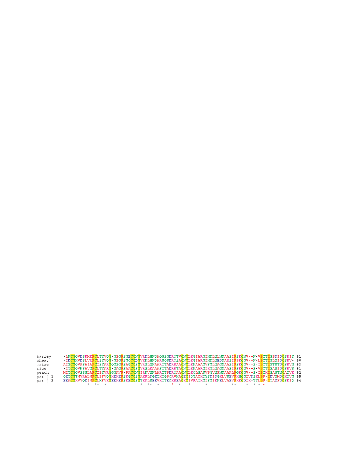
similar pattern of four disulfide links (Fig. 1).
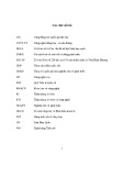
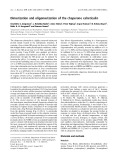
 The identification, isolation and characterization of proteins responsible for IgE-mediated allergy is a nec- essary task for improving both the diagnosis and treatment of this important increasing clinical disor- der. The knowledge of the biochemical role of novel allergens can improve the strategy for their purifica- tion and characterization and, more importantly, it can help to explain the relationships among biological function, protein structure and allergenic activity [14]. Unfortunately, a relatively small number of allergens have been biochemically characterized among the pol- len allergens. Several members of the plant ns-LTP family have been identified as relevant allergens in foods [15]. This allergen family is particularly impor- tant in the Mediterranean area. In addition to foods, allergens of the LTP family have also been identified in other plant sources, such as latex of Hevea brasili- ensis [16] and some pollens. In the latter, LTPs from Ambrosia artemisiifolia [17], Olea europaea [18], Arte- misia vulgaris [19], Arabidopsis thaliana [20], Plat- anus acerifolia [21] and P. judaica pollens [22,23] have been described.
R. Gonza´ lez-Rioja et al.
Two Parietaria allergens behave as ns-LTPs
A
B
although and Par j 2,
to the basic surface compared
C
D
relevant structural peculiarity of One dissimilar overall feature of Par j proteins with respect to ns-LTPs is the net charge. In general, plant ns-LTPs are basic proteins (pI 8–10). By contrast, Par j 1 containing many charged residues (17 positive and 16 negative side chains for Par j 2 versus eight positive and two nega- tive side chains for maize ns-LTP), show almost neu- 
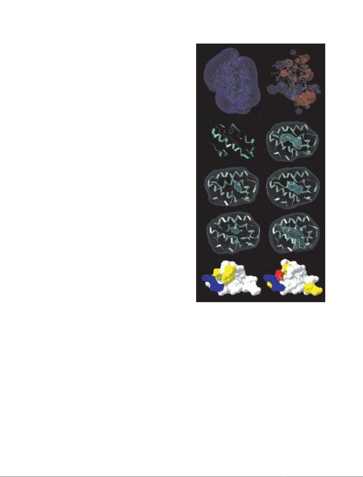
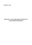
tral isolectric points. The views of the electrostatic surface potential reveal an amphipathic overall Par j 1 structure of ns-LTPmaize (Fig. 2A,B). This seems to be a common feature of allergens in that they appear to contain more charged residues compared to their non-allergic counterparts. The most
E
F
H
G
hydrophobic cavities empty and
I
J
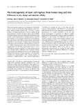
Fig. 2. (A) Electrostatic surface charge potential calculated for ns-LTPmaize (1mzl.pdb) and (B) Par j 1. (C) Ribbon diagram of ns-LTPmaize complexed with palmitate (1mzm.pdb). Tyr81 and Arg46 are shown as a ball and stick model. Surface of the cavities from ns-LTPmaize–palmitate complex (1mzm.pdb) (D), Par j 1 (E) and Par j 2 (F) models, unligated ns-LTPrice (1rzl.pdb) (G) and ns-LTPrice– (palmitate)2 complex (1uvc.pdb) (H), and van der Waals surface rep- resentations of residues facing the cavity of ns-LTPmaize (I) and Par j 1 (J). Hydrophobic residues on the surface are shown in white, polar residues are shown in yellow, negative residues are shown in red and positive residues are shown in blue.
the ns-LTP family is the internal cavity that works as the binding site for different lipidic molecules. In the pres- ent study, voidoo software was used to calculate the van der Waals volumes of the hydrophobic cavities found in the modeled structures. The volume calcu- lated for the cavity found in Par j 1 is 73 A˚ 3 (Fig. 2E) and 200 A˚ 3 in Par j 2 (Fig. 2F). Inspection of known structures shows that a palmitate molecule fills a 600 A˚ 3 cavity in ns-LTPmaize (1mzm.pdb; Fig. 2D), and two molecules the same lipid span throughout the ns-LTPrice molecule occupying an open tunnel of 1345 A˚ 3 (1uvb.pdb; Fig. 2H). On the other hand, the empty cavity of ns-LTPrice has 249 A˚ 3 in the unligated form (1uva.pdb) [28]. Apparently, the volumes of the differ filled significantly with respect to several structures. More- over, ns-LTPs are able to accommodate a wide range of lipidic ligands with little specificity due to the elas- ticity of the C-terminal loop (residue numbers 77–92), which points toward the hydrophobic cavity and blocks the lipid binding pocket in the free form [28] (Fig. 2G,H). According to this observation, it can be inferred that the volume of the empty cavity should not be critical in discriminating between potential ligands. the cavity Conversely,
FEBS Journal 276 (2009) 1762–1775 ª 2009 The Authors Journal compilation ª 2009 FEBS
1764
Tyr81 (number according to maize sequence) that are present in all the plant ns-LTPs. Both residues form hydrogen bonds with the carboxylate groups of fatty acids [29–31] and also act by filling the empty cavity, lipid binding. Arg46 is shifting significantly after in residues delineating ns-LTPs could be considered to be the functionally relevant moieties. Therefore, the character of the side chains lining the cavities of Par j 1 and Par j 2 could provide more revealing insights into the proteins func- tion than the cavity size. An asterisk in Fig. 1 indi- cates residues contacting the lipid in the ns-LTPmaize (1mzm.pdb). Most of these residues have a hydropho- bic nature in all ns-LTPs and also in Par j 1 and Par j 2 sequences, which is consistent with their potential function as lipid binding proteins. Although apolar interactions provide the majority of contacts, there are two important exceptions in Arg46 and
R. Gonza´ lez-Rioja et al.
Two Parietaria allergens behave as ns-LTPs
sensitive to environmental changes, in the absence of tryptophan residues, tyrosine provides an alternative intrinsic fluorophore. Indeed, Tyr81 (according to the maize numbering) fluorescence had been previously [11], used to monitor lipid biding to ns-LTPmaize ns-LTPbarley [33] and ns-LTPwheat [30,34,35].
lie at the nonpolar side of two Tyr residues, Tyr101 and Tyr102,
found in Par j 1 and substituted by a lysine in Par j 2, whereas Tyr81 is absent in both Parietaria sequences. The cavity of maize ns-LTP is highly polarized and mainly hydrophobic on one side, and polar and positively charged on the opposite side, where Arg46 and Tyr81 are located close to each other (Fig. 2I). This polarization appears to be ideally suited for an amphipathic negative molecule within the cavity. Tyr60, the single tyrosine residue found in the polar end as Par j 1 sequence does not expected, but at the cavity (Fig. 2J). Moreover, the net charge of the cavity is neutral due to the presence of Asp37 that compen- sates the charge of Arg46.
CD fluorescence remain
similar spectra respectively). Very
As indicated above, neither Par j 1, nor Par j 2 con- tain a Tyr residue at the corresponding position. How
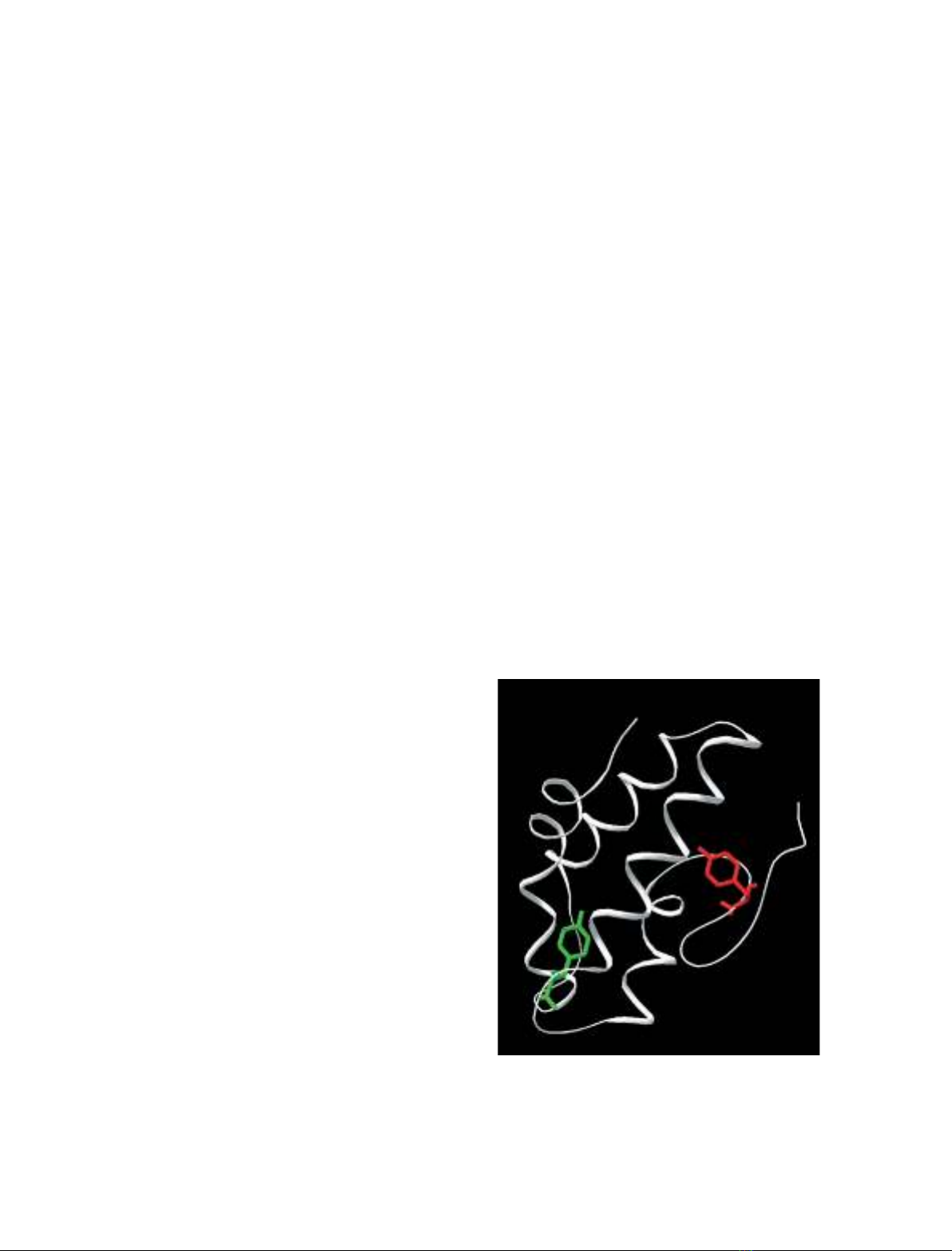
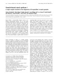
- in the model described for Par j 1, Tyr60 is ever, facing the cavity and, in principle, it can be expected to be sensitive to lipid binding (Fig. 3). Par j 2 con- tains that occupy the last two positions of the sequence. If the proposed models are correct, and these Parietaria proteins bind lipids, a saturable transition should be observed for Par j 1 with the addition of lipid, whereas unchanged. should Par j 2 Figure 4 shows the results obtained for this experi- ment. The titration was performed with 1-oleoyl-2- hydroxy-sn-glycero-3-phosphocholine (OLPC) because this lipid can be suspended in water and does not cause major changes in sample turbity when added sequentially to the protein preparation, unlike other lipids (e.g. oleic acid, also tested in the present study). Tyrosine fluorescence increased significantly in Par j 1 with the addition of OLPC (scattered light contribu- tion of the lipid had been subtracted), whereas only
The overall structure of the ns-LTPs known to date is a four helix bundle with a long C-terminal loop. To control the correct folding of both proteins after purification, CD spectroscopy was performed. CD spectra obtained for the natural mixture were com- pared with those of individual recombinant Par j 1 and Par j 2 expressed in Pichia pastoris (rPar j 1 and rPar j 2, are rPar j 2 and natural Par j 1–Par j 2, obtained for showing a minimum at 208 nm, a well defined shoul- der at 222 nm, and a maximum at 190 nm. The ratio of intensities obtained at 222 and 208 nm, however, for all-a are significantly lower than those typical proteins, suggesting that b or ⁄ and unordered confor- mations are also present in significant amounts. The content of a-helix, b-sheet and unordered structure in Par j 2, as determined by the Fasman protocol [32], was 47%, 11% and 42%, respectively, in good aggrement with secondary structure content in the Par j 2 model; 49 out of 102 residues adopt a helical conformation. The far-UV CD spectrum of rPar j 1 reveals a higher content in unordered conformations. Difference spectra of protein molar ellipticities indi- cate that the 37 extra residues of rPar j 1 are in an unordered conformation and could account for this deviation.
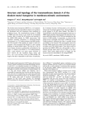
Fig. 3. Ribbon representation of maize ns-LTP structure (1mzl.pdb). Tyr81 is shown in red stick, whereas Tyr60 of Par j 1 is super- imposed in green.
Lipid binding assayed through tyrosine intrinsic fluorescence
FEBS Journal 276 (2009) 1762–1775 ª 2009 The Authors Journal compilation ª 2009 FEBS
1765
Tryptophan fluorescence is frequently used as a means to test protein conformational changes induced by unfolding, ligand binding and other protein transitions. rPar j 1, nor Similarly to plant ns-LTPs, neither rPar j 2 contain tryptophan residues. Although the tyrosine fluorescence quantum yield is lower and less
R. Gonza´ lez-Rioja et al.
Two Parietaria allergens behave as ns-LTPs
A
B
recorded after
the addition of
Fig. 4. Tyrosine intrinsic fluorescence data (excitation at 270 nm, emission at 310 nm) increasing amounts of an aqueous stock solution of OLPC 2 mM to a 1.5 lM protein (filled circles, Par j 1; open circles, Par j 2) preparation in 20 mM NaCl ⁄ Pi. Contributions of identical additions of lipid in the absence of protein are subtracted. Lines correspond to data fitting to Eqn (1).
Fig. 5. 1-Pyrenedodecanoic acid fluorescence data (excitation at 345 nm emission at 375 nm) for increasing concentrations of the probe in the presence of ns-LTPpeach in (A), and the natural mixture nPar j 1–nPar j 2 (open circles), rPar j 1 (squares) and rPar j 2 (dia- monds) in (B), at 0.15 lM protein concentration in 20 mM NaCl ⁄ Pi.
and site an (1), found for the
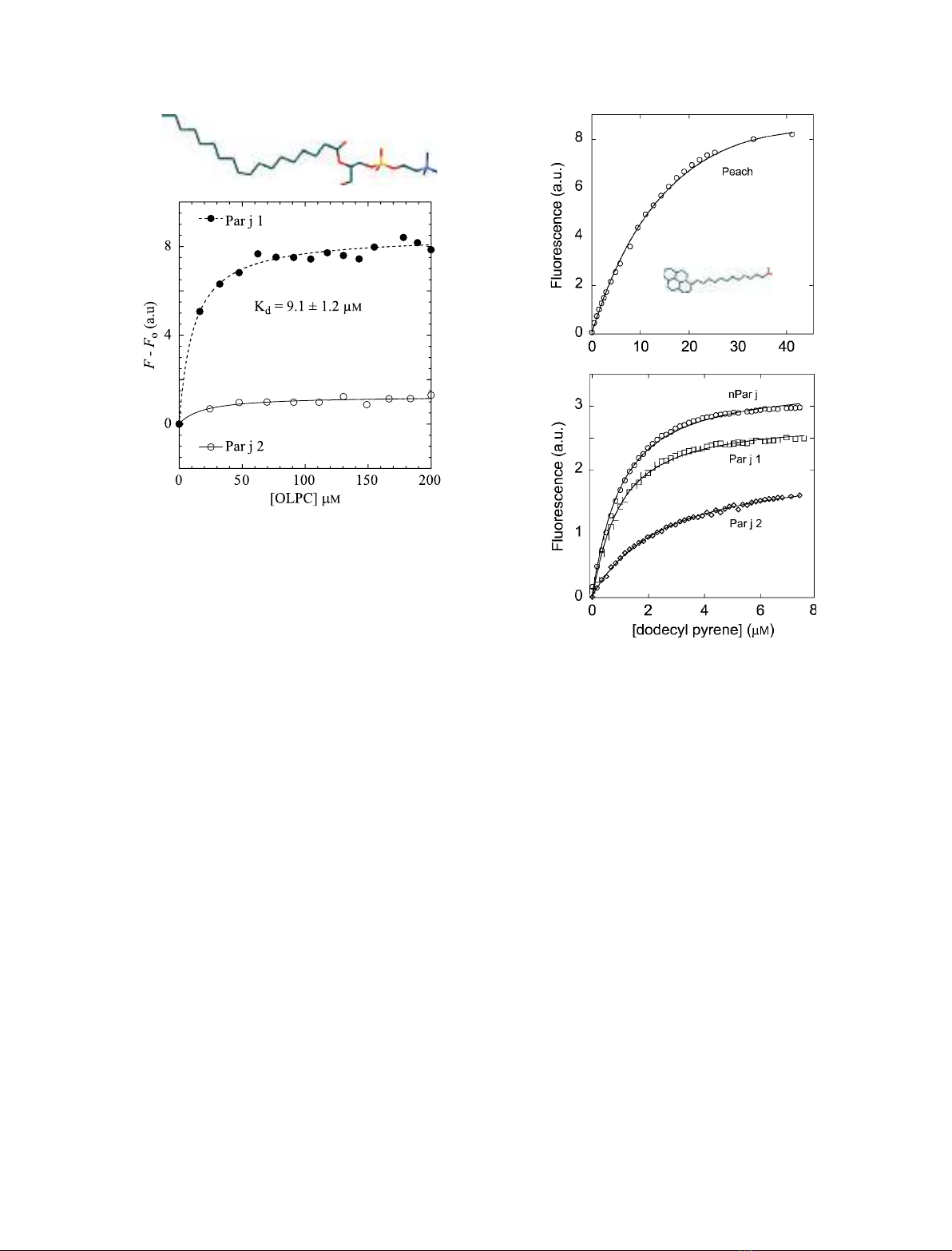
minor changes were observed for the fluorescence cor- responding to the two remote tyrosine residues in the Par j 2 sequence. Data could be fitted to a single bind- estimated ing using Eqn Kd = 9.1 ± 1.2 lm was complex rPar j 1–OLPC. An identical result was obtained when Eqn (2) was used for fitting (n = 1).
0.82 ± 0.03 lm for Lipid binding assayed with a fluorescent lipid probe
natural mixture of 0.15 lm protein in 20 mm sodium phosphate (pH 7.0), are shown in Fig. 5 (lower panel). Equation (2) is used to fit (F ) F0) for calculation of the Kd. Very similar values are obtained for the three proteins: nPar j 1–Par j 2, 0.76 ± 0.03 lm for rPar j 1 and 1.6 ± 0.06 lm for rPar j 2. These Kd values are comparable to those calculated for the binding of other ns-LTPs to monoa- cylated lipids [11] and much lower than the Kd = 27.9 ± 0.03 lm observed for the binding of 1-pyrenedo- decanoic acid to ns-LTPpeach, as also measured in the present study (Fig. 5A).
Lipid transfer activity
FEBS Journal 276 (2009) 1762–1775 ª 2009 The Authors Journal compilation ª 2009 FEBS
1766
Pyrene is an extrinsic fluorophore that exhibits fluores- cence emission maxima at 375 and 395 nm (excitation at 345 nm), attributed to a monomeric pyrene moiety. In addition, it displays an additional fluorescence emis- sion peak at longer wavelengths ((cid:2) 470 nm), which occurs only when two pyrene rings reside within 10 A˚ of each other and form an excited state dimer, usually called an excimer. In the present study, the fluores- cence of 1-pyrenedodecanoic acid was monitored for increasing concentrations of the ligand in the presence of the Par j proteins. Fluorescence data, measured in the titration of the two recombinant proteins and the Large unilamelar liposomes (LUVs) preformed with pure 1-hexadecanoyl-2-(1-pyrenedecanoyl)-sn-glycero-3- phosphoglycerol (b-py-C10-HPG) at 9 lm concentration
R. Gonza´ lez-Rioja et al.
Two Parietaria allergens behave as ns-LTPs
A
the
B
(donor vesicles) were preincubated with 360 lm LUVs of 1,2-dioleoyl-sn-glycero-3-phosphoglycerol (DOPG, acceptor vesicles) for at least 100 s in the fluorescence cuvette before the addition of 0.15 lm protein. Fluore- sence signal of pyrene moiety was registered (kex = 344; kem = 397 nm) for some minutes (Fig. 6B). The same experiment was performed with the neutral phosphoch- oline derivatives with 1-hexadecanoyl-2-(1-pyrenedeca- noyl)-sn-glycero-3-phosphocholine (b-py-C10-HPC) LUVs as donors and 1,2-dioleoyl-sn-glycero-3-phos- phocholine (DOPC) LUVs as acceptors (Fig. 6, lower panel). The activity calculated for canonical ns-LTPpeach was 35 nmolÆmin)1Æmg)1 protein, similar to the reported ns-LTPmaize activity [36]. The values obtained for Parietaria proteins are somewhat lower: 10.3 and 7 nmolÆmin)1Æmg)1 protein for rPar j 1 and rPar j 2, respectively. Activity values calculated with neutral phospholipids are two orders of magnitude lower. However, rPar j 2 transfers lipids more efficiently than ns-LTPpeach when the neutral b-py-C10-HPC ⁄ DOPC pair is used.
Thermal behaviour of native proteins
can be demonstrated by the
Time (s)
Fig. 6. (A) Time trace of fluorescence signal (excitation at 345 nm, emission at 375 nm) after the addition of 0.15 lM ns-LTPpeach (squares), Par j 1 (triangles) and Par j 2 (circles) to two populations of preformed liposomes with 9 lM b-py-C10-HPG and 360 lM DOPG in 50 mM Hepes buffer. (B) Time trace of fluorescence signal as in (A) but with 9 lM b-py-C10-HPC and 360 lM DOPC.
thermal
Figure 8 illustrates the effect of 15 h of incubation of the nPar j 1–Par j 2 with 1, 5 or 10 mm dithithreitol. Reduction induced a dramatic fall in ordered second- ary structures in all cases. The decrease in the CD signal at 222 nm was higher for Par j 2 than for Par j 1. Incubation with 10 mm dithithreitol almost completely destroyed any ordered structure in Par j 2; a flat trace was obtained for the reduced denatured protein under these conditions. Reduced Par j 1, on the other hand, still retained 50% CD sig- nal at 222 nm. This reminiscent structure is lost after thermal melting and is not recovered after cooling down to 10 (cid:2)C.
FEBS Journal 276 (2009) 1762–1775 ª 2009 The Authors Journal compilation ª 2009 FEBS
1767
The natural mixture is assumed to contain both Par j 2 and Par j 1, although the presence of other iso- As indicated above, a Kd constant could not be calcu- lated for the complex rPar j 2–OLPC, due to the absence of tyrosine residues in the hydrophobic cavity of this protein. Alternatively, the binding of a sub- strate stabilization induced on the protein. In the absence of reducing agents, temperature scans of nPar j 1–Par j 2 prepara- tion up to 90 (cid:2)C failed to show a complete melting transition (Fig. 7); instead, a typical baseline shift together with a steep slope at high temperature was observed. The same behaviour was observed for rPar j 1 and rPar j 2 (data not shown). The CD signal at 222 nm changed by less than 5% upon heating to 90 (cid:2)C. After cooling down to the original tempera- ture, an identical spectrum was obtained and a second temperature scan rendered a perfectly superimposable trace. This result suggests that the three protein prep- arations are in an oxidized thermoresistant, native form. Although significant conformational changes are observed in the C-terminal loop of some ns-LTPs upon lipid binding, these do not imply variation in the balance of regular versus non regular structure. In agreement with this, OLPC addition to 170 mm did not exert any changes in the far-UV CD spectra of Par j proteins (Fig. 7). Thus, the addition of a reduc- ing agent appears to be essential for comparing the these proteins and the effect of thermostability of OLPC binding. Par j proteins contain eight cysteine residues potentially involved in four disulfide bridges.
R. Gonza´ lez-Rioja et al.
Two Parietaria allergens behave as ns-LTPs
–5000
40 000
A
B
20 000
–10 000
) g e d m
) g e d m
(
(
0
D C
D C
–15 000
–20 000
–30 000
–20 000
20
40
80
90
10
190
200
240
250
60
220 Wavelength (nm)
Temperature (ºC)
–5000
40 000
C
D
20 000
–10 000
) g e d m
) g e d m
(
(
0
D C
D C
–15 000
–20 000
–30 000
–20 000
60
250
40
90
200
10
20
240
80
190
220 Wavelength (nm)
Temperature (ºC)
Fig. 7. CD spectra (A, C) and temperature scans recorded at 222 nm (B, D) obtained for nPar j 1–Par j 2 in 0 mM (continuous), 1 mM (dashed), 5 mM (dotted) and 10 mM dithiothreitol (dash-dotted) in the absence (A, B) and presence (C, D) of OLPC 170 lM (20 mM NaCl ⁄ Pi).
forms of similar molecular weight cannot be discarded. The behaviour observed in the present study was inter- mediate between those of Par j 1 and Par j 2, compati- ble with the above assumption.
thermal denaturation of
form that Par j 1, Par j 2 became more protected in the presence of OLPC, with a significant preservation of secondary structure. More remarkable is the effect in which 10 mm observed in the natural mixture, dithiothreitol caused only minor changes in secondary structure at 10 (cid:2)C and the thermal transition shifted by almost 10 (cid:2)C, whereas the protein was still ther- in 1 mm dithiothreitol moresistant (Fig. 7). The degree of protection induced by 170 lm OLPC, calcu- lated as the ratio of [dithiothreitol]1 ⁄ 2 concentrations in the presence and absence of the ligand, is 8.5, 13 and 87 for rPar j 1, rPar j 2 and nPar j 1–Par j 2, respectively.
its
FEBS Journal 276 (2009) 1762–1775 ª 2009 The Authors Journal compilation ª 2009 FEBS
1768
Although the Kd value could only be accurately determined for the complex Par j 1–OLPC by the titra- tion monitored by tyrosine fluorescence, the CD results suggest that OLPC binds with a higher affinity to Par j 2 and results in a very stable complexes with the natural mixture nPar j 1–Par j 2. The same experiment, conducted in the presence of 170 lm OLPC, revealed a significant protection versus protein reduction, suggesting effective complex for- mation. Additionally, the partially or totally reduced samples took place at higher temperatures. Figure 8 summarizes the reminis- cent CD signal collected at 222 nm at 10 (cid:2)C after incubation with various dithiothreitol concentrations for 15 h. Figure 8A shows that 1 mm dithiothreitol had the same reducing power for the free Par j 1 as a 10-fold concentration had for complex with OLPC. A 5 (cid:2)C shift of the melting transition was observed for this protein upon lipid binding (data not shown). The effect was more pronounced for Par j 2; although being more sensitive to reduction in its free
R. Gonza´ lez-Rioja et al.
Two Parietaria allergens behave as ns-LTPs
20 000
A
15 000
10 000
5000
two pollen allergens to bind lipids was ability of investigated. The relative sequence homology with ns- LTP together with the functional characterization would confirm Par j 1 and Par j 2 as being members of this protein family.
B
20 000
) 1 – l o m d
15 000
10 000
· 2 m c · g e d ( y t i c i t p
i l l
l
e r a o M
-
5000
20 000
with observed transition
C
15 000
10 000
5000
0
2
4
8
10
12
6 [dithiothreitol] (mM)
Fig. 8. CD signal recorded at 222 nm and 10 (cid:2)C after 15 h of incu- bation with varying concentrations of the reducing agent dithio- threitol in the absence (filled circles) and presence (open circles) of 170 lM OLPC: (A) rPar j 1, (B) rPar j 2 and (C) nPar j 1–Par j 2.
In the present study, we have shown that a monoa- cylated lipid such as OLPC is able to alter the fluores- the intrinsic probe Tyr60 in the Par j 1 cence of the purified proteins sequence and also stabilizes against reduction. Intrinsic fluorescence was used to monitor lipid binding to ns-LTPs and the observed sig- increase fitted known binding models. A single nal a is well-resolved Kd = 9.1 ± 1.2 lm compatible with a 1 : 1 complex. This value compares well with the Kd calculated for lysophospholipids and other ns-LTPs. Kd values of 10.1 lm and 1.9 lm were obtained for lyso-C16 (1-pal- mitoyl-l-a-lysophosphatidylcholine) with ns-LTPwheat and ns-LTPmaize, respectively [11], although values of 28.9 lm have also been reported for the complex of lyso-C16 with ns-LTPwheat [35]. Dissociation constants of approximately 0.5 lm were measured for the com- plexes of ns-LTPwheat and lysophospholipids and phospholipids with side chains from C14 to C18, inde- pendent of the presence of one or two insaturations [30]. Furthermore, a Kd = 7.5 lm was reported for the interaction of dimyristoyl phosphatidylglycerol small [34]. Although Par j 2 liposomes with ns-LTPwheat intrinsic fluorescence is insensitive to lipid binding and a Kd could not be measured, the CD experiments sug- gested that Par j 2 is binding with a higher affinity to OLPC than Par j 1.
Discussion
structural protein families
superfamily storage (cereal
FEBS Journal 276 (2009) 1762–1775 ª 2009 The Authors Journal compilation ª 2009 FEBS
1769
Allergens are found in only 2% of all sequence-based and 5% of all [37]. Sequences encoded in plant genomes, included in the prolamin proteins, nsLTPs, 2S storage albumins and inhibitors of trypsin and a-amilase), account for 65% of plant food aller- gens [38]. ns-LTPs bind a variety of lipidic molecules from fatty acids to phospholipids and are able to the transport lipids in vitro. In the present study, The Kd for the interaction between 1-pyrenedodeca- noic acid and Par j 1 and Par j 2 was also measured. A value in the micromolar range was calculated for the interaction with the three protein preparations assayed in the present study: Par j 1: 0.76 ± 0.03 lm, 1.6 ± 0.06 lm nPar j 1–Par j 2: and Par j 2: 0.82 ± 0.03 lm. Zachowski et al. [39] showed that ns-LTPwheat and ns-LTPmaize. can bind two molecules of 1-pyrenedodecanoic acid by means of the fluores- cence quenching of pyrene that followed the first signal increase. By contrast to OLPC, the colocalization of two molecules of analogues in the binding site would induce a fluorescence quenching. A Kd could not be calculated for ns-LTPwheat and ns-LTPmaize, although there are data available [39,40] suggesting that the affinity is much higher than for OLPC, with a Kd in the submicromolar range. No apparent decrease in fluoresence signal was observed after the first satura- suggesting tion in the titration of Par j proteins, that Par j proteins offer a single binding site for 1-pyrenedodecanoic acid. ns-LTPpeach has also been
R. Gonza´ lez-Rioja et al.
Two Parietaria allergens behave as ns-LTPs
assayed in the present study for comparison (Fig. 5). A much higher Kd = 27.8 ± 4 lm was obtained in this case.
important residues are absent (a)
[33], ns-LTPbarley ns-LTPwheat [39,40]. Moreover, An analysis of the structural details of the molecular models of Par j 1 and Par j 2 revealed a notable resem- blance with other ns-LTPs, together with some distinct structural details that could be relevant for protein function: in the sequence of Parietaria proteins; (b) Par j 1 and Par j 2 are markedly less basic than other ns-LTPs; and (c) their internal hydrophobic cavities seem to be smaller and less polarized compared to other members of the family. Comparison of
conserved hydrophobic position of the other ns-LTPs [44]. Accordingly, it appears that minor sequence dif- ferences are able to switch from one binding mode to the other. Both orientations can even coexist in the same complex in ns-LTPrice [28], ns-LTPwheat [29] and ns-LTPpeach [45]. In both conformations, the apolar part of the lipid would be in the interior of the cav- ity, whereas the polar head can be to either extremes of the cavity, providing two modes of binding exactly opposite to each other. Likewise, two binding sites have also been proposed from spectroscopic studies [30,40] and for in some known ns-LTPmaize complexes, the lipidic chain stretches out of the bind- ing pocket with the polar head group protruding out, facing the solvent [45], with no interactions with the protein. This may explain why the calculated Kd val- ues for lipid complexes of Par j proteins rank in the same order as their homologues, despite the absence of the highly conserved Tyr81.
efficiently that
Another molecular distinct feature that is shown to have little effect is the net protein charge as also illustrated by the surface electrostatic potential. Of the two monoacylated lipidic derivatives used in the present study, the negative 1-pyrenedodecanoic acid binds with a higher affinity than the zwitterionic OLPC to Par j 1. Also, negative phospholipids are than neutral homo- transferred more logues from LUVs. These results suggest the loss of the net positive charge of the protein is not related to a marked preference for neutral lipids. This overall feature is more likely to be related to the location where these proteins exert their in vivo than any specificity for particular function rather lipids.
FEBS Journal 276 (2009) 1762–1775 ª 2009 The Authors Journal compilation ª 2009 FEBS
1770
the binding modes of different ns-LTPs suggests that, although the sequences and the 3D structures are very similar among plant ns-LTPs, the binding modes of these proteins differ substantially. The binding site of ns-LTPs is a hydro- phobic groove in the globular helical structure, which is covered by the C-terminal peptide. The majority of the residues lining the cavity are hydrophobic, with few exceptions. A major role has been conferred to these polar side chains. For some plant ns-LTP com- plexes, the highly conserved Arg46 and Tyr81 form hydrogen bonds with the carboxylate groups of fatty acids [29–31]. They also act by filling the empty cavity and they both shift significantly after lipid binding. This highly conserved tyrosine among ns-LTPs is absent in Par j 1 and Par j 2. The ns-LTP tunnel has a wide opening in one end and a narrow opening in the other end [41]. The wide opening is considered to be the entrance. Most polar residues reside in this side and it is also where the carboxylate binds. The narrow opening is considered to be a closed exit. This is where the methyl group binds, surrounded by hydrophobic side chains from the protein. Thus, the binding site results in a polarized cleft in the interior of a basic container with two main openings to bulk solvent, which appears to be ideally suited for fitting small molecule. Lipid– an negative amphipathic [31,41], protein 1 : 1 for ns-LTPmaize complexes indicate that [29,42] and ns-LTPrice [28] ns-LTPwheat this appears to be the preferred mode of binding and ligand orientation. However, the opposite orientation has been observed in complexes with ns-LTPbarley [43,44]. With ns-LTPbarley, it has been shown that the fatty acid or fatty acylCoA adopts a different orienta- tion within the protein cavity and Tyr81 is involved only in hydrophobic interaction with the aliphatic chain, whereas no hydrogen bond can be formed with the lipid polar head group. The inversion of the coor- dination of the ligand in ns-LTPbarley has been related to the charged Lys9 replacing the corresponding Nonetheless, the experimental data obtained in the present study explain certain differences that were visi- ble in the models. The small cavity detected by voidoo software is shown experimentally to provide a single binding site for monoacylated lipids under conditions where other ns-LTPs are able to bind two lipid mole- cules. The tunnel volume of Par j 1 and Par j 2 models are rather small compared to other ns-LTPs, mainly due to some bulky side chains together with a one and two residue insertion, respectively, in the C-terminal loop. It is demonstrated that ns-LTPs have a consider- able capability of expansion. The present results, how- ever, suggest that the Par j 1 and Par j 2 cavities do not appear to be able to spread out to accommodate two lipidic ligands. It is possible that Parietaria pro- teins are more specialized in monoacylated lipids, whereas other plant ns-LTPs are designed to accept bigger ligands, such as diacylated phospholipids. This
R. Gonza´ lez-Rioja et al.
Two Parietaria allergens behave as ns-LTPs
Experimental procedures
allergens
Purification of natural and recombinant P. judaica major allergens
Natural (natural Par j 1–Par j 2 mix) were immunopurified from defatted pollen from P. judaica (Iber- pollen, Ma´ laga, Spain) using polyclonal rabbit anti- Par j 1–Par j 2 sera coupled to a CNBr-activated sepharose 4B column, as described previously [52]. Coding regions of Par j 1 and Par j 2 were amplified and cloned in pPIC9 and expressed in the methylotrophic yeast P. pastoris as described previously [53]. Purification of the recombinant proteins was carried out by immunoaffinity chromatogra- phy as described previously [53]. Protein concentration was determined using the method of Gill and Von Hippel [54]. ns-LTPpeach was obtained as previously described [55].
is partially demonstrated when the affinities for mono- acylated lipids and activities for diacylated lipids are compared for Parietaria proteins and the canonical ns-LTPpeach. 1-Pyrenedodecanoic acid binds with a higher affinity to Parietaria proteins. Conversely, ns-LTPpeach is able to transfer b-py-C10-HPG with greater efficiency.
[56]. Ns-LTPmaize
Molecular modeling of Par j 1 and Par j 2
The homology model of the N-terminal region of Par j 1 and Par j 2 (Fig. 1) was generated using swiss-model at the expasy molecular biology server (http://www.expasy.ch/ (Pro- swissmod/SWISS-MODEL.html]) tein Databank code: 1mzl) was selected as modeling tem- plate (33% and 34% identity with Par j 1 and Par j 2, respectively. Par j 1 is 52% identical to Par j 2). Despite of the low sequence identity, the four cystines impose powerful restraints, largely assisting homology modeling. The final total energy of the calculated model is 1946 KJÆmol)1. The lowest energy structure was subject to 100 cycles of unre- strained Powell minimization using cns [57].
experimentally determined an
Cavity volumes within Par j 1 and Par j 2 were computed with voidoo software [58] using a probe radius of 1.4 A˚ , and were visualized with the o software [59].
Cavity volume calculations and display
Plant LTPs are prefixed nonspecific (ns) because they show very broad specificity. In the present study, four very dissimilar lipid derivatives are shown to be able to bind to these P. judaica allergens, with affini- ties similar to other ns-LTPs. However, the Kd calcu- lated for 1-pyrenedodecanoic acid is one order of magnitude lower than for OLPC, and rPar j 2 trans- fers b-py-C10-HPG 10-fold more efficiently than b-py- C10-HPC (i.e. this ratio is 40-fold for rPar j 1 and suggests a certain 100-fold for ns-LTPpeach). This degree of specificity for monoacylated negative phos- pholipids for Parietaria proteins. On the other hand, Par j 2 binds and transfers neutral phospholipid better than Par j 1, whereas Par j 1 works better with nega- tive lipids. The bigger cavity of 200 A˚ 3 found in the model or Par j 2 compared to the 73 A˚ 3 cavity of Par j 1 may explain this preference because phospho- choline is bigger than the phoshoglycerol moiety. However this cannot be stated clearly in the absence of 3D structure, because, in general, the lipid molecules interact with the ns-LTPs binding cavity mainly through hydropho- bic interactions. Although some known complexes exhibit definite hydrogen bonds between protein side chains with the carboxylate group of fatty acids or the hydroxyl group of the glycerol phospholipid back- bone, in other complexes, the polar head group is not in contact with the protein [44]. Regretfully, crystalli- zation trials with rPar j 1 and rPar j 2 have so far proved unsuccessful, most probably due to flexibility of C-terminal extensions.
Fluorescence spectroscopy
Titration experiments with OLPC were conducted at 25 (cid:2)C with a SLM Bowman Series 2 luminiscence spectrometer (Aminco, Lake Forest, CA, USA). Tyrosine fluorescence was monitored with excitation and emission wavelengths at 275 and 310 nm, with 2 and 4 nm bandwidth, respectively. Buffer contributions were corrected and inner filter effect than was negligible, with a sample absorbance lower 0.05 units. One microliter aliquotes of 200 lm to 20 mm OLPC preparations in 20 mm sodium phosphate (pH 7.0) were added stepwise to a cuvette containing 100 lL of a 1.5 lm Par j 1 solution in the same buffer. Volume changes were also taken into account (i.e. a maximum increase of 15% at the end of the titration).
features essential
FEBS Journal 276 (2009) 1762–1775 ª 2009 The Authors Journal compilation ª 2009 FEBS
1771
These versatile, malleable and nonspecific proteins are able to bind hydrophobic molecules in different cellular contexts. It is not expected that discriminating structural for ligand binding will readily become apparent. Conversely, binding modes and clues appear to be redundant and unspecialized, which, together with the coexistence of isoforms [46– 48], suggests that promiscuity is probably of major functional relevance. The present study provides some evidence that Par j 1 and Par j 2 are structurally and functionally related to this group of proteins and are able to transfer lipids in vitro. LTPs have been usually identified from in vitro activities. Only recently has strong evidence become available for lipid transfer in living cells [49–51].
R. Gonza´ lez-Rioja et al.
Two Parietaria allergens behave as ns-LTPs
The binding of a fluorescent lipid derivative, 1-pyrenedo- decanoic acid, was also tested. The lipid was added from a concentrated stock solution in ethanol to 0.15 lm protein preparations in 20 mm sodium phosphate (pH 7.0). Emis- sion signals at 375 nm with excitation at 345 nm through 2–4 nm slits were collected at 25 (cid:2)C. A baseline experiment was carried out in the absence of protein under the same conditions.
evaporated to dryness under a stream of nitrogen. Traces of solvent were removed by evacuating the samples under high vacuum for at least 2 h. The samples were hydrated at 45 (cid:2)C in 50 mm Hepes (pH 7.4), helping dispersion by stir- ring with a glass rod. The solution was frozen in liquid nitrogen and defrozen at 45 (cid:2)C 10 times. LUVs were pre- pared by the extrusion method [60], using polycarbonate filters with a pore size of 0.1 lm (Nuclepore, Pleasanton, CA, USA). Vesicle sizes were determined by dynamic light scattering using a Malvern Zetasizer instrument (Malvern Instruments, Malvern, UK). The average vesicle diameter was 90–100 nm. Four independent liposomes populations were prepared with b-py-C10-HPC, b-py-C10-HPC, DOPG and DOPC in 50 mm Hepes.
It is possible to determine the Kd by monitoring tyrosine intrinsic fluorescence changes induced by lipid binding. When maximum bound lipid is considerably lower than Kd, a major fraction of the added ligand remains free in solu- tion. The following equation can be used for those cases:
ð1Þ
F ¼ F0 þ
ðFmax (cid:3) F0Þ (cid:4) ½lipid(cid:5)free Kd þ ½lipid(cid:5)free
For the lipid transfer experiments, 0.15 lm protein was added to a mixture of 9 lm b-py-C10-HPC liposomes and 360 lm DOPC liposomes in 50 mm Hepes. The same was carried out with a mixture of 9 lm b-py-C10-HPG lipo- somes and 360 lm DOPG liposomes. The increase in pyrene fluorescence signal was measured at 375 nm after excitation at 345 nm through 2 and 4 nm slits, respectively, in an SLM Bowman Series 2 luminiscence spectrometer (Aminco) at 25 (cid:2)C.
Dissociation constant determination
where F is the fluorescence value recorded at 275 nm excita- tion and 310 nm emission, F0 is the fluorescence of the free protein before lipid addition, Fmax is the fluorescence value at saturating lipid concentrations and [lipid]free is the con- centration of lipid that remains unbound. Particularly for low Kd values, [lipid]free should be calculated by substract- ing bound lipid from the total added concentration:
CD
acid. The
single binding
a
where n is the number of binding sites per protein mole- cule and [lipid]T is the total concentration of added lipid. A 1 : 1 ratio has been contemplated here for Par j proteins transition is with OLPC because observed.
Far-UV (190–250 nm) CD spectra were recorded with a Jasco J-810 spectropolarimeter (Jasco Analitica Spain S.L., Madrid, Spain), which was previously calibrated with d-10-camphorsulphonic device was equipped with a Jasco PTC-423S temperature controller and cuvettes were thermostatted at 20 (cid:2)C. The protein concentration was 0.035 mgÆmL)1 in 20 mm NaCl ⁄ Pi (pH 7.0) in a 0.2 cm cuvette. Proteins were diluted to the final concentrations required for CD analysis in the pres- ence of the desired additives and incubated overnight to allow the reduction reaction to proceed completely. Just
Alternatively, Kd can be calculated using the equation:
ð2Þ ½lipid(cid:5)free ¼ ½lipid(cid:5)T (cid:3) n (cid:4) ½protein(cid:5) (cid:4) ðF (cid:3) F0Þ ðFmax (cid:3) F0Þ
where all symbols are as indicated above. Curve fitting was carried out using kaleidagraph software (Sinergy Soft- ware, Reading, PA, USA).
q Þ ðn (cid:4) ½protein(cid:5) þ Kd þ ½lipid(cid:5)TÞ (cid:3) ffiffiffiffiffiffiffiffiffiffiffiffiffiffiffiffiffiffiffiffiffiffiffiffiffiffiffiffiffiffiffiffiffiffiffiffiffiffiffiffiffiffiffiffiffiffiffiffiffiffiffiffiffiffiffiffiffiffiffiffiffiffiffiffiffiffiffiffiffiffiffiffiffiffiffiffiffiffiffiffiffiffiffiffiffiffiffiffiffiffiffiffiffiffiffiffiffiffiffiffiffiffiffiffiffiffiffiffiffiffiffi ðn (cid:4) ½protein(cid:5) þ Kd þ ½lipid(cid:5)TÞ2 (cid:3) ð4 (cid:4) n (cid:4) ½protein(cid:5) (cid:4) ½lipid(cid:5)T ð3Þ F ¼ F0 þ ðFmax (cid:3) F0Þ (cid:6) 2 (cid:4) n (cid:4) ½protein(cid:5)
b-py-C10-HPC and b-py-C10-HPG were purchased from Molecular Probes (Eugene, OR, USA). DOPG and DOPC were obtained from Avanti Polar Lipids Inc. (Alabaster, AL, USA).
before measurement, samples were centrifuged for 15 min at 14 000 g in an Eppendorf microcentrifuge at 4 (cid:2)C. All the spectra were subtracted by the appropriate back- ground and converted to mean residue ellipticity. Second- ary structure content was determined in the spectral range 190–240 nm by means of several methods of analysis, compiled in dicroprot software [61]. Thermally induced unfolding was monitored by CD at 222 nm in 0.2 cm pathlength cuvettes in the temperature range 277–363 (cid:2)K. The temperature was increased stepwise by 0.2 (cid:2)K at a rate of 60 (cid:2)KÆh)1, and the ellipticity was recorded with a 1 nm bandwidth and a 2 s response. Melting temperatures
For liposome preparation, phospholipids were dissolved in chloroform : methanol (2 : 1, v ⁄ v), and the mixture was
FEBS Journal 276 (2009) 1762–1775 ª 2009 The Authors Journal compilation ª 2009 FEBS
1772
Vesicle preparation
R. Gonza´ lez-Rioja et al.
Two Parietaria allergens behave as ns-LTPs
(Tm) were calculated as the maxima of the first derivatives of the temperature transition curves.
Acknowledgements
13 Poznanski J, Sodano P, Suh SW, Lee JY, Ptak M & Vovelle F (1999) Solution structure of a lipid transfer protein extracted from rice seeds. Comparison with homologous proteins. Eur J Biochem 259, 692–708. 14 Aalberse RC (2000) Structural biology of allergens.
J Allergy Clin Immunol 106, 228–238.
15 Salcedo G, Sa´ nchez-Monge R, Diaz-Perales A, Garcia- Casado G & Barber D (2004) Plant non-specific lipid transfer proteins as food and pollen allergens. Clin Exp Allergy 34, 1336–1341.
R. Gonza´ lez-Rioja is indebted to the Departamento de Industria, Comercio y Turismo and the Departamento de Educacio´ n, Universidades e Investigacio´ n (Gobi- erno Vasco) for a predoctoral fellowship.
References
1 Yamada M (1992) Lipid transfer proteins in plants and
16 Beezhold DH, Hickey VL & Kostyal DA (2003) Lipid transfer protein from Hevea brasiliensis (Hev b 12), a cross-reactive latex protein. Ann Allergy Asthma Immu- nol 90, 439–445.
microorganisms. Plant Cell Physiol 33, 1–6.
17 Hiller KM, Lubahn BC & Klapper DG (1998) Cloning and expression of a ragweed allergen Amb a 6. Scand J Immunol 48, 26–36.
2 Kader JC (1997) Lipid transfer proteins: a puzzling family of plant proteins. Trends Plant Sci 2, 66–70. 3 Kader JC (1996) Lipid-transfer proteins in plants. Annu
18 Tejera ML, Villalba M, Batanero E & Rodrı´ guez R
Rev Plant Physiol Plant Mol Biol 47, 627–654.
(1999) Identification, isolation, and characterization of Ole e 7, a new allergen of olive tree pollen. J Allergy Clin Immunol 104, 797–802.
4 Douliez JP, Pato C, Rabesona H, Molle D & Marion D (2001a) Disulfide bond assignment, lipid transfer activity and secondary structure of a 7-kDa plant lipid transfer protein, LTP2. Eur J Biochem 268, 1400– 1403.
5 Garcı´ a-Olmedo F, Molina A, Segura A & Moreno M (1995) the defensive role of non-specific lipid–transfer proteins. Trends Microbiol 3, 72–74.
6 Ge X, Chen J, Sun C & Cao K (2003) Preliminary
19 Dı´ az-Perales A, Lombardero M, Sa´ nchez-Monge R, Garcı´ a-Selles FJ, Pernas M, Ferna´ ndez-Rivas M, Barber D & Salcedo G (2000) Lipid transfer proteins as potential plant panallergens, cross-reactivity among proteins of Artemissia pollen, Castanea nut and Rosaceae fruits, with different IgE-binding capacities. Clin Exp Allergy 30, 1403–1410.
20 Chardin H, Mayer C & Senechal H (2003) Lipid trans- fer protein 1 is a possible allergen in Arabidopsis thali- ana. Int Arch Allergy Immunol 131, 85–90.
21 Lauer I, Miguel-Mocin MS, Abel T, Foetisch K,
study on the structural basis of the antifungal activity of a rice lipid transfer protein. Protein Eng 16, 387–390. 7 Buhot N, Gomes E, Milat ML, Ponchet M, Marion D, Lequeu J, Delrot S, Coutos-Thevenot P & Blein JP (2004) Modulation of the biological activity of a tobacco LTP1 by lipid complexation. Mol Biol Cell 15, 5047–5052.
Hartz C, Fortunato D, Cistero-Bahima A, Vieths S & Scheurer S (1997) Identification of a plane pollen lipid transfer protein (Pla a 3) and its immunological relation to the peach lipid-transfer protein, Pru p 3. Clin Exp Allergy 37, 261–269.
8 Carvalho AO & Gomes VM (2007) Role of plant lipid transfer proteins in plant cell physiology. Peptides 28, 1144–1153.
9 Vega-Maray AM, Ferna´ ndez-Gonza´ lez D, Valencia-
22 Costa MA, Colombo P, Izzo V, Kennedy H, Venturella S, Cocchiara R, Mistrello G, Falagiani P & Geraci D (1994) cDNA cloning, expression and primary structure of Par j 1, a major allergen of Parietaria judaica pollen. FEBS Lett 341, 182–186.
Barrera R, Polo F, Seoane-Camba JA & Sua´ rez-Cerve- ra M (2004) Lipid transfer proteins in Parietaria judaica L. pollen grains: immunocytochemical localization and function. Eur J Cell Biol 83, 493–497.
10 Blein JP, Coutos-Thevenot P, Marion D & Ponchet M (2002) From elicitins to lipid-transfer proteins: a new insight in cell signalling involved in plant defence mech- anisms. Trends Plant Sci 7, 293–296.
23 Duro G, Colombo P, Costa MA, Izzo V, Porcasi R, Di Fiore R, Locotorondo G, Mirisola MG, Cocchiara R & Geraci D (1996) cDNA cloning, sequence analysis and allergological characterizacio´ n of Par j 2.0101, a new major allergen of the Parietaria judaica pollen. FEBS Lett 399, 295–298.
24 Colombo P, Bonura A, Costa MA, Izzo V, Passantino R, Locorotondo G, Amoroso S & Geraci D (2003) The allergens of Parietaria. Int Arch Allergy Immunol 130, 173–179.
25 Colombo P, Kennedy D, Ramsdale T, Costa MA, Duro
G, Izzo V, Salvadori S, Guerrini R, Cocchiara R,
11 Gomar J, Petit MC, Sodano F, Sy D, Marion D, Kader JC, Vovelle F & Ptak M (1996) Solution structure and lipid binding of a nonspecific lipid transfer protein extracted from maize seeds. Protein Sci 5, 565–577. 12 Lee JY, Min K, Cha H, Shin DH, Hwang KY & Suh SW (1998) Rice non-specific lipid transfer protein: the 1.6 A˚ crystal structure in the unliganded state reveals a small hydrophobic cavity. J Mol Biol 276, 437–448.
FEBS Journal 276 (2009) 1762–1775 ª 2009 The Authors Journal compilation ª 2009 FEBS
1773
R. Gonza´ lez-Rioja et al.
Two Parietaria allergens behave as ns-LTPs
families and possess a restricted number of biochemical functions. J Allergy Clin Immunol 121, 847–852.
Mirisola MG et al. (1998) Identification of an immuno- dominant IgE epitope of the Parietaria judaica major allergen. J Immunol 160, 2780–2785.
38 Jenkins JA, Grifffiths-Jones S, Shewry PR, Breiteneder H & Mills ENC (2005) Structural relatedness of plant food allergens with specific reference to cross-reactive allergens: an in silico analysis. J Allergy Clin Immunol 115, 163–170.
26 Asturias JA, Go´ mez-Bayo´ n N, Eseverri JL & Martı´ nez A (2003) Par j 1 and Par j 2, the major allergens from Parietaria judaica pollen, have similar immunoglobulin E epitopes. Clin Exp Allergy 33, 518–524.
27 Amoresano A, Pucci P, Duro G, Colombo P, Costa
39 Zachowski A, Guerbette F, Grosbois M, Jolliot-Cro- quin A & Kader JC (1998) Characterisation of acyl binding by a plant lipid-transfer protein. Eur J Biochem 257, 443–448.
MA, Izzo V, Lamba D & Geraci D (2003) Assignment of disulphide bridges in Par j 2.0101, a major allergen of Parietaria judaica pollen. Biol Chem 384, 1165–1172.
40 Guerbette F, Grobois M, Jolliot-Croquin A, Kader JC
28 Cheng HC, Cheng PT, Peng P, Lyu PC & Sun YJ
(2004) Lipid binding in rice nonspecific lipid transfer protein-1 complexes from Ozyva sativa. Protein Sci 13, 2304–2315.
& Zachowski A (1999) Comparison of lipid binding and transfer properties of two lipid transfer proteins from plants. Biochemistry 38, 14131–14137.
41 Shin DH, Lee JY, Hwang KY, Kim KK & Suh SW (1995) High resolution crystal structure of the non- specific lipid-transfer protein from maize seedlings. Structure 3, 189–199.
42 Tassin-Moindrot S, Caille A, Douliez J-P & Vovelle F
29 Charvolin D, Douliez JP, Marion D, Cohen-Addad C & Pebay-Peyroula E (1999) The crystal structure of a wheat nonspecific lipid transfer protein (ns-LTP1) complexed with two molecules of phospholipid at 2.1 A˚ resolution. Eur J Biochem 264, 562–568.
30 Douliez JP, Michon T & Marion D (2000) Steady-state tyrosine fuorescence to study the lipid-binding proper- ties of a wheat non-specific lipid-transfer protein (nsLTP1). Biochim Biophys Acta 1467, 65–72.
(2000) The wide binding properties of a wheat nonspecific lipid-transfer protein. Solution structure of a complex with prostaglandin B2. Eur J Biochem 267, 1117–1124. 43 Lerche MH, Kragelund BB, Bech LM & Poulsen FM (1997) Barley lipid-transfer protein complexed with palmitoyl CoA: the structure reveals a hydrophobic binding site that can expand to fit both large and small lipid-like ligands. Structure 5, 291–306.
31 Han GW, Lee JY, Song HK, Chang C, Min K, Moon J, Shin DH, Kopka ML, Sawaya MR, Yuan HS et al. (2001) Structure basis of non-specific lipid binding in maize lipid–transfer protein complexes revealed by high- resolution x-ray crystallography. J Mol Biol 308, 263– 278.
44 Lerche MH & Poulsen FM (1998) Solution structure of barley lipid transfer protein complexed with palmitate. Two different binding modes of palmitate in the homologous maize and barley nonspecific lipid transfer proteins. Protein Sci 7, 2490–2498.
45 Pasquato N, Berni R, Folli C, Folloni S, Cianci M,
Pantano S, Helliwell J & Zanotti G (2006) Crystal structure of peach Pru p 3, the prototypic member of the family of plant non-specific lipid transfer protein pan-allergens. J Mol Biol 356, 684–694.
32 Venyaminov SY, Yang JT & Fasman GD (eds) (1996). Circular Dichroism and the Conformational Analysis of Biomolecules, pp. 69–107. Plenum, New York, NY. 33 Douliez JP, Je´ gou S, Pato C, Molle D, Vinh Tran V & Marion D (2001) Binding of two mono-acylated lipid monomers by the barley lipid transfer protein, LTP1, as viewed by fluorescence, isothermal titration calorimetry and molecular modelling. Eur J Biochem 268, 384–388.
46 Tsuboi S, Osafune T, Nishimura M & Yamada M
34 Sodano P, Caille A, Sy D, de Person G, Marion D &
Ptak M (1997) 1H NMR and fluorescence studies of the complexation of DMPG by wheat non-specific lipid transfer protein. Global fold of the complex. FEBS Lett 416, 130–134.
35 Lullien-Pellerin V, Devaux C, Ihorai T, Marion D,
(1992) Nonspecific lipid transfer protein in castor bean cotyledon cells: subcellular localization and a possible role in lipid metabolism. J Biochem 111, 500–508. 47 Pyee J, Yu H & Kolattukudy PE (1994) Identification of a lipid transfer protein as the major protein in the surface wax of broccoli (Brassica oleracea) leaves. Arch Biochem Biophys 331, 460–468.
48 Takishima K, Watanabe S, Yama da M, Suga T &
Pahin V, Joudrier P & Gautier MF (1999) Production in Escherichia coli and site-directed mutagenesis of a 9-kDa nonspecific lipid transfer protein from wheat. Eur J Biochem 260, 861–868.
Mamiya G (1988) Amino acid sequences of two nonspe- cific lipid-transferproteins from germinated castor bean. Eur J Biochem 177, 241–249.
49 Hanada K, Kumagai K, Tomishige N & Kawano M
(2007) CERT and intracellular trafficking of ceramide. Biochim Biophys Acta 1771, 644–653.
36 Moreau F, Davy de Virville J, Hoffelt M, Guerbette F & Kader JC (1994) Use of a fluorimetric method to assay the binding and transfer of phospholipids by lipid transfer proteins from maize seedlings and Arabidopsis leaves. Plant Cell Physiol 35, 267–274.
50 D’Angelo G, Polishchuk E, Di Tullio G, Santoro M,
Campli AD, Godi A, West G, Bielawski J, Chuang CC,
37 Radauer C, Merima B, Wagner S, Mari A & Breitene- der H (2008) Allergens are distributed into few protein
FEBS Journal 276 (2009) 1762–1775 ª 2009 The Authors Journal compilation ª 2009 FEBS
1774
R. Gonza´ lez-Rioja et al.
Two Parietaria allergens behave as ns-LTPs
van der Spoel AC et al. (2007) Glycosphingolipid syn- thesis requires FAPP2 transfer of glucosylceramide. Nature 449, 62–67.
56 Arnold K, Bordoli L, Kopp J & Schwede T (2006) The SWISS-MODEL Workspace: a web-based environment for protein structure homology modelling. Bioinformat- ics 22, 195–201.
57 Bru¨ nger AT, Adams PD, Clore GM, DeLano WL,
51 Halter D, Neumann S, van Dijk SM, Wolthoorn J, de Mazie` re AM, Vieira OV, Mattjus P, Klumperman J, van Meer G & Sprong H (2007) Pre- and post-Golgi translocation of glucosylceramide in glycosphingolipid synthesis. J Cell Biol 179, 101–115.
52 Arilla MC, Gonzalez-Rioja R, Ibarrola I, Mir A,
Gros P, Grosse-Kunstleve RW, Jiang JS, Kuszewski J, Nilges M, Pannu NS et al. (1998) Crystallography and NMR system (CNS): a new software system for macro- molecular structure determination. Acta Crsytallogr D 54, 905–921.
58 Kleywegt GJ & Jones TA (1994) Detection, delineation, measurement and display of cavities in macromolecular structures. Acta Crystallogr D 50, 178–185.
Monteseirin J, Conde J, Martinez A & Asturias JA (2006) A sensitive monoclonal antibody-based enzyme- linked immunosorbent assay to quantify Parietaria juda- ica major allergens, Par j 1 and Par j 2. Clin Exp Allergy 36, 87–93.
59 Jones TA, Zou JY, Cowan SW & Kjeldgaard M (1991) Improved methods for building protein models in elec- tron density maps and the location of errors in these models. Acta Crystallogr A 47, 110–119.
60 Mayer LD, Hope MJ & Cullis PR (1986) Vesicles of
53 Gonza´ lez-Rioja R, Ferrer A, Arilla MC, Ibarrola I, Viguera AR, Andreu C, Martı´ nez A & Asturias JA (2007) Diagnosis of Parietaria judaica pollen allergy using natural and recombinant Par j 1 and Par j 2 aller- gens. Clin Exp Allergy 37, 243–250.
variable sizes produced by a rapid extrusion procedure. Biochim Biophys Acta 858, 161–168.
54 Gill SC & von Hippel PH (1989) Calculation of protein extinction coefficients from amino acid sequence data. Anal Biochem 182, 319–326.
61 Deleage G & Geourjon C (1993) An interactive graphic program for calculating the secondary structure content of proteins from circular dichroism spectrum. Comput Appl Biosci 9, 197–199.
55 Pastorello EA, Farioli L, Pravettoni V, Ortolani C, His- pano M, Monza M, Baroglio C, Scibola E, Ansaloni R, Incorvaia C et al. (1999) The major allergen of peach (Prunus persica) is a lipid transfer protein. J Allergy Clin Immunol 103, 520–526.
FEBS Journal 276 (2009) 1762–1775 ª 2009 The Authors Journal compilation ª 2009 FEBS
1775