
Open Access
Available online http://ccforum.com/content/9/6/R677
R677
Vol 9 No 6
Research
Tezosentan reduces the microvascular filtration coefficient in
isolated lungs from rats subjected to cecum ligation and puncture
Vladimir Kuklin1, Mikhail Sovershaev2, Thomas Andreasen3, Vegard Skogen4, Kirsti Ytrehus5 and
Lars Bjertnaes6
1Research fellow, Department of Anaesthesiology, Faculty of Medicine, University of Tromsø, MH building, 9037 Tromsø, Norway
2Research fellow, Department of Physiology, Faculty of Medicine, University of Tromsø, MH building, 9037 Tromsø, Norway
3Departmental engineer, Department of Physiology, Faculty of Medicine, University of Tromsø, MH building, 9037 Tromsø, Norway
4Associate professor, Department of Internal Medicine, University Hospital of Tromsø, MH building, 9037 Tromsø, Norway
5Professor, Department of Physiology, Faculty of Medicine, University of Tromsø, MH building, 9037 Tromsø, Norway
6Professor, Chairman of the Department of Anaesthesiology, Faculty of Medicine, University of Tromsø, MH building, 9037 Tromsø, Norway
Corresponding author: Lars Bjertnaes, Lars.Bjertnaes@fagmed.uit.no
Received: 7 Jul 2005 Revisions requested: 16 Aug 2005 Revisions received: 8 Sep 2005 Accepted: 27 Sep 2005 Published: 18 Oct 2005
Critical Care 2005, 9:R677-R686 (DOI 10.1186/cc3882)
This article is online at: http://ccforum.com/content/9/6/R677
© 2005 Kuklin et al.; licensee BioMed Central Ltd.
This is an Open Access article distributed under the terms of the Creative Commons Attribution License (http://creativecommons.org/licenses/by/
2.0), which permits unrestricted use, distribution, and reproduction in any medium, provided the original work is properly cited.
Abstract
Introduction We recently demonstrated that the non-selective
endothelin-1 (ET-1) receptor blocker tezosentan antagonizes
ovine acute lung injury (ALI) following infusion of endotoxin or
ET-1 by reducing the enhanced lung microvascular pressure,
although we could not exclude the possibility of a simultaneous
decline in microvascular permeability. In the present study, our
aim was to find out if tezosentan reverses the rise in
microvascular filtration coefficient (Kfc) in rat lungs that have
been isolated and perfused 12 h after cecum ligation and
puncture (CLP) or infusion of ET-1.
Methods Wistar rats (n = 42) were subjected to CLP.
Postoperatively, rats were randomized to a CLP group (n = 7)
and a CLP + tezosentan group (n = 7); the latter received
tezosentan 30 mg/kg. A sham-operated group (n = 5)
underwent laparotomy without CLP. Twelve hours
postoperatively, the lungs were isolated and perfused with
blood from similarly treated rats that also were used to assess
plasma concentration of ET-1 and protein kinase Cα (PKCα)
in lung tissue. Additionally, isolated blood perfused lungs from
healthy rats were randomized to a control group (n = 8), an
ET-1 group (n = 7) subjected to pulmonary arterial injection of
ET-1 10 nM, and an ET-1 + tezosentan group (n = 7) that
received tezosentan 30 mg/kg. All lung preparations received
papaverine 0.1 µg/kg added to the perfusate for vasoplegia.
Pulmonary hemodynamic variables, Kfc and lung compliance
(CL) were assessed.
Results After CLP, the plasma concentration of ET-1 increased.
Papaverine abolished the vasoconstrictor response to ET-1 and
the pulmonary vascular pressures remained close to baseline
throughout the experiments. Both CLP and injection of ET-1
caused significant changes in Kfc and CL that were prevented in
tezosentan-treated rats. Compared to sham-operated animals,
CLP increased the content of PKCα by 50% and 70% in the
cytosolic and the membrane fractions of lung tissue
homogenates, respectively. Tezosentan prevented the
upregulation of PKCα in the membrane fraction.
Conclusion In rat lungs isolated and perfused after CLP,
tezosentan precludes both the increase in Kfc and the
upregulation of PKCα in the membrane fraction of lung tissue.
Introduction
The potent vasoconstrictor peptide endothelin-1 (ET-1) is
released in response to sepsis and endotoxemia [1,2]. Recent
investigations have shown that in rats subjected to cecum liga-
tion and puncture (CLP) the plasma concentration of ET-1
increases until a maximum has been reached 10 to 12 h after
the surgical intervention [3,4].
When administered to the pulmonary circulation of healthy
rats, ET-1 causes leukocyte adhesion, platelet aggregation
ALI = acute lung injury; CLP = cecum ligation and puncture; ET-1 = endothelin-1; Kfc = microvascular filtration coefficient; PAW = airway pressure;
PEEP = positive end-expiratory pressure; PKCα = protein kinase C alpha; PLA = left atrial pressure; Pmv = pulmonary microvascular pressure; PPA =
pulmonary arterial pressure; VTD = tidal volume.

Critical Care Vol 9 No 6 Kuklin et al.
R678
and histological changes consistent with interstitial lung
edema [5,6]. In isolated rat lungs in which the vasculature has
been paralyzed with papaverine, injection of ET-1 into the pul-
monary artery provokes pulmonary edema, but the mecha-
nisms involved are not fully understood [7].
In the cell, activation of protein kinase C alpha (PKCα) is sup-
posed to be an integral part of the signal transduction system
of ET-1 [8-10]. Studies in vitro have revealed that activation of
PKCα, which includes translocation from cell cytosol to the
membrane, contributes to increased endothelial permeability
[11,12]. Based on these observations, investigators have
hypothesized that in the lungs activation of PKCα might cause
changes that could result in acute lung injury (ALI) [13]; how-
ever, to our knowledge this hypothesis has not been tested in
any study of lungs from septicemic animals.
We recently reported experiments in sheep in which the ET-1
receptor antagonist tezosentan attenuates endotoxin-induced
ALI, as evaluated by a decline in extravascular lung water [14].
In that investigation, tezosentan reduced extravascular lung
water by lessening the pulmonary microvascular pressure.
Additionally, we noticed that tezosentan decreases the slope
of the regression line between extravascular lung water and
microvascular pressure, but its effect on microvascular perme-
ability could not be determined [15]. We also found that
tezosentan prevents the activation of PKCα in lung tissue [15].
Thus, we speculate whether tezosentan, in addition to its
dampening effect on lung microvascular pressure, also coun-
teracts the increase in microvascular permeability by prevent-
ing activation of PKCα in lung endothelial cells.
The aims of the present study were: first, to investigate if rats
subjected to CLP respond with increased plasma levels of ET-
1, alterations in PKCα in lung tissue and an enhanced lung
fluid filtration coefficient (Kfc); second, to find out if administra-
tion of ET-1 to blood perfused lungs isolated from healthy rats
induces the same kind of changes; and finally to find out if
tezosentan attenuates the observed changes in PKCα and Kfc
induced by CLP or administration of ET-1.
Methods
The study was performed according to the Helsinki Conven-
tion for Use and Care of Animals and with the approval of the
Norwegian Experimental Animal Board.
Surgical procedures
Male Wistar rats (n = 154) weighing 250 to 350 g were used.
For surgical intervention, rats were anesthetized with a combi-
nation of fentanyl and fluanisone (Hypnorm®, Janssen Pharma-
ceutica, Beerse, Belgium) and midazolam (Dormicum®, F
Hoffman-La Roche AG, Basel, Switzerland) at a dose of 0.01
to 0.05 mg per 100 g and 1.0 to 1.75 mg per 100 g, respec-
tively. Three experimental groups were used. In the CLP group
(n = 7), rats underwent CLP as previously described [16,17].
Briefly, cecum was isolated via a midline laparotomy, ligated at
a point corresponding to 35% of its average length, punctured
twice with a 13-gauge needle, and compressed to extrude
bowel contents into the peritoneum. The abdominal wound
was closed in two layers and infiltrated with bupivacaine (Mar-
cain®, AstraZeneca AS, Oslo, Norway) 1 ml (2.5 mg) for post-
operative analgesia. Postoperatively, saline (3 ml per 100 g
body weight) was injected subcutaneously. In the CLP +
tezosentan group (n = 7), rats were additionally treated with
tezosentan (Actelion Ltd, Allschwil, Switzerland) 30 mg/kg
dissolved in saline (3 ml per 100 g body weight). The sham-
operated group (n = 5) only underwent laparatomy. The
laparotomy was closed as described above and saline was
given as for the CLP groups. In each experiment, we used four
similarly treated animals. After 12 h with free access to food
and water, one rat underwent lung isolation and perfusion and
two were used as blood donors. The fourth was used for
determination of PKCα in lung tissue homogenates and sam-
pling of blood for testing of bacterial growth and analysis of the
plasma concentration of ET-1.
Lung isolation
Lungs of all the three groups were prepared as previously
described [7,18]. Briefly, rats were anesthetized, tracheot-
omized and ventilated at 70 inflations/minute employing tidal
volumes (VTD) of 2 ml and positive end-expiratory pressure
(PEEP) of 2.0 cmH2O. The chest was opened with a median
sternotomy. Heparin (Nycoheparin®, Leo Pharma AS, Oslo,
Norway) 250 IU dissolved in 1.0 ml saline was injected into the
right ventricle. Then, the heart-lung preparation was removed,
cannulated, and perfused at constant flow inside a ther-
mostated chamber (38°C) using a roller pump (2115 Multiper-
pex LKB, Bromma, Sweden). Air was evacuated by perfusing
briefly with Krebs-Ringer solution, which was subsequently
replaced by 20 ml of autologuous whole blood obtained by
heart puncture of two similarly treated rats. Heparin 100 IU
was added to each 10 ml of blood. The perfusate was pumped
from a reservoir via the pulmonary artery, and re-circulated via
a cannula in the left atrium. The cannula was connected to a
ladder-like tube allowing left atrial outflow pressure to be inter-
mittently raised. Pulmonary arterial pressure (PPA) and left
atrial pressure (PLA) were measured with pressure transducers
(Transpac III; Abbott, North Chicago, IL, USA) via T-shaped
side-ports in the pulmonary artery cannula and in the left atrial
cannula, distal to the ladder, as described previously [18]. Per-
fusate flow was increased gradually until a pulmonary artery
pressure of approximately 20.5 cmH2O was reached corre-
sponding to a constant flow of 10 to 15 ml/minute, as deter-
mined at the end of the experiment.
Ventilation was with the same settings as above, and airway
pressure (PAW) was monitored with a pressure transducer
(Transpac III; Abbott). All the pressures were recorded on a
Gould 6600 polygraph (Gould Instruments, Valley View, OH,

Available online http://ccforum.com/content/9/6/R677
R679
USA). Gas containing 21% oxygen, 5% carbon dioxide save
nitrogen was supplied from a Douglas bag.
Measurements and calculations
Lungs were suspended in a weight transducer (FT 30C, Grass
Instruments, Quincy, MA, USA) that was connected to the pol-
ygraph to allow continuous measurement of the lung weight.
The Kfc was determined as described by previous investiga-
tors [19]. Briefly, after an isogravimetric state was obtained,
lungs were subjected to an elevation of PLA of 7.88 cmH2O by
clamping the lower step of the ladder for a period of 6 minutes
every 30 minutes during the 120 minute experiment to provide
conditions for fluid filtration. Pulmonary microvascular pres-
sure (Pmv) was measured during elevation of PLA and at base-
line using the double vascular occlusion method [20]. The
resulting increase in Pmv (∆Pmv) was calculated as the differ-
ence between Pmv during elevation of PLA and at baseline. The
weight gain curve displayed a biphasic pattern, with an initial
steep part, which is due to a rise in intravascular blood volume
during elevation of PLA, followed by a flatter part, which is
caused by fluid filtration [21]. The rate of weight gain (in g/
minute) during elevation of PLA was averaged over the last 4
minutes of the lung weight gain curve and used to calculate
Kfc according to the formula Kfc = ∆W/4/∆Pmv. All Kfc values
were normalized to 100 g predicted lung weight (PLW), which
was based on body weight (BW) according to PLW = 0.0053
BW - 0.48 and expressed as ml/minute/cmH2O per 100 g
[19,22]. Total vascular resistance (RT) was calculated as RT =
(PPA - PLA)/Q (where Q is perfusate flow (ml/minute)) and lung
compliance (CL) as CL = VTD/PAW – PEEP.
Experimental protocols
To verify vascular paralysis, isolated blood-perfused lungs
from healthy rats (n = 4) were subjected to injections of ET-1
10 nM (Sigma Chemical, St Louis, MO, USA) into the pulmo-
nary arterial tubing before and after the injection of papaverine
0.1 µg/kg (Norges Apotekerforening AS, Oslo, Norway).
All the lung preparations isolated from CLP- and sham-oper-
ated rats received a pulmonary arterial injection of papaverine
0.1 µg/kg from the onset of perfusion. The CLP + tezosentan
group additionally received tezosentan 30 mg/kg added to the
perfusate. The other groups received a corresponding volume
of the solvent.
To study the effect of tezosentan on ET-1-induced lung injury,
isolated blood-perfused lungs from healthy rats received
papaverine 0.1 µg/kg and were subsequently randomized to:
a control group (n = 8); an ET-1 group (n = 7), which received
an injection of ET-1 10 nM into the pulmonary artery; an ET-1
+ tezosentan group (n = 7) subjected to injection of ET-1 as
above, and with the addition of tezosentan 30 mg/kg after 5
minutes. The preparations underwent the same elevations of
PLA, and measurements and calculations were the same as
described above. After termination, lungs were stored in liquid
nitrogen for later assessment of PKCα.
Microbiology
Right ventricular blood (1 ml) was collected aseptically, inocu-
lated in standard blood culture bottles (aerobic and anaerobic)
and incubated in an automated system (BacT ALLERT 3D,
Organon Technica, Durham, NC, USA). Identification of micro-
bial growth was performed according to standard methods.
Western blotting
PKCα was assessed as previously described [15]. Briefly,
samples were homogenized in ice-cold extraction buffer (250
mmol/l sucrose, 1 mmol/l EDTA, 1 mmol/l EGTA, 20 mmol/l
Tris-HCl pH 7.5, 10 mmol/l 2-mercaptoethanol, 20 mmol/l
dithiothreitol and 1 tablet of Complete® EDTA-free protease
inhibitor cocktail (Roche Diagnostics GmbH, Mannheim, Ger-
many) per 10 ml), centrifuged at 200 × g to remove debris fol-
lowed by 100,000 × g for 60 minutes at 4°C. The supernatant
was collected (cytosolic fraction), and the pellet resuspended
by sonication in buffer supplemented with 1% TritonX-100
and centrifuged at 25,000 × g for 15 minutes at 4°C to obtain
the soluble membrane fraction. For SDS-PAGE, 10% polyacr-
ylamide gels were loaded with 10 mg of protein per lane. Mem-
branes were probed with anti-PKC-α primary antibodies
(Santa Cruz Biotechnology, CA, USA). A ChemiLucent detec-
tion kit (Chemicon, Temecula, CA, USA) was used in combi-
nation with a Kodak Image Station 1000 (Kodak, Rochester,
NY, USA) for densitometry readings.
Determination of ET-1
Plasma concentrations of ET-1 were determined with ELISA
(R&D Systems Inc., Minneapolis, MN, USA) according to the
manufacturer's instructions.
Statistical analysis
Data are expressed as mean ± standard error of the mean
(SEM). The data were assessed by two-factor ANOVA for
repeated measurements using SPSS 11.0 for Windows
(LEAD Technologies Inc, Chicago, IL, USA). If F value was sta-
tistically significant, Scheffe's test was used for post hoc inter-
group analysis. Test of contrasts was used to evaluate differ-
ences within groups towards baseline (time 0 minute). One-
way ANOVA was used to evaluate differences in PKCα
between groups. P < 0.05 was considered statistically
significant.
Results
Polymicrobial Gram-positive and/or Gram-negative bacterial
growth was found in six of seven blood cultures from the CLP
group and five of seven rats in the CLP + tezosentan group.
No growth was found in blood cultures from sham-operated
rats.
Critical Care Vol 9 No 6 Kuklin et al.
R680
Vascular reactivity to ET-1
Injection of ET-1 into the pulmonary arterial tubing increased
PPA by 115% from baseline (p < 0.05; Figure 1). Administra-
tion of papaverine restored PPA to a level close to baseline
(time 0). Further injections of ET-1 did not cause any signifi-
cant changes in PPA.
CLP-induced pulmonary edema
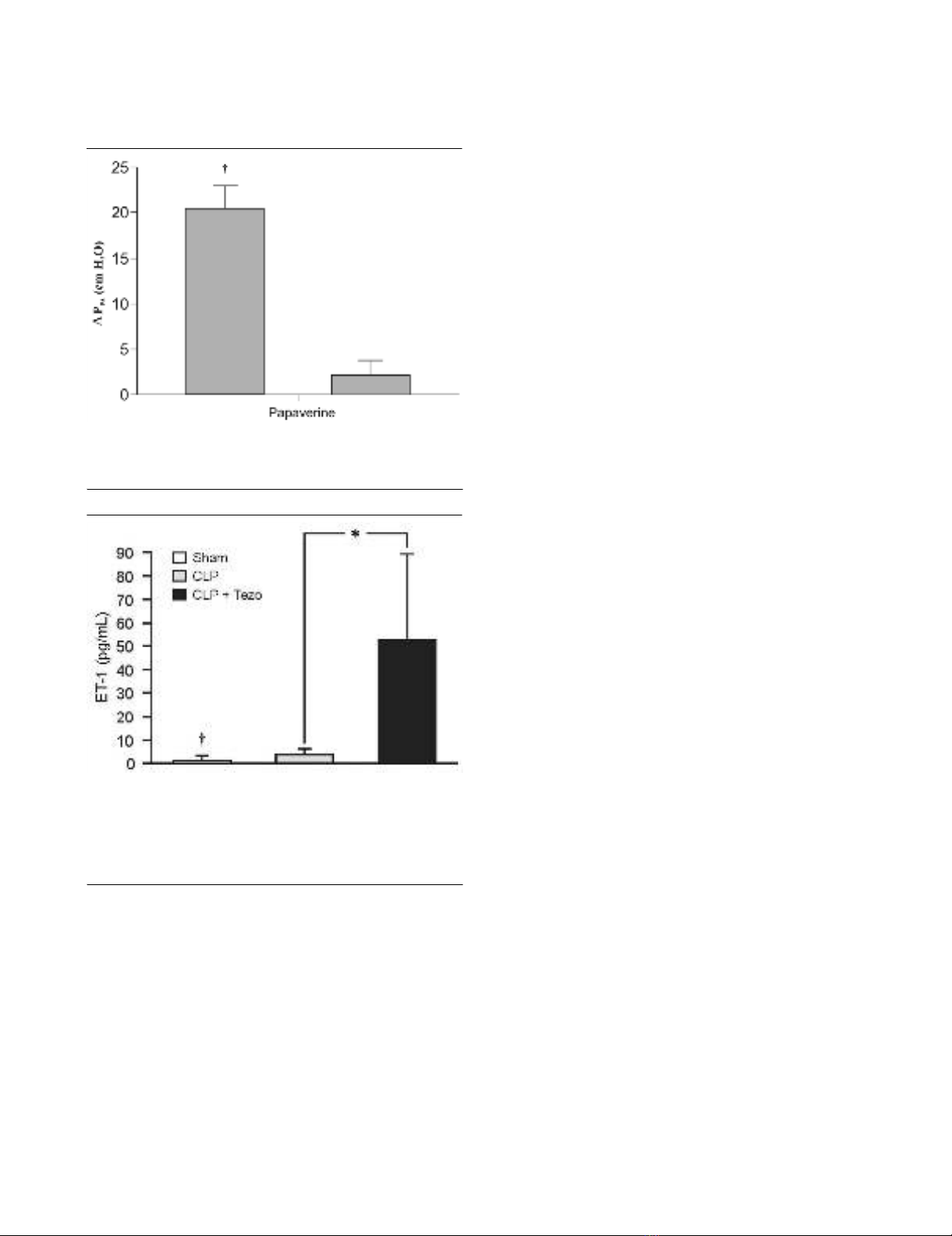
CLP induced a fourfold increase in the plasma concentration
of ET-1 compared to sham-operated rats (p < 0.05; Figure 2).
However, in the CLP + tezosentan group, the plasma level of
ET-1 was 10 to 15 times higher than with CLP alone (p <
0.05).
At baseline, we found no differences in hemodynamic varia-
bles between sham-operated rats and the CLP groups (Table
1). Because of the papaverine-induced vascular paralysis,
hemodynamics displayed no intra- or inter-group differences
throughout the experiments. In sham-operated rats, Kfc dis-
played no difference between groups at baseline and
remained unchanged throughout the experiment (Figure 3). At
variance, a threefold increase was noticed in the CLP group.
Concomitantly, CL decreased fourfold in parallel with increas-
ing pulmonary edema beyond 30 minutes. All preparations
deteriorated with visible fluid secretion into the airways after
90 minutes of perfusion (p < 0.05; Figure 3). In contrast, in the
CLP + tezosentan group, Kfc remained unchanged from base-
line throughout the experiment and CL displayed no significant
difference from sham-operated animals.
ET-1-induced pulmonary edema
Injection of ET-1 into the pulmonary arterial tubing caused a
significant rise in Kfc at 90 minutes, which was completely pre-
vented by tezosentan (p < 0.05; Figure 4). All preparations
exposed to ET-1 alone, except for one, were completely
destroyed after 90 minutes due to alveolar flooding. Adminis-
tration of tezosentan maintained Kfc at baseline level through-
out the experiments. Correspondingly, CL fell in all three
groups. In the ET-1 group, CL decreased fivefold compared to
the intra-group baseline (p < 0.05; Figure 4). The decrease
was significantly dampened in the ET-1 + tezosentan group
and did not differ from control lungs. Hemodynamic variables
revealed no significant differences between the groups (Table
2).
PKCα in lung tissue after CLP or ET-1
In the CLP group, the immunoreactivity of PKCα reached a
mean of 50% to 70% above sham in both tissue fractions (p
< 0.05; Figure 5). Tezosentan completely prevented the rise in
the cell membrane fraction of PKCα (Figure 5b).
In lungs isolated from healthy rats, acute administration of ET-
1 decreased the cytosolic fraction of PKCα by 60% (p < 0.05;
Figure 6a) and correspondingly tended to increase (not signif-
icant) the cell membrane fraction compared to controls (Figure
6b). Moreover, tezosentan prevented the reduction of the
cytosolic fraction of PKCα (p < 0.05; Figure 6a).
Discussion
The present study demonstrates that in rats CLP induces a
significant rise in the plasma concentration of ET-1 in parallel
with an increase in the PKCα content of lung tissue. Lungs iso-
lated and perfused with blood 12 h after CLP displayed visible
edema fluid in the trachea before 120 minutes had elapsed.
Correspondingly, in lungs isolated from healthy rats, pulmo-
nary arterial injection of ET-1 produced massive edema within
60 minutes of the start of blood perfusion. Tezosentan pre-
cluded the development of pulmonary edema induced by both
CLP and ET-1. As judged by western blotting, tezosentan also
Figure 1
Pulmonary arterial pressure responses (∆PPA) to endothelin-1 (ET-1) before and after papaverine administration in isolated lungs
Pulmonary arterial pressure responses (∆PPA) to endothelin-1 (ET-1)
before and after papaverine administration in isolated lungs. Data are
presented as mean ± SEM. †p < 0.05 from baseline.
Figure 2
Plasma concentrations of endothelin-1 (ET-1) in rats determined 12 h after surgical interventionsPlasma concentrations of endothelin-1 (ET-1) in rats determined 12 h
after surgical interventions. Data are presented as mean ± SEM. Sham,
sham-operated group (n = 5); CLP, cecum ligation and puncture group
(n = 7); CLP+Tezo, cecum ligation and puncture + tezosentan group (n
= 7). *p < 0.05 between CLP and CLP+Tezo groups; †p < 0.05
between Sham and CLP groups.

Available online http://ccforum.com/content/9/6/R677
R681
prevented the increase in PKCα in lung tissue after CLP. Thus,
we speculate that ET-1-binding to the endothelin receptor
could be responsible either for promoting PKCα gene expres-
sion and protein synthesis, or for inhibiting PKCα degradation.
When assessing changes in microvascular permeability in
response to ET-1 or other vasoconstrictors, papaverine is
used to deprive the lungs of their vasoconstrictor ability, which
implies that the Pmv can be kept constant [7,18,23]. The con-
trol group confirmed that papaverine had no effect on lung
microvascular permeability per se as previously demonstrated
[7,23]. Consistent with these findings, papaverine prevented
ET-1-induced changes in pulmonary arterial pressure, but did
not preclude the evolvement of pulmonary edema. In lungs
from sham-operated or healthy rats, in which no intervention
had taken place except for the injection of papaverine, Kfc
remained unchanged for the whole 120 minute perfusion time.
Other investigators have noticed significant increments in Kfc
and protein concentration in lung lavage fluid 18 h after CLP
in isolated rat lungs [24]. There is, however, no general agree-
ment about what factors determine the morbidity and mortality
after CLP. Some investigators argue that mortality depends on
the size of the punctured holes in the cecum [16]. Others
claim that increased length of the cecum distal to the ligature
raises the plasma concentrations of tumor necrosis factor-α
and interleukin-6, both factors that might contribute to the high
mortality during the first 16 to 24 h [17]. By combining the two
techniques, we expected that changes in Kfc would develop
at a higher pace. Consistently, we found that rats subjected to
our modification of CLP appeared ill and less vigorous in com-
parison with sham-operated animals. Moreover, the modified
CLP, but not sham-operation, displayed growth of Gram-neg-
ative and Gram-positive microorganisms in rat blood.
Several factors might contribute to the development of pulmo-
nary edema after CLP in rats [24,25]. Both experimental and
clinical studies have shown that transient increases in the
plasma concentrations of ET-1 might be associated with
development of pulmonary edema [2,14,26-29]. In patients
diagnosed with ALI, derangement of pulmonary function was
exacerbated by elevated plasma concentrations of ET-1,
whereas clinical improvement was associated with a signifi-
cant fall in concentrations of ET-1, indicating that ET-1 could
act as a marker of ALI [26-28]. In other species, however, ET-
1 participates in several other pathophysiological mechanisms
besides being a marker of vascular injury [29,30]. In rats, con-
tinuous infusion of ET-1 resulted in escape of 125I-labelled
albumin to liver, heart and lungs while hematocrit increased
[31]. At doses of 5 to 10 nM, ET-1 caused pulmonary edema
Table 1
Hemodynamic variables in rat lungs isolated 12 h after surgical interventions
Hemodynamic variable Time (minutes)
0 306090120
PPA, cmH2O
Sham 26.0 ± 3.1 24.6 ± 3.1 24.6 ± 2.7 26.0 ± 3.0 26.0 ± 3.0
CLP 21.9 ± 1.5 23.2 ± 1.3 23.2 ± 1.6 24.6 ± 2.3
CLP+Tezo 24.6 ± 0.9 23.2 ± 0.9 23.2 ± 1.1 21.9 ± 1.2 24.6 ± 1.7
PLA, cmH2O
Sham 14.6 ± 0.6 14.7 ± 0.6 14.9 ± 0.9 15.6 ± 0.9 14.9 ± 0.9
CLP 13.8 ± 0.4 13.8 ± 0.4 13.8 ± 0.4 13.4 ± 0.8
CLP+Tezo 14.9 ± 0.4 14.9 ± 0.4 14.7 ± 0.4 15.4 ± 0.4 15.2 ± 0.4
RT, cmH2O/ml/min
Sham 0.89 ± 0.1 0.80 ± 0.1 0.83 ± 0.1 0.84 ± 0.1 0.82 ± 0.1
CLP 0.57 ± 0.1 0.65 ± 0.1 0.67 ± 0.1 0.67 ± 0.1
CLP+Tezo 0.75 ± 0.0 0.67 ± 0.0 0.67 ± 0.1 0.67 ± 0.1 0.71 ± 0.1
∆Pmv, cmH2O
Sham 7.2 ± 0.2 7.2 ± 0.3 7.5 ± 0.3 7.2 ± 0.3 7.5 ± 0.3
CLP 6.7 ± 0.5 6.5 ± 0.4 6.4 ± 0.4 6.5 ± 0.6
CLP+Tezo 7.6 ± 0.4 7.6 ± 0.4 7.5 ± 0.4 8.0 ± 0.4 7.6 ± 0.5
Data are presented as mean ± SEM. Sham, sham-operated group (n = 5); CLP, cecum ligation and puncture group (n = 7); CLP+Tezo, cecum
ligation and puncture + tezosentan group (n = 7). PLA, left atrial pressure; PPA, pulmonary artery pressure; ∆Pmv, difference between pulmonary
microvascular pressure determined prior to and during a standardized elevation of PLA; RT, total vascular resistance.


%20--%3e%3cdefs%3e%3cstyle%3e%20.st0%20{%20fill:%20%23fff;%20}%20.st1%20{%20fill:%20%237800fa;%20}%20%3c/style%3e%3c/defs%3e%3cpath%20class='st1'%20d='M117.78,12.18H43.11c2.9,3.47,4.65,7.94,4.65,12.82,0,5.6-2.3,10.66-6.01,14.29h76.02l7.22-13.56-7.22-13.56Z'/%3e%3cg%3e%3cpath%20class='st0'%20d='M53.58,26.17h-.59v-1.46h.59v-4.96h2.83c1.78,0,2.67.94,2.67,2.82v5.76c0,1.87-.89,2.81-2.67,2.81h-2.83v-4.96ZM55.36,21.37v3.34h1.1v1.46h-1.1v3.34h1.01c.61,0,.91-.37.91-1.1v-5.93c0-.74-.3-1.1-.91-1.1h-1.01Z'/%3e%3cpath%20class='st0'%20d='M65.99,31.14h-1.8l-.31-2.07h-2.19l-.31,2.07h-1.64l1.82-11.39h2.62l1.82,11.39ZM65.28,18.04c-.25.46-.51.77-.75.94-.21.15-.47.22-.79.22-.26,0-.57-.07-.92-.22l-.38-.15c-.14-.05-.26-.07-.37-.07-.3,0-.53.18-.71.54l-.91-.68c.25-.46.51-.77.75-.94.21-.14.48-.21.79-.21.26,0,.57.07.92.21l.38.15c.14.05.26.07.37.07.3,0,.53-.18.71-.54l.91.68ZM61.91,27.52h1.73l-.87-5.76-.87,5.76Z'/%3e%3cpath%20class='st0'%20d='M74.53,26.89v1.52c0,1.91-.89,2.86-2.67,2.86s-2.67-.95-2.67-2.86v-5.93c0-1.91.89-2.86,2.67-2.86s2.67.95,2.67,2.86v1.11h-1.69v-1.22c0-.75-.31-1.12-.93-1.12s-.93.37-.93,1.12v6.15c0,.74.31,1.11.93,1.11s.93-.37.93-1.11v-1.63h1.69Z'/%3e%3cpath%20class='st0'%20d='M81.4,31.14h-1.8l-.31-2.07h-2.19l-.31,2.07h-1.64l1.82-11.39h2.62l1.82,11.39ZM75.9,19.2l1.52-1.91h1.71l1.51,1.91h-1.61l-.76-.95-.75.95h-1.61ZM77.32,27.52h1.73l-.87-5.76-.87,5.76ZM83.1,15.99l-1.76,1.91h-1.26l1.17-1.91h1.86Z'/%3e%3cpath%20class='st0'%20d='M84.86,19.75c1.78,0,2.67.94,2.67,2.82v1.48c0,1.87-.89,2.81-2.67,2.81h-.85v4.28h-1.79v-11.39h2.64ZM84.01,21.37v3.86h.85c.58,0,.87-.36.87-1.08v-1.71c0-.71-.29-1.07-.87-1.07h-.85Z'/%3e%3cpath%20class='st0'%20d='M93.51,19.75c1.78,0,2.67.94,2.67,2.82v1.48c0,1.87-.89,2.81-2.67,2.81h-.85v4.28h-1.79v-11.39h2.64ZM92.66,21.37v3.86h.85c.58,0,.87-.36.87-1.08v-1.71c0-.71-.29-1.07-.87-1.07h-.85Z'/%3e%3cpath%20class='st0'%20d='M98.8,31.14h-1.79v-11.39h1.79v4.88h2.03v-4.88h1.83v11.39h-1.83v-4.88h-2.03v4.88Z'/%3e%3cpath%20class='st0'%20d='M105.36,24.55h2.46v1.62h-2.46v3.34h3.09v1.63h-4.88v-11.39h4.88v1.63h-3.09v3.18ZM108.17,17.29l-1.76,1.91h-1.26l1.17-1.91h1.86Z'/%3e%3cpath%20class='st0'%20d='M112.2,19.75c1.78,0,2.67.94,2.67,2.82v1.48c0,1.87-.89,2.81-2.67,2.81h-.85v4.28h-1.79v-11.39h2.64ZM111.35,21.37v3.86h.85c.58,0,.87-.36.87-1.08v-1.71c0-.71-.29-1.07-.87-1.07h-.85Z'/%3e%3c/g%3e%3ccircle%20class='st1'%20cx='25'%20cy='25'%20r='20'/%3e%3cpath%20class='st0'%20d='M32.78,19.27c2.92,0,4.43,2.55,5.28,5.33l.71,2.17c.14.38-.33.75-.71.75h-5.61c.19-.33.24-.71.09-1.08l-.75-2.45c-.43-1.32-.99-2.64-1.79-3.77.75-.57,1.65-.94,2.78-.94h0ZM25,18.38c3.25,0,4.9,2.78,5.89,5.89l.76,2.45c.14.42-.33.8-.8.8h-11.69c-.42,0-.94-.38-.8-.8l.75-2.45c.99-3.11,2.64-5.89,5.89-5.89h0ZM25,11.35c1.74,0,3.11,1.37,3.11,3.11s-1.37,3.11-3.11,3.11-3.11-1.41-3.11-3.11,1.41-3.11,3.11-3.11h0ZM17.27,19.27c1.08,0,1.98.38,2.73.94-.8,1.13-1.37,2.45-1.74,3.77l-.8,2.45c-.14.38-.05.75.09,1.08h-5.56c-.42,0-.9-.38-.75-.75l.71-2.17c.9-2.78,2.41-5.33,5.33-5.33h0ZM17.27,12.91c1.51,0,2.78,1.27,2.78,2.83s-1.27,2.83-2.78,2.83-2.83-1.27-2.83-2.83,1.27-2.83,2.83-2.83h0ZM32.78,12.91c1.56,0,2.78,1.27,2.78,2.83s-1.23,2.83-2.78,2.83-2.83-1.27-2.83-2.83,1.27-2.83,2.83-2.83h0ZM27.07,28.56v.09c0,.57-.24,1.08-.61,1.46h0v.05c-.38.33-.9.57-1.46.57s-1.08-.24-1.46-.61h0c-.38-.38-.61-.9-.61-1.46v-.09h1.41v.09c0,.19.05.38.19.47v.05c.09.09.28.19.47.19s.38-.09.47-.19v-.05c.14-.09.24-.28.24-.47t-.05-.09h1.41ZM30.99,28.56v.09c0,1.65-.66,3.16-1.74,4.24-1.08,1.08-2.59,1.79-4.24,1.79s-3.16-.71-4.24-1.79l-.05-.05c-1.04-1.08-1.7-2.55-1.7-4.2v-.09h1.41v.09c0,1.27.47,2.4,1.27,3.25h.05c.85.85,1.98,1.37,3.25,1.37s2.4-.52,3.25-1.37c.85-.8,1.37-1.98,1.37-3.25v-.09h1.37ZM34.99,28.56v.09c0,2.78-1.13,5.28-2.92,7.07-1.79,1.79-4.29,2.92-7.07,2.92s-5.23-1.13-7.07-2.92c-1.79-1.79-2.92-4.29-2.92-7.07v-.09h1.41v.09c0,2.4.94,4.53,2.5,6.08,1.56,1.56,3.72,2.5,6.08,2.5s4.52-.94,6.08-2.5c1.56-1.56,2.5-3.68,2.5-6.08v-.09h1.41Z'/%3e%3c/svg%3e)