
Open Access
Available online http://ccforum.com/content/11/4/R81
Page 1 of 9
(page number not for citation purposes)
Vol 11 No 4
Research
Vasopressin improves survival in a porcine model of abdominal
vascular injury
Karl H Stadlbauer1, Horst G Wagner-Berger1, Anette C Krismer1, Wolfgang G Voelckel1,
Alfred Konigsrainer2, Karl H Lindner1 and Volker Wenzel1
1Department of Anaesthesiology and Critical Care Medicine, Innsbruck Medical University, Anichstrasse, 6020 Innsbruck, Austria
2Department of Surgery, Eberhard-Karls Unversity, Hoppe-Seyler-Straße, 72076 Tübingen, Germany
Corresponding author: Karl H Stadlbauer, karl-heinz.stadlbauer@i-med.ac.at
Received: 8 Mar 2007 Revisions requested: 13 Apr 2007 Revisions received: 19 Jul 2007 Accepted: 23 Jul 2007 Published: 23 Jul 2007
Critical Care 2007, 11:R81 (doi:10.1186/cc5977)
This article is online at: http://ccforum.com/content/11/4/R81
© 2007 Stadlbauer et al; licensee BioMed Central Ltd.
This is an open access article distributed under the terms of the Creative Commons Attribution License (http://creativecommons.org/licenses/by/2.0),
which permits unrestricted use, distribution, and reproduction in any medium, provided the original work is properly cited.
Abstract
Introduction We sought to determine and compare the effects
of vasopressin, fluid resuscitation and saline placebo on
haemodynamic variables and short-term survival in an abdominal
vascular injury model with uncontrolled haemorrhagic shock in
pigs.
Methods During general anaesthesia, a midline laparotomy was
performed on 19 domestic pigs, followed by an incision (width
about 5 cm and depth 0.5 cm) across the mesenterial shaft.
When mean arterial blood pressure was below 20 mmHg, and
heart rate had declined progressively, experimental therapy was
initiated. At that point, animals were randomly assigned to
receive vasopressin (0.4 U/kg; n = 7), fluid resuscitation (25 ml/
kg lactated Ringer's and 25 ml/kg 3% gelatine solution; n = 7),
or a single injection of saline placebo (n = 5). Vasopressin-
treated animals were then given a continuous infusion of 0.08 U/
kg per min vasopressin, whereas the remaining two groups
received saline placebo at an equal rate of infusion. After 30 min
of experimental therapy bleeding was controlled by surgical
intervention, and further fluid resuscitation was performed.
Thereafter, the animals were observed for an additional hour.
Results After 68 ± 19 min (mean ± standard deviation) of
uncontrolled bleeding, experimental therapy was initiated; at
that time total blood loss and mean arterial blood pressure were
similar between groups (not significant). Mean arterial blood
pressure increased in both vasopressin-treated and fluid-
resuscitated animals from about 15 mmHg to about 55 mmHg
within 5 min, but afterward it decreased more rapidly in the fluid
resuscitation group; mean arterial blood pressure in the placebo
group never increased. Seven out of seven vasopressin-treated
animals survived, whereas six out of seven fluid-resuscitated and
five out of five placebo pigs died before surgical intervention
was initiated (P < 0.0001).
Conclusion Vasopressin, but not fluid resuscitation or saline
placebo, ensured short-term survival in this vascular injury model
with uncontrolled haemorrhagic shock in sedated pigs.
Introduction
For haemodynamic stabilization of critically injured patients
with uncontrolled haemorrhagic shock, current advanced
trauma life support guidelines recommend infusion of crystal-
loid or colloid solutions. Interestingly, there appears to be no
evidence either for or against early or large amounts of intrave-
nous fluid administration in uncontrolled haemorrhage [1,2].
Although these findings on fluids in uncontrolled haemor-
rhagic shock are inconclusive, the strategy of delaying fluid
resuscitation must be deemed unconfirmed as well, because
the only study to examine the efficacy of this approach yielded
barely significant findings.
One way to promote vasoconstriction may be to inject vaso-
pressin, which has potent vasoconstrictive effects, even in
severe acidosis and developed vasoplegia. In porcine models
of uncontrolled haemorrhagic shock after liver trauma, vaso-
pressin was superior to fluid resuscitation, adrenaline (epine-
phrine), and saline placebo in terms of blood loss,
haemodynamic variables, and survival [3-5]. In agreement with
these experiments, vasopressin reduced blood loss and stabi-
lized arterial blood pressure in patients who had suffered trau-
matic or nontraumatic injury with haemorrhagic shock that was
refractory to catecholamines [6-9]. Although beneficial effects
of vasopressin may seem realistic in a parenchymatic organ
such as the liver, pronounced vascular injury may impose

Critical Care Vol 11 No 4 Stadlbauer et al.
Page 2 of 9
(page number not for citation purposes)
limitations with regard to vasoconstriction-mediated reduc-
tions in blood loss. This may be important because patients
suffering multiple trauma often sustain complex injuries, with
bleeding from multiple sources. A clinical trial comparing vaso-
pressin against placebo as an adjunct to standard shock treat-
ment in unstable multiple trauma patients is currently in
preparation [10], but further information about underlying
mechanisms is needed.
The purpose of the present study was to compare the effects
of vasopressin with those of fluid resuscitation and saline pla-
cebo on haemodynamic variables and short-term survival in an
abdominal vascular injury model of uncontrolled haemorrhagic
shock. Our null hypothesis was that there would be no differ-
ences in study end-points.
Materials and methods
Surgical preparations and measurements
The project was approved by the Austrian Federal Animal
Investigational Committee, and the animals were managed in
accordance with the American Physiological Society institu-
tional guidelines and the Position of the American Heart Asso-
ciation on Research Animal Use, as adopted on 11 November
1984. Animal care and use were performed by qualified indi-
viduals and under the supervision of veterinarians, and all facil-
ities and transportation complied with current legal
requirements and guidelines. Anaesthesia was used in all sur-
gical interventions, any unnecessary suffering was avoided
and research was terminated if unnecessary pain or stress
resulted. Our animal facilities meet the standards of the Amer-
ican Association for Accreditation of Laboratory Animal Care.
This study was conducted in 19 healthy swine, aged 12 to 16
weeks and weighing 30 to 40 kg. The animals were fasted
overnight, but they had free access to water. The pigs were
pre-medicated with azaperone (4 mg/kg intramuscularly) and
atropine (0.1 mg/kg intramuscularly) 1 hour before surgery,
and anaesthesia was induced with propofol (1 to 2 mg/kg
intravenously). After intubation during spontaneous respira-
tion, the pigs were ventilated using a volume controlled venti-
lator (Draeger EV-A, Lübeck, Germany) with 35% oxygen at
20 breaths/minute, 5 cmH2O positive end-expiratory pressure,
and tidal volume adjusted to maintain normocapnia. Anaesthe-
sia was maintained with propofol (6 to 8 mg/kg per hour) and
a single injection of piritramide (30 mg) [11]. Muscle paralysis
was achieved with 0.2 mg/kg per hour pancuronium after intu-
bation, in order to facilitate laparotomy. Lactated Ringer's solu-
tion (250 ml) and a 3% gelatine solution (250 ml; Gelofusin®,
B. Braun, Melsungen, Germany) were administered during the
preparation phase. A standard lead II electrocardiograph was
used to monitor cardiac rhythm; depth of anaesthesia was
judged according to blood pressure, heart rate and electroen-
cephalography (Neurotrac; Engström, Munich, Germany).
If cardiovascular variables or electroencephalography indi-
cated reduced depth of anaesthesia, then additional propofol
and piritramide were given. Body temperature was maintained
between 38.0°C (100.4°F) and 39.0°C (102.2°F). A 7-Fr cath-
eter was advanced into the descending aorta via a femoral cut-
down for withdrawal of arterial blood samples and measure-
ment of arterial blood pressure. A 7.5-Fr pulmonary artery
catheter was placed via cut-down in the neck for measurement
of right atrial and pulmonary artery pressures. Blood pressure
was measured using a saline-filled catheter attached to a pres-
sure transducer (model 1290A; Hewlett Packard, Böblingen,
Germany), which was calibrated to atmospheric pressure at
the level of the right atrium. Pressure tracings were recorded
using a data acquisition system (Dewetron port 2000 [Dewet-
ron, Graz, Austria] and Datalogger [custom software]). Blood
gases were measured using a blood gas analyzer (Rapidlab
865; Chiron, Walpole, MA, USA); end-tidal carbon dioxide
was measured using an infrared absorption analyzer (Multicap,
Datex, Helsinki, Finland).
Experimental protocol
Figure 1 provides a summary of the experimental protocol.
After assessing baseline haemodynamic values, a midline
laparotomy was performed. Propofol infusion was adjusted to
2 mg/kg per hour, and infusion of lactated Ringer's and gela-
tine solution was stopped before initiation of the experiment.
During uncontrolled haemorrhage and experimental therapy,
tidal volume was not adjusted. An incision (width 5 cm and
depth 0.5 cm) was made across the mesenterial shaft. When
mean arterial blood pressure was below 20 mmHg and heart
rate had declined by more than 30% of its peak value, pharma-
cological support was provided for 30 min to simulate a pre-
hospital phase before surgical intervention.
At that point, the 19 animals were randomly assigned to
receive one of the following: 0.4 U/kg vasopressin (Pitressin®;
Parke-Davis/Pfizer, Karlsruhe, Germany) diluted to 20 ml with
saline (n = 7); fluid resuscitation (25 ml/kg lactated Ringer's
Figure 1
Flow chart of the experimental protocolFlow chart of the experimental protocol. BL, baseline; MAP, mean arte-
rial pressure.
Available online http://ccforum.com/content/11/4/R81
Page 3 of 9
(page number not for citation purposes)
and 25 ml/kg 3% gelatine solution; n = 7); or a single injection
of 20 ml saline placebo (n = 5). Fluid resuscitation was initially
set at about 2 ml/kg per min over the first 10 min. If this
approach failed to restore arterial blood pressure, then fluid
resuscitation was enhanced to about 8 ml/kg per min. We
used a combination of Ringer's lactate and gelatine solution
for fluid resuscitation, because it is the usual strategy in
Europe. Vasopressin-treated animals were then given a contin-
uous infusion of 0.08 U/kg per min vasopressin, whereas the
remaining two groups received saline placebo at an equal rate
of infusion.
After initiating experimental therapy, pigs were ventilated with
100% oxygen. The investigators were blinded as to the treat-
ment given to each pig. To achieve blinding, we employed
three-way stopcocks, which directed experimental treatment
either into a covered bucket or into the central venous line. The
infusion rate of fluids was set in all groups to 2 ml/kg at the
beginning of the experimental therapy. A three-way stopcock
determined whether subsequent experimental therapy was
directed into the central venous line (fluid-resuscitated ani-
mals) or into a covered bucket (vasopressin-treated and pla-
cebo-treated animals). Hence, our vasopressin-treated and
placebo-treated animals received no additional fluid during
experimental therapy. The infusion rate was enhanced to 8 ml/
kg per min after 10 min of experimental therapy if mean arterial
pressure did not increase to above 40 mmHg. When mean
arterial blood pressure reached aortic hydrostatic pressure
(about 10 mmHg) and end-tidal carbon dioxide was 10 mmHg
or less, the animals were declared dead and administered an
overdose of fentanyl, propofol and potassium chloride.
After 30 min of experimental therapy, bleeding was controlled
by surgical intervention in all surviving pigs. Additionally, fluid
therapy (25 ml/kg lactated Ringer's and 25 ml/kg 3% gelatine
solution) was started, and haemodynamic variables and blood
gases were measured over an additional observation period of
60 min. Afterward, the pigs were killed as described above.
Statistical analysis
Values are expressed as mean ± standard deviation. The com-
parability of baseline data was verified using one-way analysis
of variance. Survival rates were compared using Kaplan-Meier
methods with log rank (Mantel Cox) comparison of cumulative
survival by treatment group, and were corrected using the
Bonferroni method for multiple comparisons. Differences with
a two-tailed P value < 0.05 were considered significant.
Because of rapidly changing number of surviving animals in
the experimental therapy phase, we did not perform further sta-
tistical analysis.
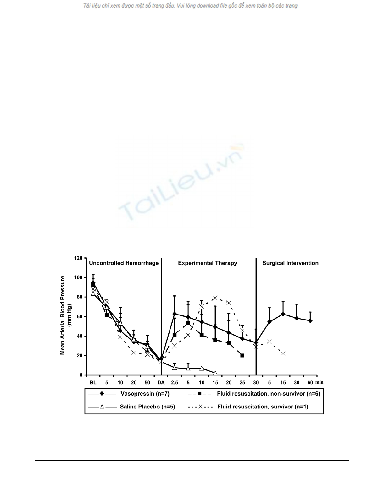
Figure 2
Mean arterial blood pressureMean arterial blood pressure. Values are expressed as mean (± standard deviation) arterial blood pressure before, during and after administration of
a 0.4 U/kg bolus dose and 0.08 U/kg per min continuous infusion of vasopressin (n = 7), fluid resuscitation (divided into survivors [n = 1] and non-
survivors [n = 6]), and saline placebo (n = 5). 'Uncontrolled haemorrhage' indicates the non-intervention interval after vessel injury; 'experimental
therapy' indicates vasopressin treatment, fluid resuscitation, or saline placebo administration without bleeding control; and 'surgical intervention' indi-
cates surgical management of the mesenteric shaft to control bleeding. The x-axis does not reveal the true time slope. BL, baseline; DA, drug
administration.
Critical Care Vol 11 No 4 Stadlbauer et al.
Page 4 of 9
(page number not for citation purposes)
Results
Before induction of haemorrhage, there were no differences in
haemodynamic variables, blood gases, weight, or temperature
between groups. Experimental therapy was initiated after 76 ±
24 min in the vasopressin group, 63 ± 19 min in the fluid
resuscitation group and 64 ± 9 min in the saline placebo
group (not significant). At that time, mean arterial blood pres-
sure (Figure 2) and total blood loss (Figure 3) were compara-
ble between groups. Before drug administration, lactate and
arterial carbon dioxide tension were significantly lower in the
saline placebo group than in the vasopressin group (Table 1).
After initiating experimental therapy, the heart rate remained at
about 210 beats/min in the vasopressin group, but it
decreased from about 180 to about 120 beats/min in the fluid
resuscitation and saline placebo groups (Figure 4). Mean arte-
rial blood pressure increased in both vasopressin-treated and
fluid-resuscitated animals from about 15 mmHg to about 55
mmHg within 5 min of experimental therapy, but it decreased
immediately in placebo-treated animals. Mean arterial blood
pressure declined more rapidly in the fluid resuscitation group
than in vasopressin-treated swine after 5 min of experimental
therapy (Figure 2). End-tidal carbon dioxide remained at about
25 mmHg in the vasopressin-treated group, but it decreased
rapidly in the placebo group. In fluid-resuscitated animals, end-
tidal carbon dioxide increased from about 20 mmHg to 40
mmHg within 5 min, but it subsequently deteriorated (Figure
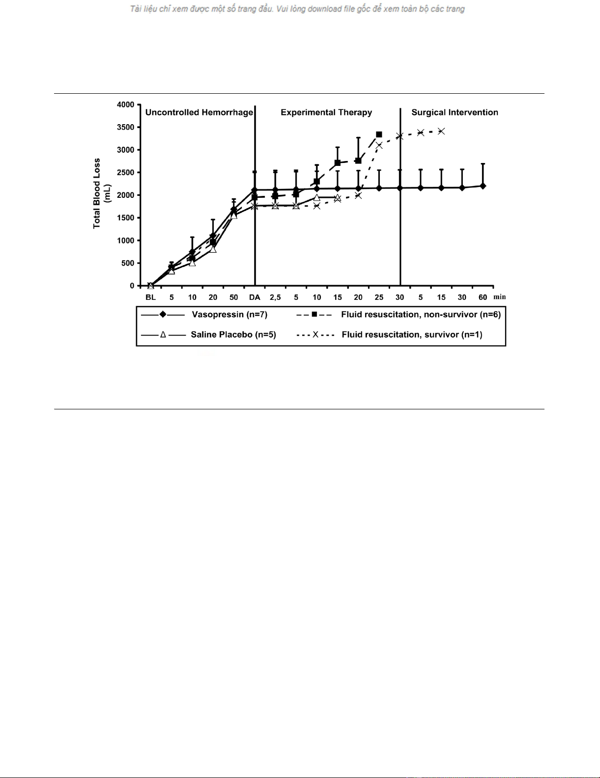
5). Total blood loss was constant in the vasopressin and saline
placebo groups, but it increased in the fluid resuscitation
group from about 2,000 ml to about 2,800 ml (Figure 3).
Within the first 5 min of experimental therapy, haemoglobin
was constant in the vasopressin and saline placebo groups,
but it decreased in the fluid resuscitation group from about 7.2
to 4.4 g/dl (Table 1). Seven out of seven vasopressin-treated
animals survived, whereas six out of seven fluid-resuscitated
and five out of five placebo-treated pigs died before surgical
intervention was initiated (Figure 6; P < 0.0001).
Discussion
In this porcine model of vascular injury of uncontrolled haem-
orrhagic shock, vasopressin maintained cardiocirculatory
function at a level that was sufficient to permit at least short-
term survival. In contrast, within 20 min of experimental ther-
apy, six out of seven fluid-resuscitated and five out of five pla-
cebo-treated animals died.
Bleeding was initiated in this model of uncontrolled haemor-
rhagic shock by an incision to the mesenteric shaft. Thus, we
simulated a blood vessel injury, which is often associated with
blunt trauma and a subsequent high mortality rate [12]. Dos-
ages employed in the experimental strategies were similar to
those in interventions in an established porcine liver trauma
Figure 3
Total blood lossTotal blood loss. Values are expressed as mean (± standard deviation) total blood loss before, during, and after administration of a 0.4 U/kg bolus
dose and 0.08 U/kg per min continuous infusion of vasopressin (n = 7), fluid resuscitation (divided into survivors [n = 1] and nonsurvivors [n = 6]),
and saline placebo (n = 5). 'Uncontrolled haemorrhage' indicates the non-intervention interval after vessel injury; 'experimental therapy' indicates
vasopressin treatment, fluid resuscitation, or saline placebo administration without bleeding control; and 'surgical intervention' indicates surgical
management of the mesenteric shaft to control bleeding. The x-axis does not reveal the true time slope. BL, baseline; DA, drug administration.

Available online http://ccforum.com/content/11/4/R81
Page 5 of 9
(page number not for citation purposes)
model involving uncontrolled haemorrhagic shock [4,5].
Accordingly, we suggest that this model of uncontrolled haem-
orrhagic shock, caused by a blood vessel injury, is a valuable
tool in which to assess the effect of experimental advanced
trauma life support on haemodynamic variables and short-term
survival.
Vasopressin has been investigated in various catecholamine-
refractory shock states, such as cardiac arrest [13-15] and
vasodilatory shock [16-18]. For example, even in normovolae-
mic but catecholamine-refractory haemorrhagic shock, vaso-
pressin effectively restored cardiocirculatory function in a
canine model [19]. That report is in full agreement with the
findings of our study, in which vasopressin rapidly increased
mean arterial blood pressure from about 15 mmHg to about
50 mmHg after approximately 65 min of uncontrolled bleeding
(total blood loss about 50 ml/kg), resulting in severe haemor-
rhagic shock. This vasopressin-mediated increase in mean
arterial blood pressure was also observed in trauma patients
with uncontrolled haemorrhagic shock that was refractory to
massive fluid resuscitation and catecholamines [7-9,20]. Thus,
vasopressin may be a simple and effective treatment for
trauma patients who do not respond to advanced trauma life
support, which may maintain arterial blood pressure at a level
sufficient to ensure vital organ perfusion [7,8,20-22].
The data reported thus far about the type of fluid resuscitation
[23-25], the target mean arterial blood pressure [23] and tim-
Table 1
Arterial blood gas variables, haemoglobin, and lactate
Parameter Group Baseline Haemorrhagic shock Experimental therapy Surgical intervention
5 min after DA 30 min after DA 15 min 60 min
7.34 ± 0.12 7.18 ± 0.10 7.08 ± 0.12 7.21 ± 0.13
Fluid resuscitation 7.50 ± 0.05 7.36 ± 0.08 7.17 ± 0.03 - - -
Saline placebo 7.52 ± 0.01 7.44 ± 0.08 7.41 ± 0.06 - - -
Paco2 (mmHg) Vasopressin 37 ± 3 31 ± 4 30 ± 3 34 ± 5 44 ± 4 41 ± 5
Fluid resuscitation 37 ± 3 30 ± 5 45 ± 7 - - -
Saline placebo 34 ± 3 24 ± 4* 24 ± 7 - - -
Pao2 (mmHg) Vasopressin 154 ± 19 132 ± 21 312 ± 134 437 ± 31 363 ± 102 262 ± 98
Fluid resuscitation 138 ± 20 123 ± 28 300 ± 168 - - -
Saline placebo 154 ± 10 116 ± 25 239 ± 131 - - -
Base excess (mmol/l) Vasopressin 5.7 ± 1.7 -9.0 ± 4.5 -8.8 ± 5.8 -14.1 ± 6.1 -15.2 ± 5.0 -10.5 ± 6.0
Fluid resuscitation 5.3 ± 3.8 -7.7 ± 3.1 -11.4 ± 1.4 - - -
Saline placebo 4.5 ± 1.8 -7.5 ± 3.4 -8.9 ± 3.3 - - -
Haemoglobin (g/dl) Vasopressin 9.0 ± 1.2 7.7 ± 0.9 7.2 ± 1.0 6.4 ± 1.3 4.2 ± 0.7 3.3 ± 1.1
Fluid resuscitation 8.5 ± 0.9 7.2 ± 1.5 4.4 ± 1.4 - - -
Saline placebo 8.4 ± 0.9 8.1 ± 0.5 7.8 ± 0.7 - - -
Lactate (mmol/l) Vasopressin 1.78 ± 0.33 9.88 ± 2.78 11.10 ± 3.11 13.10 ± 3.44 11.55 ± 3.89 9.88 ± 3.55
Fluid resuscitation 2.00 ± 1.22 7.55 ± 2.55 7.33 ± 2.66 - - -
Saline placebo 1.22 ± 0.22 5.99 ± 1.44* 8.44 ± 2.66 - - -
All variables are expressed as mean ± standard deviation. 'Baseline' indicates measurements taken before mesenteric vessel trauma; 'haemorrhagic shock' indicates
values taken at the point of experimental intervention (mean arterial pressure <20 mmHg and heart rate <30% of its peak value); and 'experimental therapy' indicates
vasopressin, fluid resuscitation, or saline placebo administration without bleeding control. Statistical comparison was only performed at baseline and haemorrhagic
shock: *P < 0.05 for placebo versus vasopressin. -, not applicable because of death in fluid-resuscitated and placebo-treated animals; DA, drug administration; Paco2,
arterial carbon dioxide tension; Pao2, arterial oxygen tension.


![Vaccine và ứng dụng: Bài tiểu luận [chuẩn SEO]](https://cdn.tailieu.vn/images/document/thumbnail/2016/20160519/3008140018/135x160/652005293.jpg)


