
THE SIR HANS KREBS LECTURE
LAT – an important raft-associated transmembrane
adaptor protein
Delivered on 6 July 2009 at the 34th FEBS Congress in Prague,
Czech Republic
Va
´clav Hor
ˇejs
ˇı
´, Pavel Ota
´hal and Toma
´s
ˇBrdic
ˇka
Institute of Molecular Genetics, Academy of Sciences of the Czech Republic, Prague, Czech Republic
Introduction
A number of immunologically important receptors,
e.g. T cell and B cell antigen receptors (TCR, BCR),
Fc-receptors, natural killer (NK) ⁄myeloid cell activat-
ing receptors, collagen receptor on platelets, some
cytokine receptors, employ common functional princi-
ples for signal transduction. These multichain receptor
complexes consist of a ligand-recognition module and
noncovalently associated signalling subunits. The sig-
nalling subunits are transmembrane proteins contain-
ing in their intracellular domains tyrosine residues that
can be phosphorylated by kinases associated constitu-
tively or, more often just very transiently, with the
receptor.
Extracellular domains of these signalling subunits
are in some cases large, sometimes contributing to
ligand binding (many cytokine receptors). In other
cases the extracellular domains are relatively small and
participate rather in interactions with the ligand-bind-
ing chains of the receptor complexes, such as the
CD3c,d,esubunits of the TCR complex [1] or
CD79a, b components of the BCR complex [2]. Some
of the receptor-associated signalling chains have only
very short extracellular segments (fchain of the TCR
complex [3], cchain of several Fc receptors [4],
DAP12 and DAP10 chains of several NK ⁄myeloid cell
activating receptors [5]).
Keywords
immunoreceptor signalling; LAT; raft;
transmembrane adaptor protein; tyrosine
phosphorylation
Correspondence
V. Hor
ˇejs
ˇı
´, Institute of Molecular Genetics,
AS CR, Vı
´den
ˇska
´1083, 142 20 Prague 4,
Czech Republic
Fax: 420 244472282
Tel: 420 241729908
E-mail: horejsi@biomed.cas.cz
(Received 8 July 2010, revised 12 August
2010, accepted 24 August 2010)
doi:10.1111/j.1742-4658.2010.07831.x
Membrane rafts are microdomains involved in a number of biologically
important processes, including immunoreceptor signalling. Among the
functionally important protein components of these microdomains are
transmembrane adaptor proteins, containing in their intracellular domains
tyrosine residues that can be phosphorylated and bind other cytoplasmic
signalling proteins. The most important leukocyte transmembrane adaptor
protein is LAT (linker for activation of T cells), which is critically involved
in T cell receptor signalling, but also plays important roles in signal initia-
tion by several other immunologically important receptors. Here we review
recent progress in the elucidation of several aspects of this protein, e.g. the
controversy concerning the importance of LAT being present in membrane
rafts, the involvement in signalling through a number of receptors other
than the T cell receptor and the puzzling phenotype of some LAT mutants.
Abbreviations
BCR, B cell receptor; cSMAC, central supramolecular activation cluster; DRM, detergent-resistant membrane complex; GPVI, glycoprotein
VI; LAT, linker for activation of T cells; NK, natural killer; PI3K, phosphatidylinositol 3-kinase; TCR, T cell receptor; TRAP, transmembrane
adaptor protein.
FEBS Journal 277 (2010) 4383–4397 ª2010 The Authors Journal compilation ª2010 FEBS 4383

Several other proteins structurally similar to the
last group (fchain-like) exist that are not directly
associated with any receptor, but also play more or
less important roles in the regulation of receptor sig-
nalling. Some of these transmembrane adaptor pro-
teins (TRAPs) are palmitoylated and targeted to
membrane rafts (LAT, NTAL, LIME, PAG), others
are found in nonraft membrane (SIT, TRIM, LAX,
GAPT) [6,7]. In our opinion, the term TRAP can also
be used for the abovementioned proteins closely asso-
ciated with receptors, i.e. f,cchains, DAP12, DAP10.
Common features of TRAPs thus include: short extra-
cellular domain, single transmembrane domain, intra-
cellular domain containing signalling-relevant motifs,
such as potentially phosphorylated tyrosine motifs,
polyproline sequences, PDZ-binding motifs, etc.
This review deals mainly with the functionally most
important TRAP, linker for activation of T cells
(LAT). We will concentrate mainly on the latest devel-
opments in the field, but will also review the literature
on rather neglected roles of LAT in non-T cells.
Several relatively recent reviews exist, dealing with
TRAPs in general or specifically with some of them
[4,5,8–16].
Membrane rafts
Membrane rafts are membrane microdomains enriched
in cholesterol, sphingolipids and glycerolipids contain-
ing mainly saturated fatty acid residues. These lipids
have a tendency to form a specific ‘ordered liquid
phase’ distinguished from the less ordered rest of the
membrane composed mainly of lipids possessing
mostly polyunsaturated fatty acids. The term ‘lipid
raft’ has been used more frequently in the literature,
but because not only lipids, but also proteins, are
essential for the formation of this type of membrane
microdomain, the term ‘membrane rafts’ has been
recommended [17] and therefore will be used through-
out this review.
Most transmembrane proteins are excluded from the
rafts, exceptions being mostly palmitoylated molecules,
such as several members of the tumour necrosis factor
(TNF) receptor family, TRAPs LAT, NTAL, PAG,
LIME or the coreceptors CD4 and CD8. Typical com-
ponents of membrane rafts are extracellularly oriented
proteins anchored in the membrane through a glyco-
lipid moiety (glycosylphosphatidylinositol) [18,19] such
as CD14, CD16b, CD24, CD48, CD52, CD55, CD58,
CD59, CD73, CD87, CD90 (Thy-1), CD108, CD109,
CD157, CD160, CD177, CD228, CD230 (prion pro-
tein), Ly-6 family. Importantly, several lipid-modified
cytoplasmic molecules are present in the rafts, e.g. Src
family kinases [20] heterotrimeric and small G-proteins
[21].
Because of the presence of important signalling mole-
cules, membrane rafts have been implicated in signal-
ling through a wide range of receptors, including
immunoreceptors, and also in many other biologically
important processes, such as antigen presentation, cell
interactions with pathogens and bacterial toxins, bud-
ding of viruses from a host cell membrane, pathogene-
sis of prion and other neurodegenerative diseases,
specific forms of endocytosis, vesicle trafficking and
establishing cell polarity [22–28].
Although native rafts are, due to their small size
and dynamic nature, difficult to observe directly, they
can be visualized using, for example, specific lipid
probes [29] or electron microscopy [30,31]. A special
type of raft microdomain, caveolae, can be readily
observed by electron microscopy [32]. ‘Elementary
rafts’ are probably quite small (diameter < 20 nm)
and dynamic and contain very few (perhaps even
single and some none at all) protein molecules
surrounded by a ‘shell’ of several hundreds of the
specific lipid molecules. These ‘elementary rafts’ may
easily coalesce into larger patches, especially after
membrane exposure to certain types of detergent or
after cross-linking of their protein or glycolipid com-
ponents by antibodies or natural multivalent ligands
[25,27,33,34].
Because of their specific lipid composition, mem-
brane rafts are, especially at low temperatures, rela-
tively resistant to solubilization by some detergents
commonly used for membrane solubilization, such as
polyoxyethylene type (Brij-series, Triton X-100), but
are readily solubilized in other detergents, such as octyl-
glucoside or SDS. The detergent-resistant membrane
complexes (DRMs) derived from the rafts can be easily
purified by density gradient ultracentrifugation or size-
exclusion chromatography [35].
There are probably several types of membrane raft
in the plasma membrane of a cell type, differing in
their lipid and protein composition. Recently we
described a novel type of raft (‘heavy rafts’) producing
upon detergent solubilization complexes that do not
flotate in a density gradient [36].
It is not clear to what extent the DRM preparations
obtained from detergent-solubilized cells correspond to
the native rafts. The detergent usually used as a stan-
dard in the raft studies, Triton X-100, is probably a
bad choice, as it may dissolve the raft membrane
essentially completely at increased temperature or after
prolonged exposure. Brij-98 appears to be a much bet-
ter alternative, as it produces much more stable and
reproducible DRMs, presumably corresponding much
LAT, a key membrane raft-associated protein V. Hor
ˇejs
ˇı
´et al.
4384 FEBS Journal 277 (2010) 4383–4397 ª2010 The Authors Journal compilation ª2010 FEBS

better to raft microdomains present in the membrane
before detergent exposure [26,37,38].
In the following text, ‘rafts’ usually refers to ‘DRMs’
derived from the native rafts. We are fully aware of
the fact that the DRMs are not identical to native
rafts, but we believe that this simplification is useful.
LAT – basic properties, roles in TCR
signalling
One of the functionally most important leukocyte raft
molecules is the TRAP LAT. LAT was originally
called pp36-38 and was of great interest as it was the
most rapidly tyrosine-phosphorylated protein upon
TCR engagement, associated with several signalling
molecules (see [39] and references therein).
Cloning of the LAT cDNA [40,41] revealed it as a
type III (leaderless) transmembrane protein of 262
amino acids (human) or 242 amino acids (mouse). A
shorter human isoform exists (233 amino acids), which
arises by alternative splicing and lacks residues 114–
142 of the long form. So far nothing is known about
the possible functional importance of this difference
between the two LAT forms.
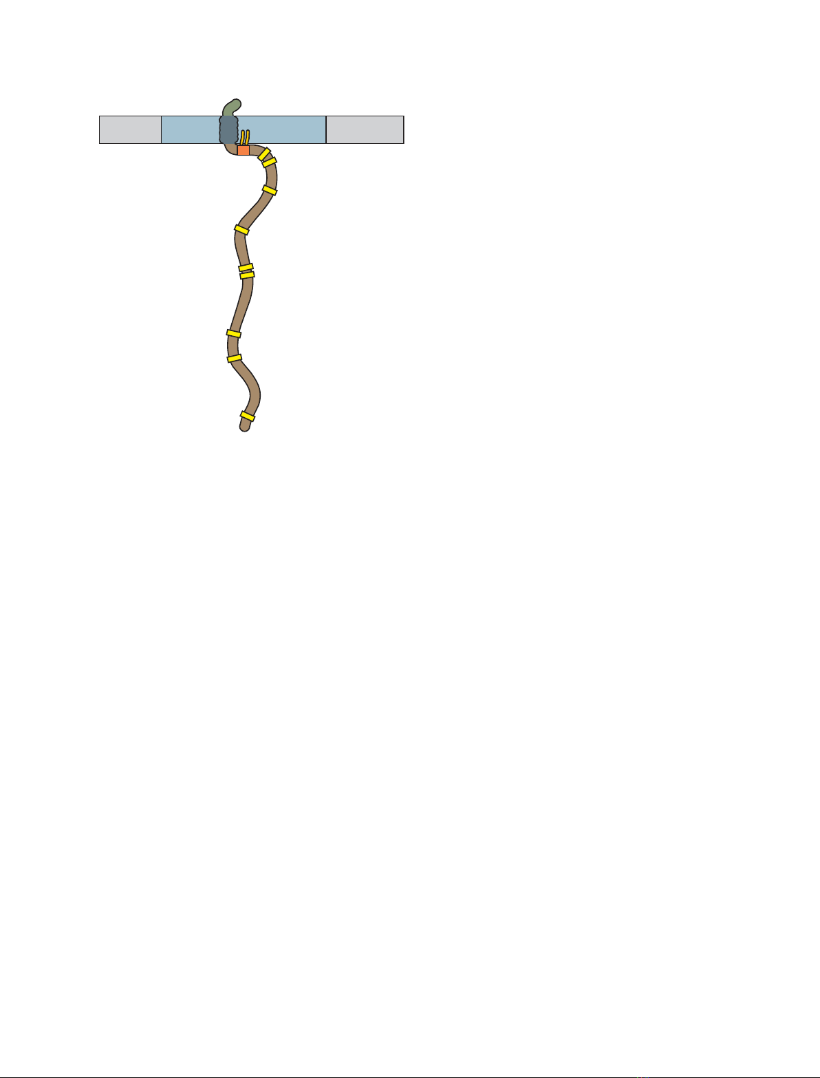
This prototypic TRAP (Fig. 1) is expressed in thymo-
cytes and T cells, NK cells and mast cells; later it was
also found in pre-B cells (but not in mature B cells)
[42,43], myeloid cells [44], megakaryocytes and platelets
[45,46].
The LAT polypeptide chain contains in its mem-
brane-proximal part two cysteine residues (C26, C29 in
humans, C27, C30 in mouse), which can be palmitoy-
lated by a so far unidentified palmitoyl transferase(s).
This post-translational modification is essential for
LAT membrane and raft association (see below);
recent data demonstrate that monopalmitoylation of
LAT on C26 is sufficient for its association with the
plasma membrane and function (however, it was not
reported whether the monopalmitoylated mutant is
present in membrane rafts to the same extent as the
double-palmitoylated wild-type protein) [47].
Upon immunoreceptor engagement of several recep-
tors [most notably TCR, FccR, FceRI, collagen recep-
tor glycoprotein VI (GPVI), but see below for more
examples] at least five of its nine tyrosine motifs can
be phosphorylated by ZAP-70 or Syk kinases [40], but
also by Itk [48] and possibly Lck [49]. Phosphorylated
LAT associates with several SH2-containing molecules
[Grb2, Gads, Grap, PLCc1, p85, phosphatidylinositol
3-kinase (PI3K), Vav], thereby organizing signalosomes
needed for the initiation of several intracellular signal-
ling pathways [40,50–52]. A key cytoplasmic adaptor,
SLP-76, is recruited to phospho-LAT via its constitutive
association with Gads [53]. It is not known how many
different phospho-LAT containing complexes exist,
differing in their composition. The formation of
phosphotyrosine-dependent multiprotein signalling
complexes organized around phospho-LAT was also
examined more rigorously in an in vitro system based
on recombinant LAT incorporated in liposomes and
recruitment of signalling protein complexes from
Jurkat cell cytosol [54].
Little is known about the structural details of differ-
ential recognition of tyrosine-phosphorylated sites in
LAT by SH2-containing ligands, an exception being the
adaptor Gads; high-resolution structures of Gads–SH2
complexed with phosphopeptides corresponding to sites
171, 191 and 226 revealed the structural basis for prefer-
ential recognition of specific phospho-LAT sites by
Gads, as well as for the related adaptor Grb2 [55].
LAT – negative regulation in TCR
signalling
LAT was reported to interact with the active (open)
form of Lck in rafts and possibly induce its transition
into the inactive (closed) conformation [56]. The inter-
action of LAT with a negative regulator of the
Ras–MAPK pathway of receptor tyrosine kinases,
Sprouty1, negatively regulates LAT phosphorylation.
A C-terminal deletion mutant of Sprouty1 is unable to
translocate to the immune synapse and interact with
LAT [57]. Cytoskeletal protein 4.1R negatively regu-
lates T cell activation by directly binding to LAT, and
thereby inhibiting its phosphorylation by ZAP-70 [58].
Tyrosine phosphatase SHP-2 is recruited to the LAT–
Gads–SLP-76 complex and regulates the phosphoryla-
tion of signalling proteins Vav1 and ADAP. This
enzyme is transiently inactivated by reactive oxygen
species produced after TCR stimulation [59]. The
inhibitory Fc receptor FccRIIB (present also on acti-
vated T cells) associates with phosphatases SHP-1,
SHP-2 and SHIP-1 inhibit TCR-mediated Ca
2+
mobi-
lization, in part through the inhibition of LAT phos-
phorylation followed by the inhibition of PI3K
activation [60]. Cellular localization and functionality
of LAT was reported to be sensitive to intracellular
redox status. Oxidative stress results in conformational
changes (the formation of intramolecular dislufidic
bridges) causing membrane displacement of LAT and
consequent hyporesponsiveness of T lymphocytes [61].
LAT, as a key component of the TCR activation
pathways, may be expected to be a target of pathogens
trying to eliminate T cell-based immune responses.
Indeed, Yersinia suppresses T lymphocyte activation
through the virulence factor YopH, a tyrosine
V. Hor
ˇejs
ˇı
´et al. LAT, a key membrane raft-associated protein
FEBS Journal 277 (2010) 4383–4397 ª2010 The Authors Journal compilation ª2010 FEBS 4385
phosphatase that dephosphorylates LAT and SLP-76
in activated T cells [62].
LAT – signalling clusters, membrane
rafts and interactions with TCR
complexes
One of the current models postulates that TCR
molecules or their clusters in the plasma membrane of
resting T cells are physically separated from raft micr-
odomains containing several important signalling mol-
ecules, e.g. Lck, Fyn, LAT, PAG, PIP2 (it is not clear
whether individual rafts contain several of the proteins
or rather there are separate LAT-containing, Lck-
containing, etc. rafts) [23]. A variant of the model
assumes that TCR clusters and a subset of rafts are
preassembled even in resting T cells and TCR ligation
just reorients them such that the raft signalling pro-
teins start to interact functionally with the TCR [37].
After TCR ligation, the TCR clusters are either mixed
or concatenated with the rafts, which may be simulta-
neously fused to form larger patches. Such processes
also apparently accompany the formation of physio-
logical immunological synapses or ‘patches’ or ‘caps’
induced by artificial cross-linking of TCR [63].
The understanding of the involvement of rafts in this
process is complicated by unresolved problems, such
as the heterogeneity of raft microdomains and techni-
cal problems in studies on the nature of apparently
highly dynamic raft assemblies. An illustration of the
raft heterogeneity is provided by the observations that
cholesterol extraction destabilizes the membrane micr-
odomains containing Lck, whereas those containing
LAT remain almost intact. As shown by electron
microscopy, following T cell activation, both LAT and
Lck colocalize in 50–100 nm microdomains, which cor-
relates with the initiation of T cell signal transduction
[64].
The involvement of LAT-containing rafts in TCR-ini-
tiated activation was demonstrated using transfectants
expressing LAT-GFP [65]. After stimulation with anti-
CD3-coated beads, LAT-GFP translocated to the area
of T cell contact with the beads. The LAT-GFP present
in the contact area was markedly immobilized compared
with the membrane outside the contact. The mobility
increased after raft disruption by cholesterol depletion,
and was also dependent on the integrity of critical bind-
ing sites (PLCc) in the cytoplasmic domain of LAT.
At present it is not entirely clear why the presence
of LAT in membrane rafts is functionally important
and how these LAT-containing rafts are related to
‘signalling clusters’ described in several papers.
Transmembrane glycoprotein CD2 involved in T cell
costimulation, LAT, and tyrosine kinase Lck were
reported to be coclustered in discrete T cell plasma
membrane microdomains. The integrity of these micr-
odomains was dependent on protein–protein interac-
tions based on phosphorylated LAT, but apparently
independent of interactions with rafts or actin [66]. In
quiescent T cells, LAT and TCR were observed in sep-
arate ‘protein islands’, which became concatenated
upon T cell activation [67]. The signalling complexes
organized around phospho-LAT and apparently vital
for intracellular signalling appear to be oligomerized
by multipoint co-operative binding of several cytoplas-
mic SH2 and SH3 domain-containing signalling pro-
teins to LAT [68–70].
The involvement of LAT in T cell activation is also
regulated by another type of membrane microdomain
heterogeneity. LAT molecules are preferentially located
in the uropod of migrating T cells. In activated T cells
forming stable immunological synapses with antigen-
loaded B cells, LAT accumulates at the contact
between the two cells (immunological synapse) [71].
LAT was reported to exist in two distinct cellular
pools, one at the plasma membrane and the other in
endocytic vesicles also containing a transferrin receptor
and the TCR fchain [72]. The plasma membrane-asso-
ciated LAT is rapidly recruited to the immune synapse,
whereas the intracellular pool is first polarized and
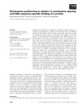
Y37
Y46
Y67
Y113
Y132
Y175 (Gads, Grb2)
Y195 (Gads, Grb2)
Y235 (Grb2)
Y136 (PLCγ)
Plasma membrane
Membrane
raft
Fig. 1. A model of the LAT molecule. A schematic representation
of mouse LAT with a palmitoylation site (orange) and the positions
of all tyrosines (yellow). The binding partners for the key phosp-
hotyrosine residues are indicated.
LAT, a key membrane raft-associated protein V. Hor
ˇejs
ˇı
´et al.
4386 FEBS Journal 277 (2010) 4383–4397 ª2010 The Authors Journal compilation ª2010 FEBS

recruited to the immunological synapse with a delay.
Critical tyrosine residues of LAT are necessary for
recruitment to the immunological synapse and a juxta-
membrane region of LAT is involved in the intracellu-
lar pool localization of LAT and T cell signalling. This
aspect was recently examined in more detail by
Purbhoo et al. [73]. The study found that the kinase
ZAP-70 and the adaptor proteins LAT and SLP-76
accumulate in separate clusters at the immunological
synapse. Importantly, a sizeable fraction of LAT was
found in vesicles that migrated to surface microclusters
containing SLP-76 and the adaptor protein GADS,
where they became temporarily immobilized. The
results suggest a surprising additional mechanism of
LAT participation in the TCR signalling process.
The involvement of LAT-containing membrane rafts
in the formation and signalling of TCR microclusters
and central supramolecular activation clusters
(cSMACs) at the immunological synapse remains con-
troversial. A recent study [74] did not find accumula-
tion of raft probes at TCR microclusters or cSMACs.
Raft association of LAT mutants was dispensable for
TCR microcluster formation. Observable accumulation
of raft probes in the cell interface actually occurred
after cSMAC formation and could rather be due to
membrane ruffling or endocytosis. The results of this
study suggest that membrane rafts may actually not
serve as a platform for T cell activation.
Is the presence of LAT in rafts
necessary for its function in TCR
signalling?
Proper functioning of LAT appeared to be dependent
on its targeting to membrane rafts [75–77]. This target-
ing was thought to be due to palmitoylation of its juxta-
membrane cysteine motif (CxxC) because the cysteine
mutants were not able to reconstitute TCR signalling in
LAT-negative T cell lines [76,77]. Furthermore, target-
ing of SLP-76 constitutively to plasma membrane rafts
in LAT-deficient Jurkat T cells largely restores the sig-
nalling defects, indicating that recruitment of SLP-76 to
the membrane raft environment via phospho-LAT is
the crucial LAT-dependent signalling event [78]. Also,
the displacement of LAT from membrane rafts was
demonstrated as a molecular mechanism responsible for
the inhibition of T cell signalling by polyunsaturated
fatty acids [79]. Furthermore, palmitoylation of LAT
was shown to be defective in anergic T cells [80].
Although fchain or ZAP-70 phosphorylation were
normal in these cells, LAT tyrosine phosphorylation
and PLCc1 activation were markedly decreased. Inhibi-
tion of T cell activation by a cytoplasmic LAT mutant
lacking the transmembrane domain is accompanied by
reduced recruitment of signalling molecules to glyco-
lipid-enriched microdomains [81].
However, the importance of LAT localization in
membrane rafts became recently doubtful as a result of
several studies. First, the nonpalmitoylated LAT cyste-
ine mutants were shown to be not only absent from
membrane rafts, but not even properly transported to
the plasma membrane and remained retained in the
endoplasmic reticulum [47,82,83]. It was suggested that
in addition to proper acylation, homotypic or hetero-
typic protein–protein interactions may also contribute
to LAT targeting to rafts [83]. Second, it was demon-
strated that a LAT construct composed of the cyto-
plasmic region of LAT fused with the extracellular and
transmembrane regions of the nonraft transmembrane
adaptor, LAX, restored TCR signalling in LAT-defi-
cient cell line and normal development of T cells from
LAT
)⁄)
haematopoietic precursors [84]. A similar con-
clusion was reached using another LAT construct (the
cytoplasmic part of LAT equipped with a membrane-
anchoring motif of Src) not targeted to membrane
rafts but yet fully functional [47].
These results, which might demolish the generally
accepted concept of the membrane raft’s importance in
immunoreceptor signalling, were recently explained by
results from our laboratory [36]. We demonstrated the
existence of a novel type of membrane raft-like micr-
odomain (‘heavy rafts’) containing a number of mem-
brane molecules, including, for example, the LAX and
the LAX-LAT chimaeric construct. The LAT con-
structs targeted to the newly identified ‘heavy rafts’ are
also able to support TCR signalling, albeit less effi-
ciently than the wild-type LAT present in ‘classical
rafts’; the least efficient are constructs targeted to non-
raft membrane. This difference may be minimized by
increased levels of LAT-construct expression in the
heavy rafts or nonraft membrane. Therefore, different
types of membrane microdomain appear to provide
environment regulating functional efficiency of signal-
ling molecules present therein.
Role of LAT in anergy induction
LAT was reported to be hypophosphorylated and
mis-localized in anergic T cells, apparently as a conse-
quence of a selective palmitoylation defect; it was
largely absent from DRM fractions corresponding to
rafts and was not normally recruited to the immuno-
logical synapse. The defects were selective for LAT,
because DRM localization and palmitoylation of Fyn
were intact. These defects were not due to enhanced
LAT degradation [80]. It should be noted that induction
V. Hor
ˇejs
ˇı
´et al. LAT, a key membrane raft-associated protein
FEBS Journal 277 (2010) 4383–4397 ª2010 The Authors Journal compilation ª2010 FEBS 4387


![Báo cáo seminar chuyên ngành Công nghệ hóa học và thực phẩm [Mới nhất]](https://cdn.tailieu.vn/images/document/thumbnail/2025/20250711/hienkelvinzoi@gmail.com/135x160/47051752458701.jpg)


