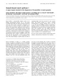
Nucleosome positioning in relation to nucleosome spacing and DNA sequence-specific binding of a protein Rama-Haritha Pusarla*, Vinesh Vinayachandran* and Purnima Bhargava
Centre for Cellular & Molecular Biology, Hyderabad, India
Keywords chromatin assembly; ionic strength; nucleosome positioning; nucleosome spacing; protein boundary
Correspondence P. Bhargava, Centre for Cellular & Molecular Biology, Uppal Road, Hyderabad-500007, India Fax: +91 40 27160591 Tel: +91 40 27192603 E-mail: purnima@ccmb.res.in
*These authors contributed equally to this work
(Received 12 October 2006, revised 2 March 2007, accepted 7 March 2007)
doi:10.1111/j.1742-4658.2007.05775.x
internucleosomal spacings of the
Nucleosome positioning is an important mechanism for the regulation of eukaryotic gene expression. Folding of the chromatin fiber can influence nucleosome positioning, whereas similar electrostatic mechanisms govern the nucleosome repeat length and chromatin fiber folding in vitro. The position of the nucleosomes is directed either by the DNA sequence or by the boundaries created due to the binding of certain trans-acting factors to their target sites in the DNA. Increasing ionic strength results in an increase in nucleosome spacing on the chromatin assembled by the S-190 extract of Drosophila embryos. In this study, a mutant lac repressor protein R3 was used to find the mechanisms of nucleosome positioning on a plas- mid with three R3-binding sites. With increasing ionic strength in the pres- the number of positioned nucleosomes in the chromatin ence of R3, decreased, whereas the positioned nucleosomes in a single register did not change. The number of the posi- tioned nucleosomes in the chromatin assembled in vitro over different plas- mid DNAs with 1–3 lac operators changed with the relative position and number of the R3-binding sites. We found that in the presence of R3, nucleosomes were positioned in the salt gradient method of the chromatin assembly, even in the absence of a nucleosome-positioning sequence. Our results show that nucleosome-positioning mechanisms are dominant, as the nucleosomes can be positioned even in the absence of regular spacing mechanisms. The protein-generated boundaries are more effective when more than one binding site is present with a minimum distance of (cid:2) 165 bp, greater than the nucleosome core DNA length, between them.
Abbreviations IEL, indirect end-labeling; IPTG, isopropyl thio-b-D-galactoside; MNase, micrococcal nuclease; NRL, nucleosome repeat length.
FEBS Journal 274 (2007) 2396–2410 ª 2007 The Authors Journal compilation ª 2007 FEBS
2396
Multifold compaction of DNA due to the presence of nucleosomes on natural templates of eukaryotic RNA polymerases results in transcriptional repression. Sev- eral studies have established that histones and nucleo- somes play an active role in regulating gene expression in eukaryotic cells [1,2]. Gene-specific, localized config- in vivo chromatin in various genome urations of regions are found due to precise positioning of nucleo- somes over the underlying DNA stretches [3,4]. Both trans-acting factors and DNA sequences can determine where histones will occupy the bound DNA. A nucleo- some is positioned translationally when its histone– DNA contacts are restricted to an identifiable stretch of DNA, giving clear boundary zones. Further preci- sion of the positioning can be achieved by restricting the rotation of DNA over the histone octamer surface (rotational setting), resulting in a defined phase ⁄ orien- tation of a particular base pair with respect to histones. Nucleosomes on certain constitutively active genes can be excluded due to rapid and tight associ- ation of trans-acting factors with promoter elements during the replication-coupled assembly of chromatin in vivo [5]. On other genes, they are removed or reshuffled through several chromatin-remodeling and
R.-H. Pusarla et al.
Mechanisms of nucleosome positioning
cells use chromatin-modifying mechanisms. Thus, nucleosome positioning as a mechanism to include or exclude the binding sites of trans-acting factors from accessible chromatin regions by passively restricting the position of nucleosomes therein [6,7]. It is now well established that a nucleosome is not necessarily repres- it can facilitate the activation of genes. sive; rather, Specific positioning of the nucleosomes allows the transcriptional machinery to work effectively in a chro- matin environment. Folding of DNA by the histones and positioned nucleosomes can bring two widely sep- arated regulatory elements into juxtaposition in space [8–10] or even orient the bound factors for productive interactions.
to its two sites, DNA-binding prokaryotic protein) 183 bp apart, was shown to result in at least five trans- lationally positioned nucleosomes in a single register on a plasmid DNA [26]. In general, boundaries gener- the ated by proteins binding to the DNA restrict randomization of nucleosome positions [27]. It was predicted that the nucleosomes close to a boundary would be precisely positioned, whereas this precision would decrease with distance from the boundary [28]. We have analyzed the effect of changing the ionic strength and the number and spacing of the binding sites on the protein-generated boundary for nucleo- some positioning in both assembly systems. We have found that the number of positioned nucleosomes in a single register changes with the ionic strength of the medium, although the spacing between them does not change. The range of positioning effects of DNA sequence-specific binding of a protein to chromatin depends on the number of sites and the distance between them.
Results
influences
All the chromatin assemblies were constructed using rat liver core histones and plasmid DNAs schematical- ly depicted in the Fig. 1, in the presence or absence of the R3 protein.
Number of positioned nucleosomes changes with ionic strength Nucleosomal repeat length (NRL) is characteristic for a species, suggesting that it is a regulated feature of the chromatin [11]. Uniform spacing of nucleosomes is proposed to promote the higher-order folding of chromatin [12]. Chromatin folding, in turn, is reported to influence nucleosome positioning [13]. The presence of positioned nucleosomes at defined and limited loca- tions may result in disruption of the uniformity in spa- it is not known whether nucleosome cing. However, spacing positioning. The nucleosome observed longer repeat lengths on inactive genes as compared to those observed on transcribed sequences [14,15] suggest that the folding of the 10 nm chromatin with beads on a string into a higher-order structure requires a minimum spacing to be maintained between core particles. Thus, nucleosome spacing and position- ing appear to be correlated.
R3 is a mutant lac repressor protein that binds the lac operator as a dimer with the same affinity as that of the wild-type lac repressor but fails to tetramerize [29]. Binding of R3 protein to a plasmid DNA pU6LNS (two lac operators at a distance of 183 bp) was reported
A
B
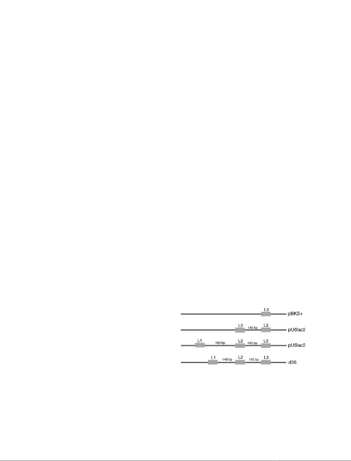
Fig. 1. Relative positions of the three lac operator sites in the plas- mid DNAs. (A) Diagrammatic representation of the plasmid con- structs with different numbers of lac operators. The solid rectangular boxes and L1, L2 and L3 denote the first, second and third lac operator sites. The distance in bp between each site is given. (B) Schematic map of plasmid d35 with the shortest dis- tance between L1 and L2.
FEBS Journal 274 (2007) 2396–2410 ª 2007 The Authors Journal compilation ª 2007 FEBS
2397
Ionic strength is reported to influence nucleosome conformations [16,17] as well as their spacings [18]. Within an array of positioned nucleosomes, the ionic strength effect dominates the sequence effect [19]. It also influences chromatin folding, presumably by modulating H1 association as well as interparticle interactions [20,21]. Of the two methods of chromatin assembly in vitro [22], the salt gradient dialysis method deposits nucleosomes in a random fashion, and has been useful for checking the ability of various DNA sequences to position nucleosomes in vitro. A Dro- sophila embryonic extract, in contrast [23], can deposit nucleosomes with regular spacing in a sequence-inde- pendent manner in the presence of ATP. The in vitro chromatin assembly carried out by cellular ⁄ nuclear extracts, giving uniformly spaced nucleosomes, is affec- ted by parameters such as ionic strength, concentration of linker histones, protein phosphorylation, and the presence of core histone tails [18,24]. Spacing of nucle- osomes is also influenced by DNA topology or histone [19,25]. Using this variants system, binding of a lac repressor R3 (a small sequence-specific mutant
R.-H. Pusarla et al.
Mechanisms of nucleosome positioning
In comparison to this, two positioned nucleosomes could be seen even at a salt concentration of 130 mm, whereas none were seen at a salt concenration of 150 mm (Fig. 2C, lanes 1 and 9). These results show that with increasing ionic strength, fewer positioned nucleosomes are aligned in a single register.
lane 5; Fig. 2C,
to result in the positioning of an array of five nucleo- somes in a single register [26]. As R3 can bind to the naked DNA at salt concentrations as high as 0.4 m (not shown), we looked at the effect of increasing ionic strength on nucleosome positioning due to R3 binding on the plasmid pU6lac3 by using the indirect end- labeling (IEL) method of chromatin structure analysis (Fig. 2). The micrococcal nuclease (MNase) digestion the naked DNA in the IEL analysis pattern of (Fig. 2A, lane 1; Fig. 2B, lane 4; Fig. 2C, lanes 5 and 6) did not change with the binding of R3 (Fig. 2A, lane 2; Fig. 2B, lanes 4 and 7), as small footprints could not be resolved in the agarose gels. The digestion pattern did not change even with the deposition of histones (Fig. 2A, lanes 3, 5 and 6; Fig. 2B, lanes 1, 6 and 7; Fig. 2C, lanes 2, 3 and 8) at every ionic strength, suggesting there are no preferred locations for nucleosome assembly on the plasmid. However, in the presence of R3, in a single register, five positioned nucleosomes at a salt concentration of 50–90 mm (Fig. 2A, lanes 4, 7 and 8; Fig. 2B, lanes 2 and 3) and three positioned nucleosomes at a salt con- centration of 110 mm (Fig. 2B, lane 8) could be seen. As positioned nucleosomes are seen in Fig. 2 only in the presence of R3, we used DNaseI footprinting to confirm the specific binding of R3 at L1 and L2 at all the salt concentrations. As no positioned nucleosomes were seen at a monovalent salt level of 150 mm in the IEL analysis of Fig. 2C, no protection was seen between L1 and L2 on the DNA subjected to chroma- tin assembly in the representative gel at this ionic strength (Fig. 3A). Chromatin assembly in our system, as judged by the generation of MNase-resistant nucleo- somal ladders, was found to be normal up to 110 mm salt, whereas only a few nucleosomal bands could be seen at higher salt levels (not shown). The binding of R3 did not disrupt the nucleosomal ladders close to the three distinct sites located at different distances from each other (Fig. 3B). Therefore, the loss of posi- tioned nucleosomes at higher ionic strengths is not due
A
B
C
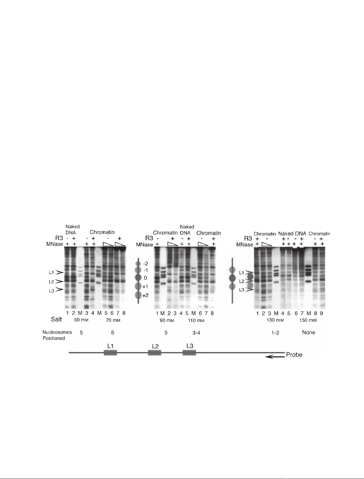
Fig. 2. Nucleosome positioning in the presence of R3 at different ionic strengths. IEL analysis of the chromatin structure of plasmid pU6lac3 assembled with S-190 extract in the absence or presence of R3 at various salt concentrations. Nucleosome positions are numbered and marked with ellipses, and arrowheads indicate the positions of lac operators, marked L1–L3. The total number of the positioned nucleo- somes in a single register is given under each salt level. The 5¢-end of the radiolabeled oligonucleotide probe (arrowhead) hybridized 709 bp downstream of the third lac operator, L3, as shown at the bottom of the figure. (A) Structure analysis of the chromatin assembled at 50 or 70 mM salt. Lanes 1 and 2 show naked DNA digestion patterns, and lanes 3–8 represent chromatin. R3 is absent in lanes 1, 3, 5 and 6, whereas chromatin was assembled at 50 mM salt for lanes 1–4, and at 70 mM salt for lanes 5–8. (B) IEL analysis for the chromatin assem- bled at 90 and 110 mM salt concentrations. Naked DNA (lanes 4 and 5) and chromatin assembled in the absence or presence of R3 protein are shown. R3 was added to lanes 2, 3, 5 and 8. (C) IEL analysis for the chromatin assembled at 130 and 150 mM salt. Naked DNA (lanes 4–7) and chromatin assembled in the absence or presence of R3 protein are shown. R3 was added to lanes 1, 4, 7 and 9.
FEBS Journal 274 (2007) 2396–2410 ª 2007 The Authors Journal compilation ª 2007 FEBS
2398
R.-H. Pusarla et al.
Mechanisms of nucleosome positioning
A
B
D
C
E
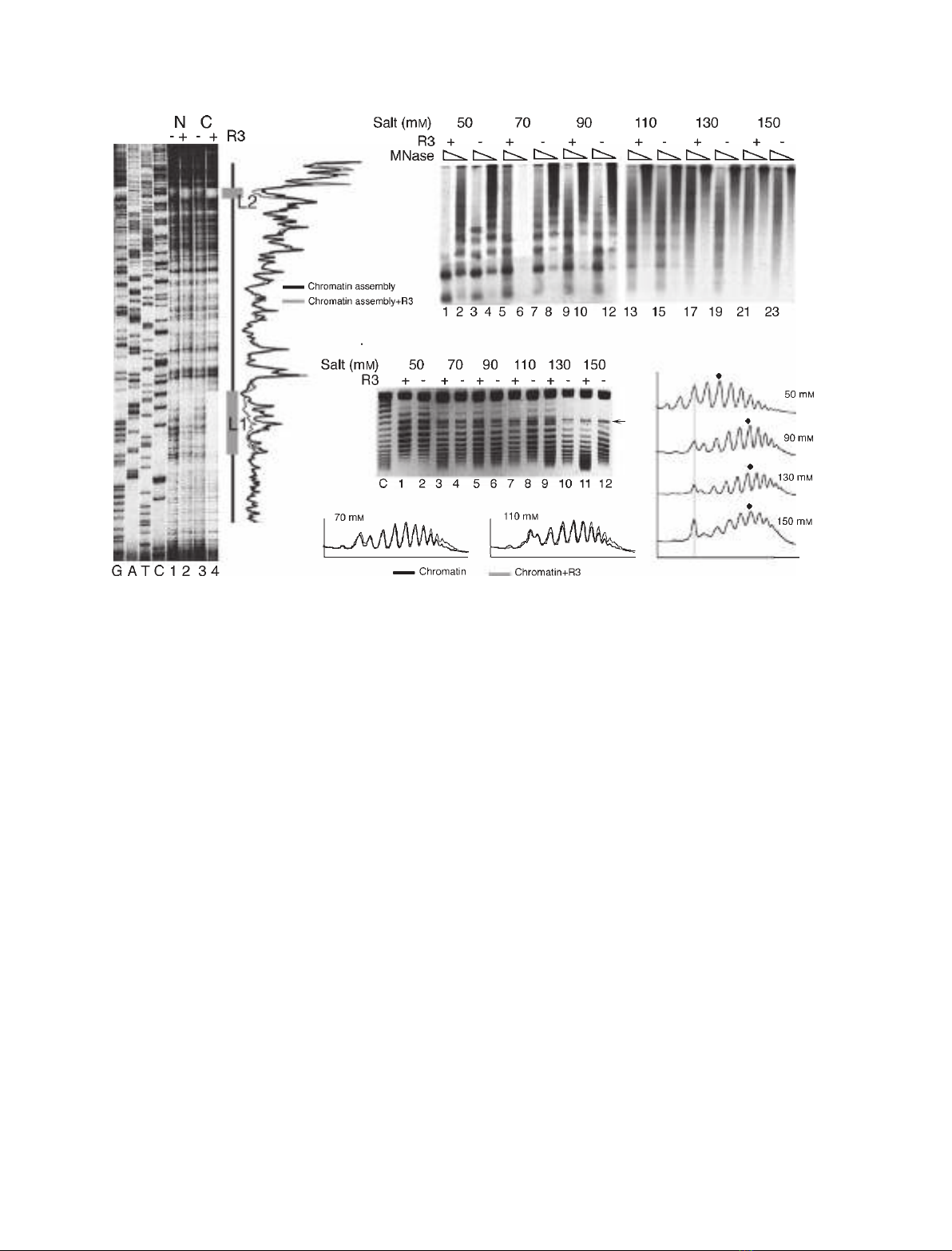
Fig. 3. Influence of ionic strength and R3 binding on chromatin assembly. (A) Binding of R3 to its sites in the chromatin. High-resolution DNaseI footprinting gel shows digestion profiles of the naked DNA (N) and chromatin assembly (C) in the presence and absence of the R3 protein. Chromatin was assembled with S-190 extract over plasmid pU6lac3 at a monovalent salt concentration of 150 mM. Comparison of the profiles of lanes 3 and 4 in the right-hand panel shows protection due to R3 at L1 and L2 (gray boxes) but not between them. The pri- mer was located 93 bp upstream of L1. GATC shows the sequencing ladders generated by the same primer. (B) MNase-resistant nucleo- some ladders from chromatin assemblies were resolved on 1.25% agarose gels, Southern transferred, and probed with a primer that hybridizes to the top strand, 53 bp downstream of L1. (C) One-dimensional supercoiling assay of chromatin assembly. Topoisomers were resolved on a 1% agarose gel with 15 lM chloroquin present in the gel as well as the tank buffer. The arrow marks a band seen in every lane, used as a reference. (C) Plasmid DNA control, which was not subjected to chromatin assembly. (D) Profiles of the topoisomer distribu- tion of chromatin from lanes 2, 6, 10 and 12 in (C). The gray vertical line shows the alignment of peaks corresponding to the band marked with an arrow in (C), and the dot marks the peak with the highest intensity in a profile. (E) Profiles of the topoisomer distribution in lanes 3 and 7 (chromatin with R3) are compared with those in lanes 4 and 8 (chromatin without R3) in (C).
FEBS Journal 274 (2007) 2396–2410 ª 2007 The Authors Journal compilation ª 2007 FEBS
2399
to the absence of R3 binding, but is probably related to the reported effects of the ionic strength on the bulk chromatin properties. As an increase in the ionic strength is reported to increase the NRL [18], the total number of uniformly spaced nucleosomes on a plasmid DNA may decrease, which would result in a change in the topological state of the plasmid. Therefore, we confirmed the ionic strength effects on nucleosomal density on plasmid DNA by the one-dimensional supercoiling assay in the presence of chloroquin. The plasmid in this assay was not relaxed prior to chroma- tin assembly, as the S-190 extract is known to have topoisomerase I activity, and assembly in this system proceeds to completion. Therefore, under the condi- tions of the gel run in Fig. 3C, all of the resolved bands may be positively supercoiled topoisomers, which differ by one linking number [30]. Comparison of the profiles of the topoisomers (Fig. 3D) showed a downward shift of the mean of the Gaussian distribu- tion at different salt concentrations, denoting an increase in the linking number of the DNA. As chloro- quin introduces positive supercoils into the DNA, this shift confirms that with increasing salt concentration, there is a change in the superhelical density, which is caused by a decrease in the number of nucleosomes deposited over the plasmid DNA. In contrast to this, the topoisomer profiles in the presence and absence of R3 protein at different salt concentrations showed a perfect overlap (Fig. 3E), suggesting that binding of R3 at these ionic strengths did not change the nucleo- some spacing further. Although the chromatin assem- bly appeared to be better in the absence of R3
R.-H. Pusarla et al.
Mechanisms of nucleosome positioning
positioned Translationally
(Fig. 3C), at 150 mm salt, the significantly different superhelical density (Fig. 3C) and presence of only sparse nucleosomes in an MNase ladder assay (not shown) suggest that the absence of positioned nucleo- somes in this case is due to inefficient chromatin assem- bly, rather than the loss of the boundary effect of R3.
Spacing between the positioned nucleosomes does not change with ionic strength
The bulk chromatin is reported to show an increase in NRL with increasing ionic strength [18]. Although this may influence the spacing even between the positioned nucleosomes, the IEL analysis in Fig. 2 suggested that the relative location of the positioned nucleosomes remains the same at every ionic strength. Numbering the nucleosome between the operators 183 bp apart as 0 (Fig. 2A), we used MNase footprinting to map the positions of nucleosomes 0, ) 1 and + 1 by using different primers to look at one nucleosome at a time (Fig. 4). nucleosomes showed a clear 145 bp protection in the profile com- parisons of the chromatin lanes with and without R3. The + 1 nucleosome was found with its 5¢ edge the lac operator L2 located 10 bp downstream of (Fig. 4D), whereas the 3¢ edge of nucleosome ) 1 was found 10 bp upstream of the lac operator L1 (Fig. 4B) at every ionic strength tested. Mapping of nucleosome 0 between the lac operators L1 and L2 showed its location to be 25 bp downstream of L1 on the 5¢ side, and 12 bp upstream of L2 on the 3¢ side. Similarly, the the + 2 and ) 2 nucleosomes did not location of change with changing ionic strength. This analysis the number of positioned nucleosomes shows that
A
C
B
D
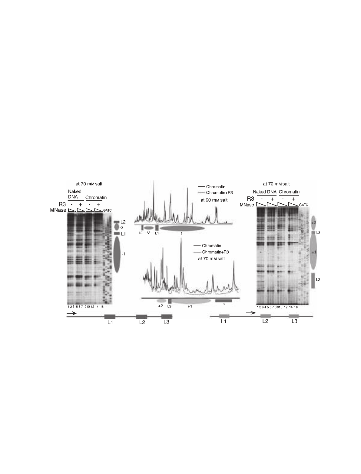
Fig. 4. Structural analysis of the pU6lac3 chromatin. High-resolution MNase footprinting was used to map the positioned nucleosomes over plasmid pU6lac3 without or with bound R3. A seven-fold molar excess of R3 over operators was added at the start of the assembly. Aliqu- ots of the same assembly were subjected to three levels of MNase digestion. Ellipses mark the nucleosomal size protections, and solid boxes represent the R3 footprint and the lac operators. The positions of nucleosomes ) 1, 0, + 1 and + 2 are marked. (A) Mapping the posi- tion of the nucleosomes in the presence of R3 by the primer extension footprinting of the chromatin assembled at 70 mM salt. The primer was located 196 bp upstream of the lac operator L1, hybridizing to the bottom strand, as depicted in the cartoon in the left-hand bottom cor- ner. Lanes 1–8 show extension products of naked DNA digestions, and lanes 9–16 show chromatin samples. R3 was added to the samples in lanes 5–8 and 13–16. GATC shows the sequencing ladder generated with the same primer. (B) Primer extension footprinting of the chro- matin reconstituted at 90 mM salt. A comparison of the profiles of the digested chromatin without and with R3 is shown. The primer was the same as in (A). (C) Higher-resolution MNase footprinting of the chromatin with R3 bound or unbound shows that the lac operator L3 gets included in the positioned nucleosome + 1. Lanes 1–8 show the naked DNA pattern with (lanes 5–8) and without (lanes 1–4) R3, and lanes 9–16 show the digestion pattern of the chromatin assembled at 70 mM salt (R3 was added to lanes 13–16). The primer was located 64 bp upstream of lac operator L2, hybridizing to the bottom strand as shown schematically in the right-hand bottom corner. GATC shows the sequencing ladder generated by using the same primer. (D) A comparison of the profiles of lanes 9 and 13 in (C) is shown. Protection of 145 bp due to a positioned nucleosome + 1 overlaps with part of L3 occupied by R3.
FEBS Journal 274 (2007) 2396–2410 ª 2007 The Authors Journal compilation ª 2007 FEBS
2400
R.-H. Pusarla et al.
Mechanisms of nucleosome positioning
with a nucleosome [31,32], and once formed, the repres- sor–DNA complex is stable [33]. The properties of bind- ing of R3 to DNA are similar to those of the wild-type protein [34]. Therefore, it appears that the observed pro- tection is due to a positioned nucleosome encroaching on half of the R3-bound L3. These results explain why the presence of the third operator L3 did not increase the range of nucleosome positioning in pU6lac3.
changes with ionic strength, but the spacings of these nucleosomes do not change with ionic strength. It is important to note that the reported increase in the measured NRL with increasing ionic strengths [18] represents the change in the average NRL of the bulk chromatin, and we did not see any disruption of the regular spacing in the presence of R3 (Fig. 3B). There- fore, the small changes in the average nucleosome spa- cing at lower ionic strengths ([18], data not shown) can easily adjust with unaltered spacing of a few posi- tioned nucleosomes (Fig. 4) in a relatively shorter region of the chromatin. At higher salt concentrations (> 110 mm), the extreme nucleosomes ) 2 and + 2, which are positioned due to alignment rather than restricted mobility, moved out of register and behaved like bulk nucleosomes, resulting in loss of positioning. As the change in ionic strength did not disturb the positioned nucleosomes at lower ionic strengths, we conclude that nucleosome positioning dominates the nucleosome-spacing effects on the bulk chromatin.
The distance between protein-binding sites decides nucleosome positioning
At every ionic strength, nucleosome 0 was positioned 25 bp from L1 and 12 bp from L2, whereas the flanking nucleosomes ) 1 and + 1 were positioned 10 bp from L1 and L2, respectively. This observation suggests that a minimum distance of (cid:2) 10 bp is maintained between the operators and the histone octamer position, giving a minimum requirement of 165 bp between the two pro- teins if they are to work as flag-ends for a positioned nu- cleosome, protecting 145 bp of core DNA length between them. The results shown in Fig. 4 suggest that the distance between the two binding sites should be more than 140 bp if they are both to work as demarca- tion boundaries. In order to find this minimum, optimal distance, we derived a set of plasmids from pU6lac3 by reducing the 183 bp distance between L1 and L2 in 5 bp increments (Fig. 1B). Reducing this distance by 10 or 20 bp (as well as 5 and 15 bp, not shown) did not result in any loss of positioning (Fig. 5A). A distance of 163 bp in plasmid d20, more than the core DNA length, shows the presence of five positioned nucleosomes in the presence of R3. In contrast, the deletion by 25, 30 or 35 bp resulted in the loss of the array of positioned nu- cleosomes, and only one positioned nucleosome show- ing less pronounced protection between L1 and L2 could be seen (lanes 3, 6 and 8) in the IEL analysis in the profiles in Fig. 5B. However, a comparison of Fig. 5C revealed the presence of three nucleosomal size protections in d30, whereas no protection could be seen in the profile comparisons for d35 (Fig. 5C). A strong nuclease sensitivity (marked by asterisks) next to the R3 bound to all three operators, suggesting an alteration in the chromatin structure, could also be seen.
FEBS Journal 274 (2007) 2396–2410 ª 2007 The Authors Journal compilation ª 2007 FEBS
2401
Positioning of five nucleosomes in a single register as a result of the binding of R3 to two lac operators in plas- mid pU6LNS [26] suggested that the bound R3 mole- cules serve as bookmarks for alignment of nucleosomes on both sides. However, as compared to pU6LNS with two lac operators, the presence of a third operator L3 did not increase the number of positioned nucleosomes in pU6lac3. Whereas the 183 bp distance between the two operators L1 and L2 in pU6LNS and pU6lac3 can easily accommodate one nucleosome, the subnucleosomal dis- tance of 140 bp between L2 and L3 in pU6lac3 did not disrupt positioning of the nucleosomes downstream of L2 (Fig. 2). Footprinting to map the two downstream nucleosomes, + 1 and + 2 in Fig. 4C, showed protec- tion at L3 contiguous with the remaining 130 bp between L2 and L3 (nucleosome + 1 is positioned 10 bp downstream of L2), suggesting that L3 becomes inclu- ded in the core particle + 1 at its 3¢ edge (Fig. 4C,D). Although there is a possibility that R3 at L3 was removed by nucleosome + 1, a total protection size of more than 145 bp DNA downstream of L2 and span- ning over L3 suggests that it was generated by the co- occupancy of R3 and nucleosome at L3. Indeed, the R3 footprint at both L3 and L2 at higher digestion levels (Fig. 4C, lane 13 versus lane 9) was clearly visible. This means that R3 is included in the positioned nucleosome rather than working as a protein block at L3, as dis- cussed later. The lac repressor, a major groove-binding small protein, is reported to form tripartite complexes The higher-resolution MNase footprinting used to ascertain protection confirmed the presence of the 145 bp protection (nucleosome 0) between L1 and L2 (Fig. 5D,E) in plasmids d25–d35. A 10 bp distance was maintained from L2 on its 3¢ edge, but the dis- tance from L1 was reduced to less than 9 bp on the 5¢ side in all three plasmids (Fig. 5D), suggesting that L2 works as the central reference point for nucleosome deposition, leaving a minimum gap of (cid:2) 9 bp from L2 on both of its sides. A distance of 158 bp between L1 and L2 in d25 allows L1 to remain excluded from 145 bp protection (middle profile, Fig. 5E), whereas the distance of 153 bp in d30 makes the 5¢ boundary
R.-H. Pusarla et al.
Mechanisms of nucleosome positioning
B
C
A
D
E
F
Fig. 5. Position of the nucleosome between two protein-binding sites changes with the distance between them. Chromatin was assem- bled using an S-190 extract of Drosophila embryos in the presence or absence of the R3 protein. Ellipses indicate the nucleosomes, and solid boxes represent the R3 footprint over the lac operator. Arrowheads and arrows mark the positions of the lac operators. The names of the plasmid DNAs are given for each panel. (A) IEL analysis of the chromatin assembled over plasmids d10 (lanes 1–7) and d20 (lanes 8 and 9). M, molecular size marker, and positioned nucleosomes are numbered. The probe was the same as that in Fig. 2. (B) IEL analysis of the chromatin assembled over plasmids d25, d30 and d35. M, molecular size marker. Lanes 1–3 represent d25, lanes 4–6 represent d30, and lanes 7–9 represent chromatin and naked DNA samples of d35. The probe was the same as that in Fig. 2. The black dot marks the spot in lane 9, which is an artefact, and asterisks denote the positions of the hypersensitivity generated due to R3 binding in lanes 3, 6 and 8. (C) Comparisons of the chromatin from (B), showing profiles of lanes 1 and 3 for d30 (upper panel) and lanes 7 and 8 for d35 (lower panel). The asterisk denotes the position of the hypersensitivity generated due to R3 binding. (D) Higher-resolution MNase footprint- ing of the chromatin assembled over d30 and d35. The 5¢ boundary of the nucleosome between L1 and L2 is close to the 3¢ edge of L1 in d30 and encroaches on it in d35. The primer hybridizes to the top strand 53 bp downstream of L2. GATC shows the sequencing ladder generated by using the same primer. (E) Comparison of digestion profiles of the chromatin without and with R3 for d10, d25 and d35 from high-resolution footprinting gels identifies the 145 bp protection due to the positioned nucleosome 0 between L1 and L2. The comparison for d35 [lanes 4 and 6 from (D)] in the lower panel shows that the protection between L1 and L2 is less than 145 bp. The black dot near L1 in all the profiles marks the small peak in the presence of R3 (d10 and d25), which is absent in the lower panel (d35). (F) Higher-resolu- tion MNase footprinting of the chromatin assembled over d35 shows the increased nuclease sensitivity of the top strand of the DNA in the presence of R3. The primer hybridizes to the bottom strand 93 bp upstream of L1. GATC shows the sequencing ladder generated by using the same primer.
FEBS Journal 274 (2007) 2396–2410 ª 2007 The Authors Journal compilation ª 2007 FEBS
2402
R.-H. Pusarla et al.
Mechanisms of nucleosome positioning
A
B
D
C
E
Fig. 6. The number of positioned nucleosomes decreases with the number of R3-binding sites. Chromatin was assembled over plasmids with two or one R3-binding sites, in the presence or absence of the R3 protein. Ellipses mark the nucleosomes, and the solid box repre- sents the R3 footprint over the lac operator. Arrowheads and arrows mark the positions of the lac operators. The asterisk denotes the posi- tion of the hypersensitivity generated due to R3 binding. (A) IEL analysis of plasmid pU6lac2 (with two lac operator sites) as chromatin and naked DNA, with and without R3. Chromatin was assembled using the S-190 extract of Drosophila embryos. The probe position was the same as that in Fig. 2. M, molecular size marker. (B) IEL analysis of plasmid pBKS+ assembled into chromatin by using the S-190 extract of Drosophila embryos in the absence and the presence of R3. The probe position was the same as that in Fig. 2. M, molecular size marker. (C) Positioned nucleosome upstream of L2 in the presence of R3 on the pU6lac2 chromatin assembled by using the S-190 extract of Dro- sophila embryos. A comparison of the profiles of MNase-digested chromatin without and with R3 from a higher-resolution footprinting gel is shown. A radiolabeled primer used for the extension reaction hybridized 53 bp downstream of L2. (D) A low-resolution structure analysis of the chromatin over plasmid pU6lac2 reconstituted by the salt gradient dilution method of nucleosome assembly. The probe position was the same as that in Fig. 2. M, molecular size marker. (E) A profile comparison of the low-resolution IEL analysis of the plasmid pBKS+ chromatin reconstituted by the salt dilution method, with and without R3, shows the single nucleosome.
repressor-binding surface is always facing away from the octamer [31,32], and binding of repressor to the operator bends it away from the repressor [35], it is possible that the phase difference of 5 bp from d30 dis- turbs the protection of the ends of the core DNA in d25 and d35. Thus, despite the clear R3 footprint on L1 and L2 in d35 (Fig. 5D,F), and the probable pres- ence of the positioned nucleosome, the higher MNase sensitivity of the only top strand between L1 and L2 and deformation of DNA at the core ends may result in the less clear protection (Fig. 5B,C, lane 8, lower panel) in the IEL analysis, which revealed the double- strand cuts by MNase. Therefore, as R3 was not removed in d35 (Fig. 5F), we conclude that R3 and histone octamer co-occupy the DNA between L1 and L2.
FEBS Journal 274 (2007) 2396–2410 ª 2007 The Authors Journal compilation ª 2007 FEBS
2403
of nucleosome 0 touch operator L1 (Fig. 5D). A fur- ther 5 bp deletion in d35 included L1 in the core parti- cle (Fig. 5D,E). In the presence of R3, the large peak marked with an asterisk (Fig. 5E) marks the 5¢ bound- ary of nucleosome 0 in d10, but nucleosomal protec- tion was seen over it in d25 and d35. Similarly, the small peak in the presence of R3, near L1 in the pro- files (Fig. 5E, black dot), coincided with the 3¢ demar- cation boundary of the protection due to R3 at L1. As compared to d10, the absence of this peak at the same position in d35 (Fig. 5E, lowest panel) showed a 145 bp protection overlapping L1, suggesting that L1 was included in the nucleosome at the 5¢ boundary. In contrast to this, the MNase footprinting on the com- plementary strand to probe nucleosome 0 in d35 from the 5¢ side did not show a nucleosomal size footprint, but R3 protection at both L1 and L2 was seen, along with slightly higher nuclease sensitivity of the DNA between L1 and L2 (Fig. 5F). As the operator is the accommodated by the nucleosome such that The IEL profile of d30 showed three positioned nucleosomes, whereas only one positioned nucleosome was confirmed by the footprint profiles on the bottom strand in d25 and d35 (Fig. 5D,E). Thus, R3 can work
R.-H. Pusarla et al.
Mechanisms of nucleosome positioning
as a true boundary when the distance between two R3- binding sites is 165 bp at a minimum, as in d20. The nucleosomes flanking the bound protein should have flexibility of leaving minimum 8–10 bp from the pro- tein-binding site when they occupy their positions. Positioning is adjusted according to the distance avail- able between two blocks, but a constraint on the flexi- bility due to a shorter distance than this minimum leads to loss of positioning.
Number of protein-binding sites decides the number of positioned nucleosomes
the three plasmids with different numbers of operators showed different numbers of translationally positioned nucleosomes in the presence of R3. Whereas no posi- tioned nucleosomes were seen downstream of L3, only one positioned nucleosome was found on the 5¢ sides of L2 in pU6lac2 (Fig. 6D) and L3 in pBKS+ (Fig. 6E) in the presence of R3. Although R3 bound to both the operators in pU6lac2, the presence of pro- tection between L2 and L3 could not be confirmed, either by the IEL profile comparisons or MNase foot- printing. Moreover, a clear 5¢ boundary of the nucleo- some upstream of L2 was not visible in Fig. 6D, probably due to its unrestricted mobility on the 5¢ side. Nevertheless, the double-stranded nature of the MNase cuts of DNA in IEL maps, and the size of this protec- tion ((cid:2) 220 bp of DNA), suggest that the protection is due to weak translational positioning or unique rota- tional phasing of multiple nucleosome positions there. Thus, these results show that at least one nucleosome gets positioned on one side of a DNA-bound R3 in the salt dilution method of chromatin assembly. The 3¢ side of the DNA occupied by R3 in the chromatin shows hypersensitivity to MNase, which gives an important structural insight into the nucleosome posi- tioning seen over these plasmids, as discussed in the next section.
Multiple translationally positioned nucleosomes due to R3 binding in the salt dilution method of chromatin assembly
In comparison,
The presence of two suboptimally placed pairs of R3-binding sites in d35, L1 at 148 bp and L3 at 140 bp from L2, resulted in disruption of the alignment of nucleosomes on both sides of L2. These results suggest that in the absence of L1, the remaining two operators, L2 and L3, separated by less than the nucleosome core length, should behave like a single operator. Therefore, we removed the lac operator L1 from pU6lac3 to obtain plasmid pU6lac2 (Fig. 1A). Chromatin was assembled on the plasmids with R3 bound at a single position (pBKS+) or at two sites 140 bp apart (pU6lac2), and IEL was used to find the presence of positioned nucleosomes on both plasmids (Fig. 6). Four positioned nucleosomes on pU6lac2 (Fig. 6A, lane 4) only in the presence of R3, two downstream of L2, probably with inclusion of L3 in the nucleosome, and two upstream of L2, confirmed by the MNase footprinting analysis (Fig. 6C, gel profile comparisons), were seen. in pBKS+, only two weakly positioned nucleosomes could be seen flanking L3 (see Fig. 6B, lanes 3 and 4). Interestingly, strong nuclease sensitivity was seen downstream of L3 in both plasmids (marked by an asterisk), which may be due to the exclusion of more nucleosomes downstream of L3, as discussed later. Therefore, the results confirm that the number of positioned nucleosomes reduces with the number of protein blocks.
Binding of R3 leads to positioned nucleosomes only on one side
FEBS Journal 274 (2007) 2396–2410 ª 2007 The Authors Journal compilation ª 2007 FEBS
2404
All chromatin assemblies in the experiments mentioned above were assisted by the various S-190 extract com- the presence of positioned ponents. Nevertheless, nucleosomes in the absence of uniform spacing and the ability of R3 to bind to its site even at high ionic strengths (Figs 2C and 3A) suggested that nucleosomes can be positioned in the presence of R3 even by the salt gradient method of chromatin assembly. Chroma- tin assemblies formed by the salt dilution method on With the two methods of assembly used in Fig. 6, R3 binding did not give the same number of positioned nucleosomes on pU6lac2 and pBKS+. However, on pU6lac3, similar to S-190-assembled chromatin, five nucleosomes were positioned in the presence of R3 (Fig. 7A; compare lanes 7–9 with lanes 5–6) in the salt gradient method of chromatin assembly. Positioning of two nucleosomes flanking both sides of L1 as well as L2 was confirmed by MNase footprinting analyses, which showed well-positioned nucleosomes on both sides of the middle operator, L2, whereas L3 was the 3¢ edge of the + 1 nucleosome included at (Fig. 7B,C). the sequence In the absence of R3, upstream of L2 in both pU6lac3 and pU6lac2 was found to give rotational but not translational position- ing of a nucleosome, whereas the sequence upstream of L3 in pBKS+ did not show even rotational posi- tioning (data not shown). However, the translationally positioned nucleosomes in Figs 6 and 7 are sequence- independent, as they could not be seen in the absence of R3 and did not show any rotational positioning in DNaseI footprinting.
R.-H. Pusarla et al.
Mechanisms of nucleosome positioning
B
D
E
A
C
F
Fig. 7. Nucleosomes are positioned in the presence of R3 on the chromatin assembled by the salt gradient dilution method. Chromatin assembled as described in Experimental procedures was subjected to MNase digestion. Nucleosomes are marked by ellipses, and the posi- tions of lac operators are indicated. (A) Low-resolution IEL analysis of the pU6lac3 chromatin assembled by the salt gradient method. The probe was the same as that in Fig. 2. (B) Mapping of the positioned nucleosomes in the presence of R3 on the pU6lac3 chromatin. High- resolution gel analysis of the extension products of the MNase-digested chromatin without and with R3, using a primer that hybridized 53 bp downstream of L2, is shown. Gray boxes denote the R3 footprint, and ellipses mark the nucleosomal protection. GATC shows the sequencing ladders generated with the same primer. (C) Comparison of the profiles of the MNase-digested chromatin without and with R3 from a high-resolution footprinting analysis of the extension products of a primer that hybridized 64 bp upstream of L2 shows that L3 gets included in a positioned nucleosome. (D) Effect of IPTG on the chromatin assembled in the presence of R3. Excess IPTG was added at the end of the chromatin assembly by the salt gradient dilution method, and incubation was continued for 30 min at 30 (cid:2)C. IEL analysis of the chromatin structure is shown. The probe was the same as that in Fig. 2. M, molecular size markers. (E) Addition of IPTG dislodges R3 from its sites. High-resolution MNase footprinting shows loss of protection at the operators while nucleosomal protection is maintained in the presence of IPTG (lanes 7–9). The primer was same as that in (B). GATC shows sequencing ladders generated with the same primer. (F) Comparison of the profiles of the MNase-digested chromatin with R3 from the gel in (E) shows that profiles in the presence or absence of IPTG perfectly match everywhere except at the lac operators L1 and L2.
was removed by IPTG but the nucleosomal protection between L1 and L2 was not lost (compare lane 9 with lanes 7–8). Comparison of profiles of digestion of chromatin with R3 to that with R3 and IPTG (Fig. 7F) showed that the profiles matched and com- pletely overlapped each other, except at the operators, where the footprint of R3 was lost in the presence of IPTG. The analysis confirmed that the nucleosomes positioned due to R3 binding at the operators stayed in place even after removal of R3.
FEBS Journal 274 (2007) 2396–2410 ª 2007 The Authors Journal compilation ª 2007 FEBS
2405
Deposition of five positioned nucleosomes on pU6lac3 in the presence of R3 can serve as a potential method to obtain the array of translationally posi- tioned nucleosomes by the salt gradient dilution method of chromatin assembly. However, the presence of R3 in the array may interfere with the use of this chromatin for further analyses and restrict the utility of the method. Therefore, we used isopropyl thio-b-d- galactoside (IPTG) to dislodge R3 from the assembled chromatin to obtain a chromatin with positioned and spaced nucleosomes by this method. IPTG is reported to quickly shatter the DNA–repressor complex. The 30 min incubation of chromatin with IPTG gives the nucleosomes ample scope for readjustment. However, the IEL analysis in Fig. 7D showed that addition of IPTG to the chromatin assembled in the presence of R3 did not disrupt the array (compare lanes 9 and 10 with lanes 7 and 8). Addition of IPTG did not inter- fere with MNase activity (Fig. 7D; compare lanes 3–6 and 11 with lanes 1–2). Further analysis of the struc- ture by MNase footprinting (Fig. 7E) showed that R3 We have previously used R3 to obtain templates with translationally positioned nucleosomes in S-190- assisted chromatin assembly. The presence of R3 on the template does not interfere with chromatin remode- ling or transcription [36]. Unlike other published proto- cols, which depend on the presence of positioning sequences of substantial length, the proposed method would require the introduction of only 20–30 bp of DNA containing a lac operator at the appropriate posi- tion. Thus, R3 can be used to generate nucleosomal arrays by the salt dilution method on any DNA.
R.-H. Pusarla et al.
Mechanisms of nucleosome positioning
strengths is conferred on it by the boundary effect of the R3 pair bound to L1 and L2.
Taken together, the results of this study have shown that the positioning of nucleosomes in the vicinity of a sequence-specific DNA-bound trans-acting protein does not require a context of uniformly spaced fewer nucleosomes. With increasing ionic strength, nucleosomes get aligned in the flanking regions. A DNA-bound protein does not always work as a demarcating flag for the positioned nucleosomes. The number of positioned nucleosomes in a single register depends on the ionic strength, the number of binding sites, and the distance between the binding sites.
Discussion
Turning a gene into an active state from a state of inactivation often involves binding of a single activator molecule to its site in a repressive chromatin structure [37]. Thus, the binding of proteins at singular sites in an array of densely packed and equally spaced nucleo- somes can cause substantial decondensation and rear- rangements in condensed chromatin regions, working as trigger for the opening of regions. Active genes with nucleosome-free regions flanked by densely packed nucleosomal regions or widely spaced but positioned nucleosomes on gene regions have been observed in vivo [38–40]. In agreement with this, the results in Figs 2 and 7 show that nucleosomes can be positioned in the absence of an array of equally spaced nucleosomes. Nucleosome spacing and positioning with changing ionic strength
Mechanism of nucleosome positioning by R3
involves interparticle interactions of
superhelical different and
Nucleosomes are nucleoprotein complexes formed due to the neutralization of the acid ⁄ base nature of their components and held together by attractive forces of opposite charges. Like those of other such complexes, the structure and stability of nucleosomes are greatly influenced by the ionic strength of the medium in vitro [16,17]. Folding of the chromatin into higher-order structures the nucleosome cores. Several charge-neutralizing mecha- nisms operating on histones or the DNA backbone similarly in vivo. may modulate these interactions Some of these modifying activities are covalent modifi- cations of the charged histone tails, DNA methylation, incorporation of histone variants, and DNA-binding basic proteins. Thus, the ionic strength of the medium in the in vitro experiments represents different charged states of genomic DNA in vivo. The previously repor- ted change in the repeat length of regularly spaced nucleosomes densities (Fig. 3C) in response to the changing ionic strength thus explain how the packing densities of nucleosomal arrays may change in response to any of the above- mentioned activities.
and
FEBS Journal 274 (2007) 2396–2410 ª 2007 The Authors Journal compilation ª 2007 FEBS
2406
It is known that the ionic strength of the medium affects the sliding of the nucleosomes. Persistence of nucleosome 0 between L1 and L2 in the presence of R3 at higher ionic strengths (Fig. 2) suggests that it is probably more stable than the flanking nucleosomes. The sequence between L1 and L2 in pU6lac3 was found to give rotationally positioned nucleosomes in the absence of R3 only in the salt gradient assembly. However, chromatin assembly in the S-190 extract is ATP-dependent sequence-independent, which ensures uniform occupancy of the full DNA length by the nucleosomes with equal spacings. Therefore, the apparent stability of nucleosome 0 at higher ionic Depending upon their location, nuclease-hypersensitive regions and DNA distortions are reported to be important factors for nucleosome positioning [41,42]. Binding of the lac repressor to DNA has been studied in great detail. Earlier studies based on gel mobility shift assays have reported that the wild-type operator does not show a detectable bend on wild-type repres- sor binding [43]. However, the crystal structure of the repressor–operator complex has shown that the bind- ing of the repressor to the 21 bp symmetric operator distorts the DNA conformation, bending it away from the repressor [35]. All three operators used in this study constitute the high-affinity, wild-type O1 oper- ator sequence of the Escherichia coli lac operon. An unidentified conformational change was reported to accompany the binding of the lac repressor to nucleo- somal DNA [32]. Similar to this, we have observed an asymmetric DNA deformation when R3 binds to the lac operator only on chromatin. The location of the operator site in nucleosomal DNA was also reported to affect the binding affinity of the lac repressor [32], which follows an asymmetric mode of logging onto the operator [35]. The MNase hypersensitivity in the pres- ence of R3, only on one side (3¢ side) of L2 and L3 in the chromatin (Figs 5 and 6), suggests a distortion of the DNA there, probably because of the asymmetric nature of the operators [35]. R3 is a mutant of the lac repressor that binds to the operator only as a dimer. Therefore, its binding to the asymmetric operator may result in only asymmetric deformation of the operator without subsequent looping of the intervening DNA that is seen with the wild-type protein. This asymmetry in the location of the DNA deformation is found to exclude the positioned nucleosomes on the 3¢ side of
R.-H. Pusarla et al.
Mechanisms of nucleosome positioning
both L2 and L3 (Fig. 6). However, chromatin assem- bly and spacing of nucleosomes in S-190 extract are energy-driven processes, which even out the effects of the DNA deformations by including them in nucleo- somes rather than excluding them. This may be the reason why different numbers of nucleosomes were positioned due to R3 in both methods of assembly.
Operator tially new method of preparing templates with posi- tioned nucleosomes in vitro. However, less efficient positioning on pU6lac2 as well as pBKS+ as com- pared to pU6lac3 suggests that the presence of two operators at more than the core nucleosome distance in pU6lac3 may be the reason for the difference. When R3 is bound to both of the sites separated by more than 145 bp, the mobility of the nucleosome between them is restricted, generating strong translational posi- tioning (e.g. in pU6lac3), which facilitates the align- ment of flanking nucleosomes also. spacing also shows
L1 in pU6lac3 is present in an opposite orientation from that of the other two operators, suggesting that a similar deformation can be expected on its 5¢ side. In contrast to the L2 and L3 pair, the distortions on the opposing and flanking sides of the L1 and L2 pair may in facilitated nucleosome deposition between result them. Owing to the 183 bp distance between L1 and L2, the R3s bound to these sites can work as boundaries, restricting the translational mobility of nucleosomes between them. Fixing the position of nucleosome 0 in this way can therefore lead to alignment of more posi- tioned nucleosomes on both of its sides. With L3 at a distance of 140 bp on the 3¢ side of L2, the nucleosome formation in this region would be possible only by inclu- ding and accommodating L3 as well as R3 bound to it in the 3¢ edge of a positioned nucleosome.
some differences between the methods of assembly. In d35, where both L1 and L3 come closer to L2, arrays are lost in the S-190-assisted assembly, whereas in pU6lac2, where L2 and L3 are at a subnucleosomal distance, arrays are lost in the salt gradient assembly. Although we have changed spacing only with reference to core DNA length, by extrapolation of the results in Fig. 5, any distance less than 130–140 bp may exclude the nucleo- some from the intervening DNA, as it may be difficult to accommodate a protein within the core as compared to at the ends. Similarly, two adjacent sites would work as single block, and nucleosomes can be posi- tioned on both of its sides. When R3 is bound to a sin- gle operator, one nucleosome can align on one or both of its sides, according to the method of assembly. Nucleosome mobility is limited only in one direction and its position is less restricted; because of this, the shows greater alignment of flanking nucleosomes flexibility, and the properties of the underlying DNA sequence can influence the positioning. These results suggest that, depending on the requirement, variation in number and distance of R3-binding sites can be used in vitro to obtain chromatin with nucleosome-free regions or with positioned nucleosomes.
The presence of lac operators in low-energy regions such as the ends of the nucleosomes as compared to the high-energy dyad axis region for conformational changes was reported to facilitate the formation of the ternary complex between the nucleosome and the lac repressor [31]. The lac operator always faces the his- tone octamer in a manner that allows repressor bind- ing on the outward surface [31]. We have previously shown that binding of a transcription factor next to the R3-positioned nucleosome leads to its upward shift and chromatin remodeling, resulting in inclusion of R3 bound to its site in a positioned nucleosome [36], rather than R3 being dislodged. Thus, it is most likely that R3 is not removed from L1 in d35 and L3 in pU6lac3 or pU6lac2, which get included in the nucleo- somal protection at the 3¢ edge in both the salt gradi- ent and S-190 extract-assisted chromatin assemblies.
R3 as a tool for positioning nucleosomes in vitro
FEBS Journal 274 (2007) 2396–2410 ª 2007 The Authors Journal compilation ª 2007 FEBS
2407
Assembly extracts, capable of depositing nucleosomes over the DNA with uniform spacings, represent an important breakthrough in the investigation of the assembly and disassembly of chromatin in the context of gene expression. We have previously reported the use of R3 to prepare templates with positioned nucle- sosomes using the S-190 extract method of assembly [36]. Significantly, the protein binding at specific sites led to positioned nucleosomes even in the salt dilution method of chromatin assembly, presenting a poten- The presence of transcription factor-binding sites is reported to regulate the presence of nucleosomes on a genomic region [4]. We have shown in this study that trans-acting proteins are the dominant determinants for nucleosome positioning and spacing. Several exam- ples of nucleosome positioning due to the binding of a transcription factor to the target site and subsequent remodeling are known. Positioned nucleosomes bring two distant sites closer and facilitate the interaction between the factors bound to them [8,9,44,45]. Posi- tioned nuclesomes are often found at the borders of hypersensitive sites in the regulatory regions of genes [6], which may have multiple factor-binding sites. On the basis of our results, we propose that any protein such as R3, showing DNA-interaction properties, can show similar effects on nucleosome positioning. It is not the size of a factor that makes it work as a
R.-H. Pusarla et al.
Mechanisms of nucleosome positioning
boundary marker for disruption or alignment of nucleo- somes next to the protein. It is, rather, the distance between the binding sites of two factors that will decide either the positioning or exclusion of nucleosomes between them. Thus, the results can be used to predict whether a positioned nucleosome would be involved in juxtaposing two known distant sites for the productive interaction of the protein factors binding to them.
Experimental procedures
octamers, prepared by using hydroxylapatite [17]. Taking the ionic strength of the S-190 extract as equivalent to 100 mm NaCl, the ionic strength of the assembly mix was increased by addition of different amounts of NaCl to give the total NaCl concentration as stated. Saturating amounts of R3 protein (7–8-fold molar excess of dimers over the operators) or its storage buffer were incubated with DNA in the presence of appropriate buffer for 30 min before the start of assembly. Aliquots of the same assembly mix were used for structural analyses using the DNA-cleavage enzymes MNase or DNaseI as described previously [8,26].
Chromatin assembled with or without R3 by using the S-190 extract of Drosophila embryos was deproteinized at the end of assembly, and 250 ng of DNA per lane was loa- ded in a 1% agarose gel containing 15 lm chloroquin. The gel was run at 60 V for 12 h in 1 · TBE buffer containing 15 lm (10 lgÆmL)1) chloroquin. Chloroquin was removed before staining the gel with ethidium bromide by shaking the gel for 10 min in deionized water with five changes.
DNA Topological analysis of chromatin assembly
Three plasmid DNA constructs with different numbers of lac operator sequence copies were used in this study (Fig. 1A). Plasmid pU6lac3 containing three copies of high- affinity lac operator sequences of type O1 was similar to plasmid pU6LNS [29] with two lac operators at a distance of 183 bp, reported previously. Plasmid pU6lac2 was derived from pU6lac3 such that L1 was deleted to give two lac operator sites, and the vector DNA Bluescript, pBKS+ (Stratagene, La Jolla, CA, USA) was used in place of the plasmid with a single lac operator site. Figure 1A shows the relative positions of the three sites in these plasmids. The distance between L1 and L2 is 183 bp, and L3 is at a distance of 140 bp from L2. The plasmids had unique sites for the restriction enzymes: AlwN1 at 709 bp from the third lac operator, L3; and Xmn1 1117 bp upstream from the first lac operator, L1. Plasmids d5, d10, d15, d20, d25, d30 and d35 were derivatives of pU6lac3 wherein the distance between L1 and L2 was reduced by deleting the intervening DNA in 5 bp increments such that the distance between L1 and L2 was reduced to 148 bp in d35 (Fig. 1B), 153 bp in d30, 158 bp in d25, and so on.
Salt gradient dilution method of chromatin assembly
For chromatin assembly, 2 lg of DNA and rat liver core histones (1 : 1.6 molar ratio of DNA to histones) were incubated with 1 lg of BSA in a 10 lL volume at a final NaCl concentration of 2 m at 37 (cid:2)C for 10 min. Serial dilu- tions were made to bring down the salt concentration slowly to 1.5, 1.0, 0.8, 0.7, 0.6, 0.5, 0.4, 0.25 and 0.1 m by adding a buffer containing 20 mm Tris ⁄ HCl (pH 8), 10 mm b-mercaptoethanol, 1 mm EDTA, 0.1 mm phenyl- methanesulfonyl fluoride and 0.33% NP-40 with a 15 min incubation at 30 (cid:2)C for each dilution, followed by a 15 min incubation at 37 (cid:2)C for the last dilution. R3 protein (12.2 pmol) was added at the 0.8 m salt step, and 120 nmol of IPTG was added at the end of the assembly. The assem- bly was incubated for 30 min with IPTG to dislodge R3. Reconstituted chromatin samples were subsequently diges- ted with MNase or DNaseI to carry out further analysis.
Preparation of R3 protein
A mutant lac repressor protein, R3, which can dimerize but not tetramerize [35], was prepared from an overexpression clone (gift from K. Matthews, Rice University, Houston, TX, USA) according to the method described therein. The activity of the protein was estimated by a gel shift assay, and saturating amounts of protein (typically, 12.2 pmol of R3 dimers to 550 fmol of plasmid DNA with three lac operator sites) were used for both the IEL and footprinting experiments.
Chromatin structure analysis
The structure of the assembled chromatin was analyzed by the low-resolution IEL method or footprinting analysis by the primer extension method as described previously [8,26]. Briefly, 125 ng of both chromatin and naked DNA samples partially digested with MNase or DNaseI were deproteinized before secondary digestion with restriction enzymes (IEL) or primer extension with Vent Exo- DNA polymerase (foot- printing). Profiles of DNA digestions resolved in Southern blots (IEL) or by electrophoresis on 6% polyacrylamide ⁄ 8 m
Chromatin with regularly spaced nucleosomes in the absence of histone H1 was assembled at an NaCl concentration of 50 mm, using an S-190 extract of Drosophila embryos and exogenously added rat liver core histones as described previously [8,26]. Rat liver core histones were salt-extracted
FEBS Journal 274 (2007) 2396–2410 ª 2007 The Authors Journal compilation ª 2007 FEBS
2408
Chromatin assembly
R.-H. Pusarla et al.
Mechanisms of nucleosome positioning
12 Fletcher TM & Hansen JC (1996) The nucleosomal array: structure ⁄ function relationships. Crit Rev Eukaryot Gene Expr 6, 149–188.
13 Thoma F & Zatchej M (1988) Chromatin folding modu- lates nucleosome positioning in yeast minichromosomes. Cell 55, 945–953.
urea gels (footprinting) were generated from the Phosphori- mager using the image gauge program of Fuji (Tokyo, Japan). All protections were ascertained by matching the profiles of the lanes with similarly digested DNA. A protec- tion of 145 bp DNA seen in footprinting gels marked the location of the positioned nucleosome, whereas a larger size protection for the same could be seen in IEL analyses.
14 De Ambrosis A, Ferrari N, Bonassi S & Vidali G
(1987) Nucleosomal repeat length in active and inactive genes. FEBS Lett 225, 120–122.
Acknowledgements
15 Berkowitz EM & Riggs EA (1981) Characterization of rat liver oligonucleosomes enriched in transcriptionally active genes: evidence for altered base composition and a shortened nucleosome repeat. Biochemistry 20, 7284–7290. 16 Dieterich AE, Axel R & Cantor CR (1979) Salt-induced changes of nucleosome core particles. J Mol Biol 129, 587–602.
17 Bhargava P (1993) Dynamics of interaction of RNA
We thank P. Vanathi for pure rat liver core histones. Financial support from CSIR, Government of India is acknowledged. Rama-Haritha and Vinesh are recipi- ents of Senior and Junior Research fellowship, respect- ively, from CSIR.
References
polymerase II with nucleosomes. I. Effects of salts. Prot Sci 2, 2233–2245.
1 Grunstein M (1990) Histone function in transcription.
18 Blank TA & Becker PB (1995) Electrostatic mechanism
Annu Rev Cell Biol 6, 643–678.
of nucleosome spacing. J Mol Biol 252, 305–313.
19 Blank TA & Becker PB (1996) The effect of nucleosome phasing sequences and DNA topology on nucleosome spacing. J Mol Biol 260, 1–8.
2 Paranjape SM, Kamakaka RT & Kadonaga JT (1994) Role of chromatin structure in the regulation of tran- scription by RNA polymerase II. Annu Rev Biochem 63, 265–297.
20 Clark DJ & Kimura M (1990) Electrostatic mechanism
3 Simpson RT (1986) Nucleosome positioning in vivo and
in vitro. Bioessays 4, 172–176.
of chromatin folding. J Mol Biol 211, 883–896. 21 Hansen JC, Ausio J, Stanik VK & van Holde KE
4 Bernstein BE, Liu CL, Humphrey EL, Perlstein EO & Schreiber SL (2004) Global nucleosome occupancy in yeast. Genome Biol 5, R62.1–R62.11.
(1989) Homogeneous reconstituted oligonucleosomes, evidence of salt-dependent folding in the absence of his- tone H1. Biochemistry 28, 9129–9136.
22 Laskey RA & Earnshaw WC (1980) Nucleosome assem-
bly. Nature 286, 763–767.
23 Lusser A & Kadonaga JT (2004) Strategies for the
5 Simpson RT, Roth SY, Morse RH, Patterton H-G, Cooper JP, Murphy M, Kladde MP & Shimizu M (1993) Nucleosome positioning and transcription. Cold Spring Harb Symp Quant Biol 58, 237–245.
6 Simpson RT (1991) Nucleosome positioning: occur-
rence, mechanisms and functional consequences. Prog Nucleic Acid Res Mol Biol 40, 143–184.
reconstitution of chromatin. Nature Methods 1, 19–26. 24 Banerjee S & Cantor CR (1990) Nucleosome assembly of Simian virus 40 DNA in a mammalian cell extract. Mol Cell Biol 10, 2863–2873.
25 Pusarla R-H & Bhargava P (2005) Histones in func-
7 Komura J-i & Ono T (2005) Disappearance of nucleo- some positioning in mitotic chromatin in vivo. J Biol Chem 280, 14530–14535.
tional diversification: core histone variants. FEBS J 272, 5149–5168.
26 Pazin MJ, Bhargava P, Geiduschek EP & Kadonaga JT (1997) Nucleosome mobility and the maintenance of nucleosome positioning. Science 276, 809–812.
27 Becker PB (2002) Nucleosome sliding: facts and fiction.
8 Shivaswamy S, Kassavetis GA & Bhargava P (2004) High-level activation of transcription of the yeast U6 snRNA gene in chromatin by the basal RNA polymer- ase III transcription factor TFIIIC. Mol Cell Biol 24, 3596–3606.
EMBO J 21, 4749–4753.
28 Kornberg RD (1981) The location of nucleosome in
chromatin is specific or statistical. Nature 292, 579–580.
9 Wolffe AP (1994) Nucleosome positioning and modifi- cation: chromatin structures that potentiate transcrip- tion. Trends Biochem Sci 19, 240–244.
29 Alberti S, Oehler S, von Wilcken-Bergmann B &
Muller-Hill B (1993) Genetic analysis of leucine heptad repeats of Lac repressor: evidence for a 4-helical bundle. EMBO J 12, 3227–3236.
10 Eisfeld K, Candau R, Truss M & Beato M (1997) Bind- ing of NF1 to the MMTV promoter in nucleosomes: influence of rotational phasing, translational positioning and histone H1. Nucleic Acids Res 25, 3733–3742.
30 Depew RE & Wang JC (1975) Conformational fluctua-
tions of DNA helix. Proc Natl Acad Sci USA 72, 4275–4279.
11 van Holde KE (1988) Higher-order structure. In Chroma- tin (Rich A, ed.), pp. 289–317. Springer-Verlag, New York, NY.
FEBS Journal 274 (2007) 2396–2410 ª 2007 The Authors Journal compilation ª 2007 FEBS
2409
R.-H. Pusarla et al.
Mechanisms of nucleosome positioning
31 Chao MV, Gralla S & Martinson HG (1980) The lac
gene boundaries of the PHO5 ⁄ PHO3 locus in yeast. EMBO J 5, 2681–2687.
39 Perez-Ortin JE, Estruch F, Matallana E & France L
operator nucleosomes. 1. Repressor binds specifically to the operator within the nucleosome core. Biochemistry 19, 3254–3260.
32 Chao MV, Gralla S & Martinson HG (1980) The lac
(1987) Fine analysis of the chromatin structure of the yeast SUC2 gene and of its changes upon derepres- sion. Comparison between the chromosomal and plasmid-inserted genes. Nucleic Acids Res 15, 6937–6956.
operator nucleosomes. 2. lac nucleosomes change con- formation to strengthen binding by lac repressor. Bio- chemistry 19, 3260–3269.
40 Yuan G-C, Liu Y-J, Dion MF, Slack MD, Wu LF,
33 Riggs AD, Suzuki H & Bourgeois S (1970) Lac repres- sor–operator interaction. I Equilibrium studies. J Mol Biol 48, 67–83.
34 Chen J, Alberti S & Mathews KS (1994) Wild-type
Altschuler SJ & Rando OJ (2005) Genome-scale identifi- cation of nucleosome positions in S. cerevisiae. Science 309, 626–630.
41 Fitzgerald DJ & Anderson JN (1999) DNA distortion
operator binding and altered cooperativity for inducer binding of lac repressor dimer mutant R3. J Biol Chem 269, 12482–12487.
factor as a factor in nucleosome positioning. J Mol Biol 293, 477–491.
35 Lewis M, Chang G, Horton NC, Kercher MA, Pace HC, Schumacher MA, Brennan RG & Lu P (1996) Crystal structure of the lactose operon repressor and its complexes with DNA and inducer. Science 271, 1247– 1254.
42 Thoma F (1986) Protein–DNA interactions and nucle- ase-sensitive regions determine nucleosome positions on yeast plasmid chromatin. J Mol Biol 190, 177–190. 43 Wu H-M & Crothers DM (1984) The locus of sequence- directed and protein-induced DNA bending. Nature 308, 509–513.
36 Shivaswamy S & Bhargava P (2006) Positioned nucleo- somes due to sequential remodeling of the yeast U6 snRNA chromatin are essential for its transcriptional activation. J Biol Chem 281, 10461–10472.
44 Thomas GH & Elgin S (1988) Protein ⁄ DNA architec- ture of the DNase I hypersensitive region of the Dro- sophila hsp26 promoter. EMBO J 7, 2191–2201.
37 Gross DS & Garrard WT (1987) Poising chromatin for
transcription. Trends Biochem Sci 12, 293–296. 38 Almer A & Horz W (1986) Nuclease hypersensitive
regions with adjacent positioned nucleosomes mark the
45 Zhao X, Pendergrast PS & Hernandez S (2001) A posi- tioned nucleosome on the human U6 promoter allows recruitment of SNAPc by the Oct-1 POU domain. Mol Cell 7, 539–549.
FEBS Journal 274 (2007) 2396–2410 ª 2007 The Authors Journal compilation ª 2007 FEBS
2410