
Point mutations associated with insecticide resistance in the
Drosophila
cytochrome P450
Cyp6a2
enable DDT metabolism
Marcel Amichot, Sophie Tare
`s, Alexandra Brun-Barale, Laury Arthaud, Jean-Marc Bride
and Jean-Baptiste Berge
´
Unite
´Mixte de Recherche 1112, Institut National de la Recherche Agronomique, Sophia Antipolis, France
Three point mutations R335S, L336V and V476L, distin-
guish the sequence of a cytochrome P450 CYP6A2 variant
assumed to be responsible for 1,1,1-trichloro-2,2-bis-(4¢-
chlorophenyl)ethane (DDT) resistance in the RDDT
R
strain
of Drosophila melanogaster. To determine the impact of
each mutation on the function of CYP6A2, the wild-type
enzyme (CYP6A2wt) of Cyp6a2 was expressed in Escheri-
chia coli as well as three variants carrying a single mutation,
the double mutant CYP6A2vSV and the triple mutant
CYP6A2vSVL. All CYP6A2 variants were less stable than
the CYP6A2wt protein. Two activities enhanced in the
RDDT
R
strain were measured with all recombinant pro-
teins, namely testosterone hydroxylation and DDT meta-
bolism. Testosterone was hydroxylated at the 2bposition
with little quantitative variation among the variants. In
contrast, metabolism of DDT was strongly affected by the
mutations. The CYP6A2vSVL enzyme had an enhanced
metabolism of DDT, producing dicofol, dichlorodiphenyl-
dichloroethane and dichlorodiphenyl acetic acid. The appar-
ent affinity of the enzymes CYP6A2wt and CYP6A2vSVL
for DDT and testosterone was not significantly different as
revealed by the type I difference spectra. Sequence align-
ments with CYP102A1 provided clues to the positions of the
amino acids mutated in CYP6A2. These mutations were
found spatially clustered in the vicinity of the distal end of
helix I relative to the substrate recognition valley. Thus this
area, including helix J, is important for the structure and
activity of CYP6A2. Furthermore, we show here that point
mutations in a cytochrome P450 can have a prominent role
in insecticide resistance.
Keywords: cytochrome P450; mutation; insecticide; resist-
ance; structure.
Many cytochrome P450 enzymes are known to be essential
for the protection of organisms against xenobiotics. In
insects, the involvement of cytochrome P450 enzymes in
plant toxin or insecticide resistance has already been
suggested or demonstrated [1–7], although high resistance
levels to insecticides still remain unexplained. To date, only
three of the cytochrome P450 enzymes linked to resistance
have been shown to be able to metabolize insecticides.
Two were cloned from the house fly: CYP6A1 metabolizes
aldrin, heptachlor [8], terpenoids [9] and diazinon [10] and
CYP12A1 metabolizes aldrin, heptachlor, diazinon and
azinphosmethyl [11]. The third is CYP6A2 from Drosophila
melanogaster. Baculovirus-directed production of wild-type
CYP6A2 showed metabolism of cyclodiene and organo-
phosphorous insecticides, but 1,1,1-trichloro-2,2-bis-(4¢-
chlorophenyl)ethane (DDT) metabolism could not be
detected [12]. In addition, sequence polymorphism of
CYP6A1 and CYP6D1 has been documented in the
house fly, but there is no link between these instances of
polymorphism and insecticide resistance [7,13,14]. These
results are in contrast with known instances of cytochrome
P450 polymorphisms in humans, which are well known to
affect the metabolism of drugs [15,16] and even pesticides
[17]. In fact, only two examples of pesticide resistance linked
to mutations in a cytochrome P450 have been described.
Single substitutions in CYP51 of Candida albicans (T315A)
[18] and of Uncinula necator (F136Y) [19] confer resistance
to the fungicides fluconazole and to triadimenol, respect-
ively. Nevertheless, the situation is qualitatively very differ-
ent from enhanced degradation of insecticides, as CYP51
is itself the target of the fungicides.
Significant information is now available on the structure
of cytochrome P450. The majority of the structures des-
cribed were those of cytochrome P450 from bacteria (for
the first descriptions see [20,21]) but two microsomal P450
structures have also been obtained [22,23] that are currently
the only two structures publicly available for eukaryotes.
Although these structures were obtained from bacteria,
rabbit or man, their overall similarity is striking. Based
on these structures and on quantitative structure/activity
relationships (QSAR) studies, several cytochrome P450 or
pharmacophore models from mammals were built either in
Correspondence to M. Amichot, Unite
´Mixte de Recherche 1112,
Institut National de la Recherche Agronomique, 400 route des
Chappes, BP 167, 06903 Sophia Antipolis, France.
Fax: + 33 492386 401, Tel.: + 33 492386 409,
E-mail: amichot@antibes.inra.fr
Abbreviations: DDA, dichlorodiphenyl acetic acid; DDD,
dichlorodiphenyldichloroethane; DDT, 1,1,1-trichloro-2,2-bis-
(4¢-chlorophenyl)ethane.
Database: The sequence of the CYP6A2vSVL allele has been
submitted to the GenBank database under the reference AY397730.
Enzyme: Monooxygenases including cytochromes P450 (EC 1.14.14.1)
(Received 10 December 2003, revised 3 February 2004,
accepted 6 February 2004)
Eur. J. Biochem. 271, 1250–1257 (2004) FEBS 2004 doi:10.1111/j.1432-1033.2004.04025.x

relation with xenobiotic metabolism or with metabolism of
endogenous compounds [24,25]. Models for CYP51 were
also built in Saccharomyces cerevisiae [26] and C. albicans
[27]. Cytochrome P450 structure modeling was found useful
to explore the functional consequences of sequence poly-
morphisms [28–30]. Many of these theoretical models
were validated by site-directed mutagenesis. The majority
of the mutagenesis studies focused in the vicinity or inside
the substrate recognition sites (SRS [31]). These areas
were proposed to interact with the substrates of cyto-
chrome P450 and thus to be responsible for the specificity of
the reactions catalyzed. In insects, little information is
available about the structure of cytochrome P450. A recent
report established some structure-activity relationships in
CYP6B1v1, an insect cytochrome P450 involved in furano-
coumarin metabolism [32].
Some years ago, we selected a D. melanogaster strain
for its resistance to DDT we called RDDT
R
.Its
resistance level (ratio of LD
50
of the strains) is extremely
high (> 10 000) [33]). We have shown that the expression
of Cyp6a2 was increased [34,35] and that several cyto-
chrome P450 associated enzyme activities were modified
(DDT, testosterone, lauric acid, ecdysone, ethoxycoumarin
and ethoxyresorufin metabolism) [33,36]. Three point
mutations (R335S, L336V and V476L) have been found
in the variant of CYP6A2 from this dithiothreitol-resistant
strain and preliminary studies suggested an effect of these
mutations on DDT metabolism [6]. We have expressed
several CYP6A2 variants in bacteria to study the effect of
these mutations on CYP6A2 function. The structure
of CYP102A1 that is the closest known P450 structure to
CYP6A2 was used to infer positional information on the
mutations.
Experimental procedures
CYP6A2 site-directed mutagenesis and bacterial
expression
Site-directed mutagenesis followed the protocol previously
described [37]. The first step of the mutagenesis on the
CYP6A2 cDNA (GenBank U78088) was the insertion of an
NdeI restriction site at the first ATG codon (oligonucleotide
CA1 5¢-AGCTACGCCATATGTTTGTT-3¢, the substi-
tuted nucleotides are in bold) to subclone the cDNA in the
pCW plasmid vector [38]. We then introduced the mutation
F2A (oligonucleotide CA3 5¢-CGCCATATGGCTGTTC
TAATA-3¢) to increase expression of CYP6A2 in E. coli
[39]. This sequence is hereafter called the wild-type enzyme
(CYP6A2wt) and was used for further mutagenesis. We
obtained four new variants: CYP6A2vS, CYP6A2vV,
CYP6A2vSV and CYP6A2vL using the oligonucleotides
SLR (CAGGACAGCCTGCGCAACGAG), RVR (CAG
GACAGGGTGCGCAACGAG), SVR (CAGGACAG
CGTGCGCAACGAG) and SLC (AGGGTATCCCTC
TGCGATACG), respectively. All the alleles were inserted
in pCW between the NdeIandXbaI restrictions sites. The
CYP6A2vSVL enzyme was built by the replacement of the
CYP6A2vSV HindIII-HindIII fragment by its homologue
from the CYP6A2vL allele which contained the mutation.
The CYP6A2wt and all the mutants obtained were verified
by sequencing. These constructions were transfected by
electroporation (Easyject, Eurogentec) in the E. coli strain
DH5a.
Cytochrome P450 extraction
The procedure was the same as described in [39]. At the end
of the production, the cultures were chilled on ice (15 min)
then centrifuged (4000 g,15min,4C). Bacteria were
resuspended by 10 mL of TSE buffer [100 m
M
Tris pH 7.6,
1m
M
EDTA, 1 m
M
phenylmethanesulfonyl fluoride,
30% (v/v) glycerol] including 250 lg of lysozyme. Cells
were lysed at 4 C for 1 h. The spheroplasts were pelleted
(4000 g, 15 min, 4 C) and kept overnight at )80 C. The
pellet was then resuspended in 10 mL of spheroplast buffer
[100 m
M
potassium phosphate pH 7.6, 6 m
M
magnesium
acetate, 20% (v/v) glycerol, 0.1 m
M
dithiothreitol] and
lysed by sonication (six series of 20 s at 50 W, 4 C).
Unlysed spheroplasts were pelleted by centrifugation
(4000 g,15min,4C). The sonication and centrifugation
steps were repeated once more. The supernatant was finally
centrifuged at 100 000 gfor 1 h (4 C) and the pelleted
membranes were resuspended in 1.25 mL of TSE buffer.
The preparations were aliquoted in 125 lL fractions and
kept at )70 C until used. Protein concentration was
measured as described in [40]. This process was also applied
to bacteria transformed with the pCW vector.
CYP6A2 concentrations
In order to assess the stability of the CYP6A2wt and mutant
enzymes, we measured the apoenzyme and the holoenzyme
amount for each one of them. The CYP6A2 apoenzyme
amount in each sample was determined by Western blotting
using anti-CYP6A2 Igs [12] and the ECL system (Amer-
sham Pharmacia Biotech). The photographic film was
scanned and submitted to densitometry analysis using
the
IMAGEJ
1.29 software (http://rsb.info.nih.gov/ij/).
The results were expressed as arbitrary units per litre. The
holoenzyme concentrations were measured spectrophoto-
metrically [41]. Data from densitometry and spectrophoto-
metry measurements for each construction were divided by
the relevant data obtained with the wild-type enzyme. These
operations, for each construction, gave normalized values
for the apo- and holoenzymes which were then used to
calculate the holoenzyme/apoenzyme ratio. This ratio
indicates the proportion of functional cytochrome P450
produced and is thus an index of its stability.
Enzyme activities
Each incubation initially contained house fly cytochrome b
5
(1 nmol) [42], house fly cytochrome P450-reductase
(1 nmol) [8], CHAPS (0.15%), dilauroylphosphatidyl cho-
line (1 mgÆmL
)1
) and 100 pmoles of cytochrome P450. In
the control experiments, we used 200 lgofmembrane
proteins from bacteria transformed with the pCW vector.
Membrane protein (200 lg) is the mean amount necessary
to get 100 pmol of CYP6A2vSVL, the least productive
enzyme. The enzyme mix was preincubated on ice for
15 min. The reaction was then started by the addition of an
NADPH regenerating system (80 m
M
glucose-6-phosphate;
200 m
M
NADP; 1 U glucose-6-phosphate dehydrogenase),
FEBS 2004 DDT metabolism by a mutant CYP6A2 (Eur. J. Biochem. 271) 1251

the substrate, i.e. either 0.5 lCi of [
14
C]4-4¢-DDT-Ring-UL
(82 mCiÆmmol
)1
, dissolved in ethanol; Amersham Bio-
sciences) or 0.25 lCi of [
14
C]testosterone (57 mCiÆmmol
)1
;
Sigma-Aldrich) and phosphate buffer (100 m
M
,pH7.4)up
to 200 lL. After 30 min incubation at 30 C, the reactions
were stopped by addition of 500 lL of methanol followed
by precipitation of the proteins and incubation at 4 Cfor
15 min. The mix was then centrifuged at 13 000 gfor
15 min at 4 C. For DDT metabolism, 150 lLofthe
supernatant were analyzed by HPLC [Column Altima C18,
5lm Alltech (250 ·4.6 mm) reverse phase]. The mobile
phase consisted of a linear gradient from 50 to 85% (v/v)
methanol in water, 0.2% (v/v) acetic acid (1.2 mL min
)1
).
DDT and its metabolites were detected with an in-line
Flow-one beta radioactivity detector (Radiomatic, Tampa,
FL, USA). We were not able to determine the K
m
and V
max
values because of the very high hydrophobicity of DDT
that did not allow an accurate determination of its effective
concentration. The testosterone metabolites were resolved
as described earlier [36] using thin layer chromatography
[silica gel 60F
254
, Merck, first migration in dichlorometh-
ane/acetone (4 : 1; v/v), second migration in chloroform/
ethyl acetate/ethanol (40 : 10 : 7; v/v/v)]. Cold markers
migrated alongside the testosterone metabolites. After
autoradiography of the thin layer chromatography plates,
the metabolites were quantified by scraping the radioactive
areas and counting with a Wallac 1410 counter.
Substrate-induced binding spectra
Spectral titrations were conducted using a double-beam
spectrophotometer (Kontron Uvikon 860) with two
CYP6A2 enzymes: the wild-type and CYP6A2vSVL – the
one found in the RDDT
R
strain. Microlitre amounts (never
more than 1% of the final volume) of a dimethylsulfoxide
solution of DDT or of testosterone were added to the
experimental cuvette and an equal volume of dimethylsulf-
oxide to the reference cuvette so the final concentrations for
each ligand ranged from 10 to 1000 m
M
.Eachcuvette
contained 100 pmol of cytochrome P450 prepared as des-
cribed above (Cytochrome P450 extraction). After the
addition of the substrate, the difference spectrum was
scanned from 375 to 500 nm. We checked that dimethyl-
sulfoxide had no effect on the spectra. The type of substrate-
induced binding spectra was determined by the positions
of the peak and the valley on the spectrum [43].
Sequence alignment
The alignments between CYP6A2, CYP2C5, CYP2C9 and
CYP102A1 were obtained with the
CLUSTALX
software. To
obtain information about the spatial positions of the
mutations, their similar positions were determined on the
structure of CYP102A1, this protein is the most similar to
CYP6A2 among those with a known structure. The soft-
ware used for this purpose was
DEEPVIEW
/
SWISS
-
PDBVIEWER
V3.7 (available at http://www.expasy.org/spdbv).
Results
Cytochrome P450 production in
E. coli
The CYP6A2 apoenzyme was produced by the bacterial
cells in lower amounts for all the enzymes than for
CYP6A2wt but the differences had not statistical significant
(Table 1). Most of the peptide was present in the membrane
fraction but 20% of the total was soluble (data not
shown). In addition, to address concerns over the produc-
tion efficiency, the Western blots showed that no apo-
enzyme degradation occurred during the preparation
process (Fig. 1). Furthermore, the apoenzymes have the
same apparent molecular mass as the apoenzyme from
Drosophila microsomes. As the antibodies are polyclonal
[12], we assume that they recognize equally the CYP6A2
enzymes tested here. Spectral analysis of the preparations
showed that there was a significant decrease in the specific
contents of holoenzyme for the CYP6A2vSV and the
CYP6A2vSVL enzymes (Table 1). The holoenzyme/apo-
enzyme ratios of the variants, considered as a figure of the
stability of the holoenzyme, are given in Table 1. Among
the variants, those with a single substitution were the most
stable (ratios ranging from 0.74 to 0.92). On the other hand,
the CYP6A2vSV and the CYP6A2vSVL enzymes have a
lower proportion of functional cytochrome P450 (ratio
values of 0.44 and 0.37, respectively). The membrane
preparations from bacteria transformed with pCW did
not reveal any cytochrome P450 after a spectral analysis.
Metabolism studies
The low stability of the CYP6A2 mutants prevented us from
purifying active CYP6A2 proteins to homogeneity and we
used membrane preparations for metabolism studies
Table 1. Cytochrome P450 production in bacteria. The apoenzyme and holoenzyme specific production of each mutant (mean ± SD, number of
experiments in parentheses) was calculated. For each mutant, the production of apoenzyme and holoenzyme was normalized relative to the wild-
type enzyme. The ratio of holoenzyme to apoenzyme normalized productions is an indication of the stability of the mutant.
Cytochrome
P450 mutant
Apoenzyme
production
(arbitrary units)
Holoenzyme
production
(nmolÆL
)1
)
Normalized
apoenzyme
(production)
Normalized holoenzyme
(production)
Holoenzyme/apoenzyme
(normalized values)
CYP6A2wt 15.2 ± 3.6 (3) 960 ± 800 (16) 1.00 1.00 1.00
CYP6A2vS 8.3 ± 5.5 (3) 390 ± 255 (5) 0.55 0.40 0.74
CYP6A2vV 12.9 ± 4.6 (3) 650 ± 250 (6) 0.85 0.67 0.79
CYP6A2vL 12.8 ± 3.8 (3) 750 ± 105 (3) 0.84 0.78 0.92
CYP6A2vSV 8.7 ± 4.2 (3) 245 ± 100 (11)* 0.57 0.25 0.44
CYP6A2vSVL 8.1 ± 6.7 (3) 190 ± 130 (8)* 0.53 0.20 0.37
* Statistically different from the reference (CYP6A2wt) (Dunnett test, P£0.01).
1252 M. Amichot et al. (Eur. J. Biochem. 271)FEBS 2004
instead. First, we used testosterone to probe the activity
of the CYP6A2 enzymes. All the variants were able to
hydroxylate testosterone to give a metabolite with no
significant differences in the specific activity for mutant
enzymes relative to CYP6A2wt (Table 2). We tentatively
identified this metabolite as 2b-hydroxy testosterone. No
metabolism of testosterone was measured in control
experiments (bacteria with no plasmid or empty pCW).
The CYP6A2wt enzyme and four mutants metabolized
DDT to dicofol, DDD and DDA. The CYP6A2vL variant
did not produce DDD in detectable amounts (Table 3).
The CYP6A2vV and CYP6A2vSV enzymes had the same
specific activity on DDT as the CYP6A2wt enzyme.
Statistical analysis showed that only two enzymes were
significantly more efficient than CYP6A2wt in the metabo-
lism of DDT: CYP6A2vS and CYP6A2vSVL. The former
had a 4.79-fold higher specific activity than CYP6A2wt but
only for dicofol production. In contrast, the CYP6A2vSVL
mutant had 8.59-, 5.81- and 21.00-fold higher specific
activities than did CYP6A2wt for the production of dico-
fol, dichlorodiphenyldichloroethane (DDD) and dichloro-
diphenyl acetic acid (DDA), respectively. Thus, only the
CYP6A2vSVL enzyme, present in the insecticide resistant
strain, was able to metabolize DDT efficiently. As observed
previously with testosterone, no metabolism of DDT was
observed in control experiments.
For each enzyme, dicofol is the major metabolite
produced. Nevertheless, a more careful analysis of the
results demonstrated that the relative production of each
metabolite varied among the enzymes. Focusing on the
enzymes with significant differences in the metabolism of
DDT, i.e. CYP6A2wt, CYP6A2vS and CYP6A2vSVL, the
ratio dicofol : DDA (specific activities) is 70.00, 251.50 and
28.62, respectively, and the ratio DDD/DDA (specific
activities) is 43.00, 62.25 and 11.90, respectively. These
variations of the ratios of the metabolites suggest modifi-
cations in the catalytic mechanism responsible for the
metabolism of DDT.
Substrate binding
The substrate induced binding spectra associated to DDT
and to testosterone are type I spectra (data not shown) and
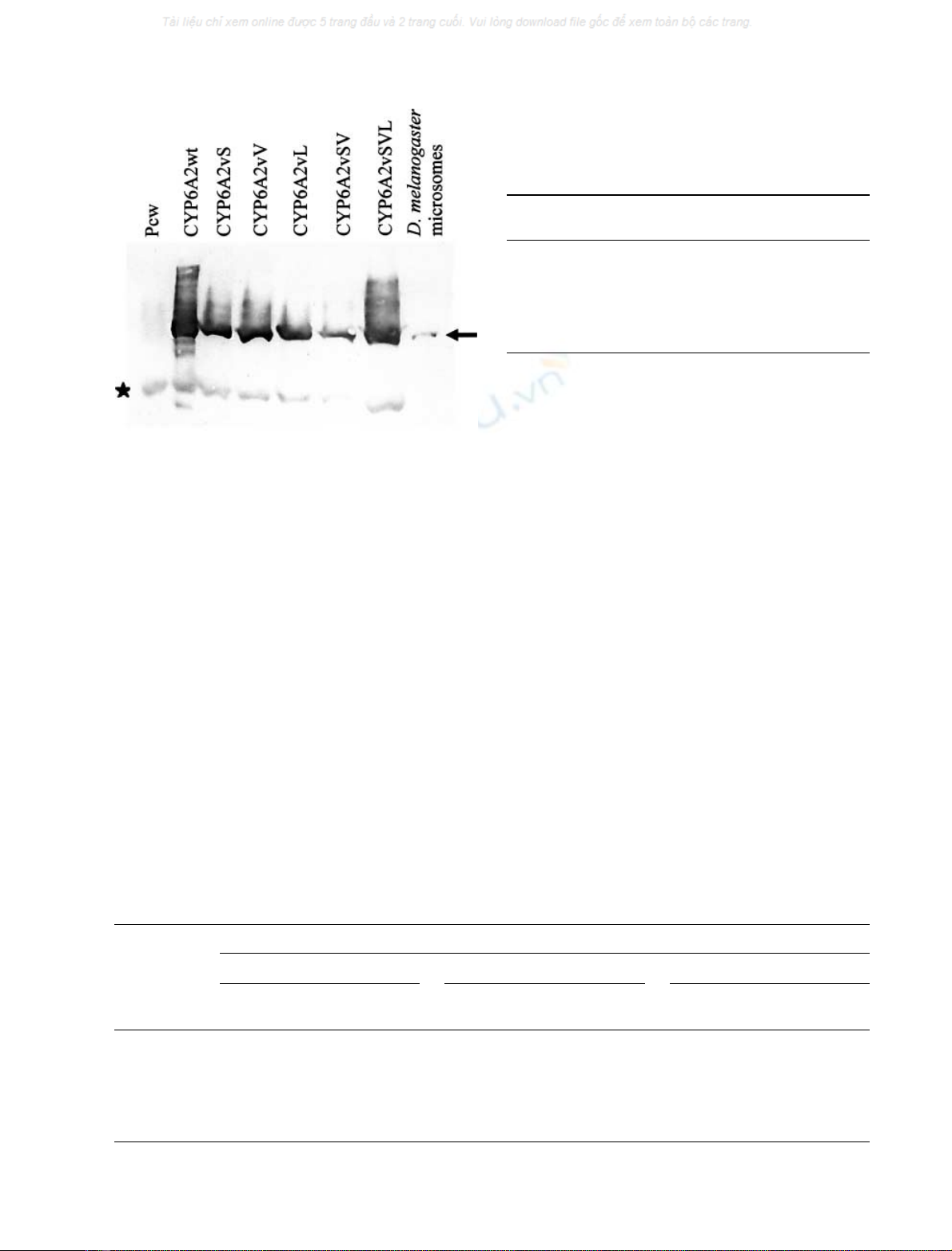
Fig. 1. Production of the CYP6A2 variants in bacteria. The lanes were
loaded with 5 mg of bacterial protein prepared as described in Cyto-
chrome P450 extraction. The arrow points to the CYP6A2 specific
signal, the star indicates unspecific signal observed in all lanes loaded
with bacterial protein. The CYP6A2 variants have the same apparent
molecularmassasCYP6A2fromD. melanogaster microsomes. The
apoenzyme production varied among the variants. No degradation
was observed for any of the apoenzymes.
Table 2. Specific production of 2b-hydroxy-testosterone by each of the
CYP6A2 variants. The mean ± SD and number of experiments (in
parentheses) are presented for each variant. No significant variation
was observed relative to the specific activity of CYP6A2wt (Dunnett
test, P>0.05).
Cytochrome P450
mutant
Hydroxy-testosterone production
(pmol per pmol P450 per 30 min)
CYP6A2wt 5.39 ± 0.50 (3)
CYP6A2vS 5.40 ± 0.15 (3)
CYP6A2vV 5.14 ± 0.12 (3)
CYP6A2vL 5.44 ± 0.40 (3)
CYP6A2vSV 3.86 ± 0.21 (3)
CYP6A2vSVL 3.79 ± 0.95 (3)
Table 3. Specific production of the DDT metabolites by each of the CYP6A2 variants. The mean ± SD and the number of experiments (in
parentheses) are presented for each metabolite and variant. Four specific activities are significantly different from the control, namely dicofol
production by CYP6A2vS and DDA, and DDD and dicofol production by CYP6A2vSVL. ND, not detected; NC, not calculated.
Membrane
preparation
DDT metabolite
DDA DDD Dicofol
pmol per pmol P450
per 30 min
Ratio to
CYP6A2wt
pmol per pmol P450
per 30 min
Ratio to
CYP6A2wt
pmol per pmol P450
per 30 min
Ratio to
CYP6A2wt
CYP6A2wt 0.03 ± 0.02 (8) 1.00 1.29 ± 0.19 (8) 1.00 2.10 ± 1.19 (8) 1.00
CYP6A2vS 0.04 ± 0.03 (9) 1.33 2.49 ± 1.28 (9) 1.93 10.06 ± 5.80 (9)* 4.79
CYP6A2vV 0.05 ± 0.04 (8) 1.66 0.97 ± 0.46 (8) 0.75 3.27 ± 2.39 (8) 1.56
CYP6A2vL 0.01 ± 0.01 (3) 0.33 ND NC 2.82 ± 2.95 (3) 1.34
CYP6A2vSV 0.04 ± 0.03 (9) 1.33 2.19 ± 1.54 (9) 1.69 4.61 ± 2.46 (9) 2.19
CYP6A2vSVL 0.63 ± 0.42 (6)** 21.00 7.50 ± 5.17 (6)** 5.81 18.03 ± 11.66 (6)** 8.59
* Significantly different from CYP6A2wt P£0.05); ** significantly different from CYP6A2wt (P£0.001; Dunnett test).
FEBS 2004 DDT metabolism by a mutant CYP6A2 (Eur. J. Biochem. 271) 1253
there was no qualitative difference between spectra obtained
with CYP6A2wt and CYP6A2vSVL. For DDT, we did not
observe significant differences between the apparent affinity
of DDT to the CYP6A2 variants (Table 4). We found the
same qualitative results for testosterone and we concluded
that CYP6A2wt and CYP6A2vSVL bind DDT or testo-
sterone with the same apparent affinity.
Sequence alignments and 3D localization
of the mutated positions
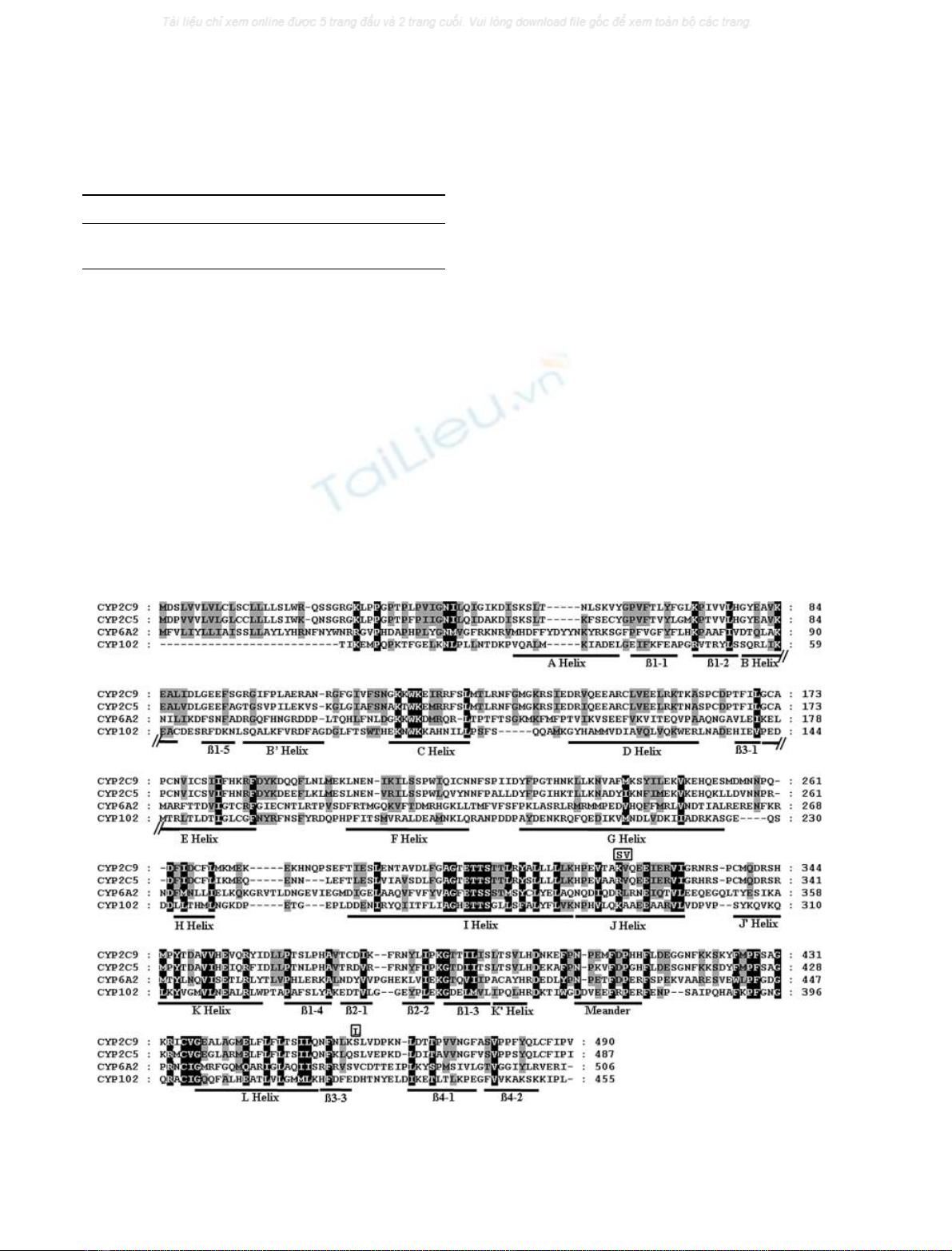
The sequence alignments of CYP6A2 with CYP2C5,
CYP2C9 and CYP102A1 are presented in Fig. 2. R335S
is at a conserved position as a positive charge is found in the
four sequences. L336V is at a position where an aliphatic
amino acid preferentially occurs, whereas, V476L is at a
nonconserved position. The R335S and L336V mutations
are located in helix J, and the V476L site is at the limit of the
b3–3 sheet. These structural elements are putative for
CYP6A2 and deduced from the sequence alignment.
These three positions of CYP6A2 are similar to K289,
A290 and D425 of CYP102A1. Strikingly, these amino acids
form a cluster distant from the active site, around the opening
of the pore containing helix I when placed on a spatial model
(Fig. 3). This cluster is located diametrically to the pole
carrying the amino acids involved in substrate binding. As
far as we know, there has been no report about structure
activity relationships in this area of the cytochrome P450s.
Discussion
The D. melanogaster insecticide resistant strain selected
in the laboratory, namely RDDT
R
, possesses a peculiar
CYP6A2 enzyme: CYP6A2vSVL carrying three mutations.
Two are contiguous (R335S and L336V) and the third one
(V476L) was found distal to the C451 which binds the heme
as described in preliminary work [6]. We expressed five
CYP6A2 mutants and the wild-type protein in bacteria to
verify whether these mutations confer to CYP6A2 the
ability to metabolize DDT. First, the enzymes’ stability was
addressed indirectly by spectrophotometry. As deduced
from the ratio of holoenzyme/apoenzyme (Table 1), the
stability of the protein was affected by each of the mutations
Table 4. Apparent affinity of DDT and testosterone for CYP6A2wt and
CYP6A2vSVL. For each apparent affinity calculated, the mean ± SD
and the number of experiments (in parentheses) are given. Statistical
analysis of the results demonstrated that there was no significant dif-
ference among the values for each compound (t-test, P>0.05).
DDT (l
M
) Testosterone (l
M
)
CYP6A2wt 146 ± 31 (5) 100 ± 36 (5)
CYP6A2vSVL 173 ± 26 (5) 106 ± 47 (5)
Fig. 2. Sequence alignments between CYP6A2, CYP102A1, CYP2C5 and CYP2C9. Identical or conserved amino acids in the four sequences are
shaded black, identical or conserved amino acids in three sequences are shaded grey. The secondary structures of CYP102A1 (labelled CYP102) are
indicated below the alignment. The mutations found in CYP6A2vSVL are indicated above the alignments and boxed.
1254 M. Amichot et al. (Eur. J. Biochem. 271)FEBS 2004


%20--%3e%3cdefs%3e%3cstyle%3e%20.st0%20{%20fill:%20%23fff;%20}%20.st1%20{%20fill:%20%237800fa;%20}%20%3c/style%3e%3c/defs%3e%3cpath%20class='st1'%20d='M117.78,12.18H43.11c2.9,3.47,4.65,7.94,4.65,12.82,0,5.6-2.3,10.66-6.01,14.29h76.02l7.22-13.56-7.22-13.56Z'/%3e%3cg%3e%3cpath%20class='st0'%20d='M53.58,26.17h-.59v-1.46h.59v-4.96h2.83c1.78,0,2.67.94,2.67,2.82v5.76c0,1.87-.89,2.81-2.67,2.81h-2.83v-4.96ZM55.36,21.37v3.34h1.1v1.46h-1.1v3.34h1.01c.61,0,.91-.37.91-1.1v-5.93c0-.74-.3-1.1-.91-1.1h-1.01Z'/%3e%3cpath%20class='st0'%20d='M65.99,31.14h-1.8l-.31-2.07h-2.19l-.31,2.07h-1.64l1.82-11.39h2.62l1.82,11.39ZM65.28,18.04c-.25.46-.51.77-.75.94-.21.15-.47.22-.79.22-.26,0-.57-.07-.92-.22l-.38-.15c-.14-.05-.26-.07-.37-.07-.3,0-.53.18-.71.54l-.91-.68c.25-.46.51-.77.75-.94.21-.14.48-.21.79-.21.26,0,.57.07.92.21l.38.15c.14.05.26.07.37.07.3,0,.53-.18.71-.54l.91.68ZM61.91,27.52h1.73l-.87-5.76-.87,5.76Z'/%3e%3cpath%20class='st0'%20d='M74.53,26.89v1.52c0,1.91-.89,2.86-2.67,2.86s-2.67-.95-2.67-2.86v-5.93c0-1.91.89-2.86,2.67-2.86s2.67.95,2.67,2.86v1.11h-1.69v-1.22c0-.75-.31-1.12-.93-1.12s-.93.37-.93,1.12v6.15c0,.74.31,1.11.93,1.11s.93-.37.93-1.11v-1.63h1.69Z'/%3e%3cpath%20class='st0'%20d='M81.4,31.14h-1.8l-.31-2.07h-2.19l-.31,2.07h-1.64l1.82-11.39h2.62l1.82,11.39ZM75.9,19.2l1.52-1.91h1.71l1.51,1.91h-1.61l-.76-.95-.75.95h-1.61ZM77.32,27.52h1.73l-.87-5.76-.87,5.76ZM83.1,15.99l-1.76,1.91h-1.26l1.17-1.91h1.86Z'/%3e%3cpath%20class='st0'%20d='M84.86,19.75c1.78,0,2.67.94,2.67,2.82v1.48c0,1.87-.89,2.81-2.67,2.81h-.85v4.28h-1.79v-11.39h2.64ZM84.01,21.37v3.86h.85c.58,0,.87-.36.87-1.08v-1.71c0-.71-.29-1.07-.87-1.07h-.85Z'/%3e%3cpath%20class='st0'%20d='M93.51,19.75c1.78,0,2.67.94,2.67,2.82v1.48c0,1.87-.89,2.81-2.67,2.81h-.85v4.28h-1.79v-11.39h2.64ZM92.66,21.37v3.86h.85c.58,0,.87-.36.87-1.08v-1.71c0-.71-.29-1.07-.87-1.07h-.85Z'/%3e%3cpath%20class='st0'%20d='M98.8,31.14h-1.79v-11.39h1.79v4.88h2.03v-4.88h1.83v11.39h-1.83v-4.88h-2.03v4.88Z'/%3e%3cpath%20class='st0'%20d='M105.36,24.55h2.46v1.62h-2.46v3.34h3.09v1.63h-4.88v-11.39h4.88v1.63h-3.09v3.18ZM108.17,17.29l-1.76,1.91h-1.26l1.17-1.91h1.86Z'/%3e%3cpath%20class='st0'%20d='M112.2,19.75c1.78,0,2.67.94,2.67,2.82v1.48c0,1.87-.89,2.81-2.67,2.81h-.85v4.28h-1.79v-11.39h2.64ZM111.35,21.37v3.86h.85c.58,0,.87-.36.87-1.08v-1.71c0-.71-.29-1.07-.87-1.07h-.85Z'/%3e%3c/g%3e%3ccircle%20class='st1'%20cx='25'%20cy='25'%20r='20'/%3e%3cpath%20class='st0'%20d='M32.78,19.27c2.92,0,4.43,2.55,5.28,5.33l.71,2.17c.14.38-.33.75-.71.75h-5.61c.19-.33.24-.71.09-1.08l-.75-2.45c-.43-1.32-.99-2.64-1.79-3.77.75-.57,1.65-.94,2.78-.94h0ZM25,18.38c3.25,0,4.9,2.78,5.89,5.89l.76,2.45c.14.42-.33.8-.8.8h-11.69c-.42,0-.94-.38-.8-.8l.75-2.45c.99-3.11,2.64-5.89,5.89-5.89h0ZM25,11.35c1.74,0,3.11,1.37,3.11,3.11s-1.37,3.11-3.11,3.11-3.11-1.41-3.11-3.11,1.41-3.11,3.11-3.11h0ZM17.27,19.27c1.08,0,1.98.38,2.73.94-.8,1.13-1.37,2.45-1.74,3.77l-.8,2.45c-.14.38-.05.75.09,1.08h-5.56c-.42,0-.9-.38-.75-.75l.71-2.17c.9-2.78,2.41-5.33,5.33-5.33h0ZM17.27,12.91c1.51,0,2.78,1.27,2.78,2.83s-1.27,2.83-2.78,2.83-2.83-1.27-2.83-2.83,1.27-2.83,2.83-2.83h0ZM32.78,12.91c1.56,0,2.78,1.27,2.78,2.83s-1.23,2.83-2.78,2.83-2.83-1.27-2.83-2.83,1.27-2.83,2.83-2.83h0ZM27.07,28.56v.09c0,.57-.24,1.08-.61,1.46h0v.05c-.38.33-.9.57-1.46.57s-1.08-.24-1.46-.61h0c-.38-.38-.61-.9-.61-1.46v-.09h1.41v.09c0,.19.05.38.19.47v.05c.09.09.28.19.47.19s.38-.09.47-.19v-.05c.14-.09.24-.28.24-.47t-.05-.09h1.41ZM30.99,28.56v.09c0,1.65-.66,3.16-1.74,4.24-1.08,1.08-2.59,1.79-4.24,1.79s-3.16-.71-4.24-1.79l-.05-.05c-1.04-1.08-1.7-2.55-1.7-4.2v-.09h1.41v.09c0,1.27.47,2.4,1.27,3.25h.05c.85.85,1.98,1.37,3.25,1.37s2.4-.52,3.25-1.37c.85-.8,1.37-1.98,1.37-3.25v-.09h1.37ZM34.99,28.56v.09c0,2.78-1.13,5.28-2.92,7.07-1.79,1.79-4.29,2.92-7.07,2.92s-5.23-1.13-7.07-2.92c-1.79-1.79-2.92-4.29-2.92-7.07v-.09h1.41v.09c0,2.4.94,4.53,2.5,6.08,1.56,1.56,3.72,2.5,6.08,2.5s4.52-.94,6.08-2.5c1.56-1.56,2.5-3.68,2.5-6.08v-.09h1.41Z'/%3e%3c/svg%3e)