
BioMed Central
Page 1 of 14
(page number not for citation purposes)
Retrovirology
Open Access
Research
Highly specific inhibition of leukaemia virus membrane fusion by
interaction of peptide antagonists with a conserved region of the
coiled coil of envelope
Daniel Lamb1, Alexander W Schüttelkopf2, Daan MF van Aalten2 and
David W Brighty*1
Address: 1The Biomedical Research Centre, College of Medicine, Ninewells Hospital, The University, Dundee, DD1 9SY, Scotland, UK and 2The
Division of Biological Chemistry and Drug Discovery, College of Life Sciences, University of Dundee, Dow Street, Dundee, DD1 5EH, Scotland, UK
Email: Daniel Lamb - d.J.Lamb@dundee.ac.uk; Alexander W Schüttelkopf - a.schuettelkopf@dundee.ac.uk; Daan MF van
Aalten - dava@davapc1.bioch.dundee.ac.uk; David W Brighty* - d.w.brighty@dundee.ac.uk
* Corresponding author
Abstract
Background: Human T-cell leukaemia virus (HTLV-1) and bovine leukaemia virus (BLV) entry into
cells is mediated by envelope glycoprotein catalyzed membrane fusion and is achieved by folding of
the transmembrane glycoprotein (TM) from a rod-like pre-hairpin intermediate to a trimer-of-
hairpins. For HTLV-1 and for several virus groups this process is sensitive to inhibition by peptides
that mimic the C-terminal α-helical region of the trimer-of-hairpins.
Results: We now show that amino acids that are conserved between BLV and HTLV-1 TM tend
to map to the hydrophobic groove of the central triple-stranded coiled coil and to the leash and
C-terminal α-helical region (LHR) of the trimer-of-hairpins. Remarkably, despite this conservation,
BLV envelope was profoundly resistant to inhibition by HTLV-1-derived LHR-mimetics.
Conversely, a BLV LHR-mimetic peptide antagonized BLV envelope-mediated membrane fusion but
failed to inhibit HTLV-1-induced fusion. Notably, conserved leucine residues are critical to the
inhibitory activity of the BLV LHR-based peptides. Homology modeling indicated that hydrophobic
residues in the BLV LHR likely make direct contact with a pocket at the membrane-proximal end
of the core coiled-coil and disruption of these interactions severely impaired the activity of the BLV
inhibitor. Finally, the structural predictions assisted the design of a more potent antagonist of BLV
membrane fusion.
Conclusion: A conserved region of the HTLV-1 and BLV coiled coil is a target for peptide
inhibitors of envelope-mediated membrane fusion and HTLV-1 entry. Nevertheless, the LHR-based
inhibitors are highly specific to the virus from which the peptide was derived. We provide a model
structure for the BLV LHR and coiled coil, which will facilitate comparative analysis of leukaemia
virus TM function and may provide information of value in the development of improved,
therapeutically relevant, antagonists of HTLV-1 entry into cells.
Published: 4 August 2008
Retrovirology 2008, 5:70 doi:10.1186/1742-4690-5-70
Received: 14 April 2008
Accepted: 4 August 2008
This article is available from: http://www.retrovirology.com/content/5/1/70
© 2008 Lamb et al; licensee BioMed Central Ltd.
This is an Open Access article distributed under the terms of the Creative Commons Attribution License (http://creativecommons.org/licenses/by/2.0),
which permits unrestricted use, distribution, and reproduction in any medium, provided the original work is properly cited.

Retrovirology 2008, 5:70 http://www.retrovirology.com/content/5/1/70
Page 2 of 14
(page number not for citation purposes)
Background
Bovine Leukemia Virus (BLV) and Human T-Cell Leuke-
mia Virus Type-1 (HTLV-1) are closely related deltaretro-
viruses that cause aggressive lymphoproliferative
disorders in a small percentage of infected individuals [1-
3]. In order to efficiently enter cells, both viruses are
dependent on a fusion event between viral and cell mem-
branes. As with other retroviruses, fusion is catalyzed by
the virally encoded Env complex, which is synthesized as
a polyprotein precursor and is subsequently cleaved to
yield the surface glycoprotein (SU) and transmembrane
glycoprotein (TM) subunits. On the surface of the virus or
infected cell, Env is displayed as a trimer, with three SU
subunits linked by disulphide bonds to a spike of three
TM subunits.
The amino-acid sequences of the HTLV-1 and BLV enve-
lope glycoproteins are strikingly similar [4] and, in com-
mon with other oncoretroviruses, share a characteristic
modular structure [4-8]. A receptor-binding domain is
located at the amino-terminal end of SU and is connected
to a C-terminal domain by a proline-rich linker [4,6,9].
The C-terminal domain includes a conserved CXCC
sequence and is required for interactions with TM [10-12].
The modular nature of envelope extends into TM, and it is
here that the homology between retroviruses and phylo-
genetically diverse viral isolates is most apparent. The
functional regions of TM include a hydrophobic fusion
peptide linked to an isoleucine/leucine heptad repeat, a
membrane spanning segment and a cytoplasmic tail of
variable length. These conserved modules identify retrovi-
ral TM proteins as members of a diverse family of virally
expressed class 1 membrane fusion proteins.
Accumulating evidence advocates a conserved mechanism
of retroviral envelope-mediated membrane fusion [13-
15]. SU binds to the cellular receptor, which is accompa-
nied by isomerisation of the disulphide linkages between
SU and TM [11,12], and triggers a conformational change
in TM. The N-terminal hydrophobic fusion peptide of TM
is then inserted into the target cell membrane, while the
C-terminus remains anchored in the viral or host cell
membrane. This transient rod-like conformation, referred
to as a "pre-hairpin" intermediate, is stabilized by the
assembly of a trimeric coiled coil composed of one alpha
helix from each of the three adjacent TM monomers. A
more C-terminal region of the TM ecto-domain, which in
HTLV-1 includes an extended non-helical leash and short
α-helix [16], then folds onto the coiled coil to generate a
six-helix bundle or trimer-of-hairpins [16-19]. These dra-
matic conformational changes draw the opposing mem-
branes together, destabilise the lipid bilayers, promote
lipid mixing and culminate in membrane fusion [13,14].
Despite the sequence homology and conserved modular
structure, there are notable differences in primary
sequence, size, and function of the HTLV-1 and BLV enve-
lope proteins. It is likely that these differences contribute
in a substantial way to the species-specificity, and the dis-
tinctive patterns of tissue tropism and pathogenesis that
are observed for these viruses [2,3]. Consequently, com-
parative analysis of the envelope glycoproteins will pro-
vide significant insight into the determinants of species-
and tissue-specific tropism, the strategies for immune
modulation, and the mechanisms of membrane fusion
that are adopted by these viruses. Information derived
from such studies will aid the development of effective
vaccines and small-molecule inhibitors of viral entry and
cell-to-cell viral transfer.
Significantly, our laboratory [20-22], and others [23],
have demonstrated that synthetic peptides that mimic the
C-terminal non-helical leash and α-helical region (LHR)
of HTLV-1 TM are inhibitory to envelope-mediated mem-
brane fusion. Prototypic α-helical TM-mimetic inhibitory
peptides have also been characterized for a number of
highly divergent enveloped viruses, including HIV and
paramyxoviruses [24-27]. The HTLV-derived peptide
binds to the coiled coil of TM and, in a trans-dominant
negative manner, blocks resolution of the pre-hairpin
intermediate to the trimer-of-hairpins, thus impairing the
fusogenic activity of TM. The potency of these inhibitors
makes them attractive leads for antiviral therapeutics.
Although the HTLV-1 peptide inhibitor also blocks viral
entry of the divergent HTLV-2 it is inactive against a vari-
ety of heterologous viral envelope proteins [20,23]. How-
ever, the molecular features that determine the target
specificity, activity, and potency of these peptide inhibi-
tors is only beginning to be understood [20-22]. In this
study, we examine the target specificity and activity of
peptide inhibitors derived from the conserved C-terminal
leash and α-helical region (LHR) of the HTLV-1 and BLV
trans-membrane glycoproteins. We demonstrate that a
synthetic peptide that mimics the BLV LHR is a potent
antagonist of BLV envelope-mediated membrane fusion.
Surprisingly, despite the high level of identity between the
HTLV-1 and BLV derived peptides, the inhibitory activity
of the peptides is limited exclusively to the virus from
which they were derived. While the peptides display
remarkable target specificity, the activity of each peptide is
nevertheless dependent upon the interaction of conserved
amino acid side chains with their respective targets. An
amino acid substitution analysis reveals that several con-
served residues within the BLV LHR play a critical role in
determining peptide potency and identifies a single
amino acid substitution within the BLV peptide that
yields a more potent inhibitor. Finally, based on homol-
ogy with HTLV-1 TM, the inhibition data and amino acid
substitution analysis support a model for the BLV trimer-
of-hairpins.

Retrovirology 2008, 5:70 http://www.retrovirology.com/content/5/1/70
Page 3 of 14
(page number not for citation purposes)
Materials and methods
Cells
HeLa and BLV-FLK (a kind gift of Dr Arsène Burny and Dr
Luc Willems; Universitaire des Sciences Agronomiques de
Gembloux, Belgium) cells were maintained in Dulbecco's
modified Eagle medium supplemented with 10% fetal
bovine serum (FBS).
Plasmids
The Plasmid HTE-1 [28] and pRSV-Rev [29] have been
described. The plasmid pCMV-BLVenv-RRE was con-
structed by replacing a fragment of the HIV-1 envelope
open reading frame in pCMVgp160ΔSA [30] with a
genomic fragment spanning the entire BLV envelope. In
brief, pCMVgp160ΔSA was digested with EcoR I, which
cuts the recipient vector after the CMV early promoter but
prior to the initiating ATG of the HIV-1 env sequences. The
vector was subsequently digested with BglII, which
removes the HIV-1 SU region but retains the HIV RRE. A
fragment encompassing the entire BLV envelope open
reading frame between a 5' Xho I site and a 3' BamH I site
(nucleotides 4347–6997 of NC_001414) was ligated into
the vector backbone using an EcoR I-Xho I linker. The
resulting plasmid encodes BLV env including the natural
BLV env stop codon placed upstream of the HIV RRE; the
transcription unit is terminated by the SV40 poly A site
and is expressed from the CMV early promoter.
Peptides
Peptides (Table 1) were synthesized using standard solid-
phase Fmoc chemistry and unless stated otherwise have
acetylated N-termini and amidated C-termini. The pep-
tides were purified by reverse-phase high-pressure liquid
chromatography and verified for purity by MALDI-TOF
mass spectrometry. All peptides were dissolved in dime-
thyl sulfoxide (DMSO), the concentration of peptide
stock solutions was confirmed where possible by absorb-
ance at 280 nm in 6 M guanidine hydrochloride and pep-
tides were used at the final concentrations indicated. For
the peptide PBLV-ΔN, peptide concentration was estimated
by Bradford assay at 5 two-fold serial dilutions from a
stock solution using the PBLV-ΔC peptide in concentra-
tions verified by absorbance at 280 nm in 6 M guanidine
hydrochloride to plot a standard curve. The HTLV-1-
derived peptides are based on the sequence of HTLV-1
strain CR and conform closely to the consensus sequence
for HTLV-1 and HTLV-2 strains, the BLV peptides conform
to the consensus sequence for most BLV isolates.
Peptide biotinylation
Peptides to be biotinylated were reduced using immobi-
lized Tris [2-carboxyethyl] phosphine (TCEP) reducing
agent (Pierce), and subsequent biotinylation was carried
out with EZ-Link® Iodoacetyl-PEO2-Biotin (Pierce), in
both cases according to the manufacturer's protocols. The
biotinylation reaction was quenched with cysteine. The
biotinylated peptide was incubated for 30 mins at room
temperature with either streptavidin-agarose (Gibco-BRL)
or amylose-agarose (New England Biolabs) in a spin-col-
umn. Unbound peptide was recovered by centrifugation,
the flow-through was re-applied to the column, and the
incubation and centrifugation was repeated. The flow-
through from the second centrifugation was used in syn-
cytium interference assays; the peptide concentration of
the amylose-agarose flow through was established by UV
spectrometry as described above, and added to tissue cul-
ture medium to produce the final assay concentrations as
indicated. In the case of the flow-through from the
streptavidin-agarose column, volumes equivalent to those
used with the amylose-agarose flow-through were added
to the wells.
Determination of relative peptide solubility
A two-fold serial dilution of peptide in DMSO was per-
formed, and added in duplicate to 96-well microplates.
Filtered PBS was added to give a total volume of 200 μl
and a final DMSO concentration of 1.5 % in all wells. The
plates were incubated at room temperature for 1 hr and
the relative solubility of peptides was established by meas-
uring forward scattered light using a NEPHELOstar laser-
Table 1: Peptides used in this study.
Peptide Amino Acid Position Sequence MW Maximum Solubility (μM)*
Pcr-400 gp21 400–429 CCFLNITNSHVSILQERPPLENRVLTGWGL 3,411 > 90.00
Pcr-400 L/A gp21 400–429 ---A---------A-----A----A----A 3,200 45.00
PBLV-391 gp30 391–419 CCFLRIQNDSIIRLGDLQPLSQRVSTDWQ 3,447 > 90.00
PBLV-ΔN gp30 400–419 S------------------- 2,312 > 90.00
PBLV-ΔC gp30 391–410 -------------------L 2,317 > 90.00
PBLV-L/A gp30 391–419 ---A---------A--A--A--------- 3,236 45.00
PBLV-L404/410A gp30 391–419 -------------A-----A--------- 3,321 > 90.00
PBLV-ΔCCF gp30 394–419 L------------------------- 3,052 11.25
PBLV-R403A gp30 391–419 ------------A---------------- 3,321 22.50
C34 gp41 627–661 GWMEWDREINNYTSLLIHSLIEESQNQQEKNEQELL 4,418 > 90.00
* Maximum solubility in aqueous solution determined by laser nephelometry.

Retrovirology 2008, 5:70 http://www.retrovirology.com/content/5/1/70
Page 4 of 14
(page number not for citation purposes)
based microplate nephelometer (BMG LABTECH). Wells
containing PBS and 1.5 % DMSO only were used as
blanks. Data analysis was carried out using ActivityBase,
and peptides giving readings up to and including 3-fold
higher than the average reading for the DMSO control
were considered to be in solution at the concentrations
specified.
Syncytium Interference Assays
Syncytium interference assays were performed by stand-
ard methods [20,31]. Briefly, HeLa cells for use as effector
cells were transfected with the envelope expression vector
pHTE-1 or with equal amounts of pCMV-BLVenv-RRE and
pRSV-Rev using the Genejuice™ transfection reagent
(Novagen) in accordance with the manufacturer's instruc-
tions. 24 h later, 3 × 10⁵ effector cells were added to 7 ×
105 untransfected HeLa target cells in six-well dishes
(Nunc). Where appropriate, the co-culture was incubated
in the presence of peptides at the concentrations specified.
To assess the ability of the peptides to inhibit fusion
induced by virally expressed BLV envelope, 2 × 10⁵ BLV-
infected FLK cells were used as effectors and added to 8 ×
105 uninfected HeLa cells. After incubation at 37°C for 16
h, cells were washed twice with PBS and fixed in PBS + 3%
paraformaldehyde. Assays were performed in triplicate
and the number of syncytia (defined as multinucleated
cells with 4 or more nuclei) from 10 low-power fields
(LPF) per replicate was scored by light microscopy; some
assays were stained using Giemsa. A syncytium formation
value of 100% is defined as the number of syncytia
formed in the absence of peptide but in the presence of
1.5% DMSO. The peptide concentration required to give
50% inhibition (IC50) of syncytium formation was calcu-
lated using GraphPad Prism 4.
Results
Amino acid residues conserved between the HTLV-1 and
BLV TM ectodomains map to the interacting surfaces of
the LHR and coiled-coil
Although there are considerable differences in the amino
acid sequence of class-1 fusion proteins from diverse viral
groups there is exceptional conservation of secondary and
tertiary structure. To compare the class-1 fusion proteins
from the related retroviruses BLV and HTLV-1, the pre-
dicted coiled-coil regions of the BLV TM were identified
using the program LearnCoil-VMF [32] and the BLV and
HTLV-1 amino acid sequences were aligned using Clustal-
W [33] (Figure 1A). The alignment revealed that for the
TM 33% of the residues are identical and a further 10% are
conservative substitutions. The homology is particularly
evident in the predicted coiled-coil region incorporating
the heptad repeat and in the LHR of the TM ectodomain
(Figure 1A), the LHR lies distal to a CX6CC motif common
to oncoretroviral fusion proteins. The crystal structure of
the HTLV-1 six-helix-bundle has been solved and the
structure spans these regions of homology [16].
Using the crystal structure of the HTLV-1 TM as a tem-
plate, we mapped on the coiled coil and LHR the location
of amino acid residues that are conserved between the
ectodomain of HTLV-1 and BLV TM (Figure 1B). Using
this approach, we observed that for the core coiled-coil
the majority of conserved residues map along the grooves
formed by the interface of each pair of interacting N heli-
ces. Importantly, these grooves act as docking sites for the
LHR as TM folds from the pre-hairpin intermediate to the
trimer-of-hairpins. Moreover, many of the conserved
amino acids of the LHR are located on the face of the LHR
that interacts with the grooves on the coiled coil. By exam-
ining the location of substituted residues on the HTLV-1
TM it becomes clear that where there are amino acid sub-
stitutions on the BLV LHR there are complimentary or
accommodating amino acid changes within the hydro-
phobic grooves of the core coiled coil (Figure 1C). For
example, leucines 413 and 419 in the HTLV-1 LHR are
conserved in BLV, and these leucines interact with eight
coiled coil residues of which seven are identical in BLV
and one is a conservative substitution (Figure 1C). In con-
trast, HTLV-1 LHR residues H409 and R416 interact with
the side chains of six residues of the coiled coil, but H409
and R416 are not conserved in BLV and of the six interact-
ing coiled coil residues four have diverged and only one
residue is semi-conserved (Figure 1C). Overall, the analy-
sis indicates that the majority of the conserved residues
occupy positions that form the interacting surfaces of the
trimer-of-hairpins. In agreement with these observations,
those residues that do not involve the interacting surfaces
of the TM are invariably solvent exposed on the trimer-of-
hairpins and are subject to the highest degree of variation
between the two viruses.
A synthetic peptide, Pcr-400, which mimics the LHR of the
HTLV-1 TM is a potent inhibitor of envelope-catalysed
membrane fusion [20]. This peptide interacts directly and
specifically with a recombinant coiled coil derived from
HTLV-1 TM and substitution of critical amino acid resi-
dues within the peptide disrupts coiled coil binding and
impairs the biological activity of the peptide [20-22].
These findings are consistent with the view that the pep-
tide blocks membrane fusion by binding to the coiled coil
of fusion-active envelope. As illustrated above, there are
remarkable similarities in the interacting surfaces of the
coiled coil and LHR between HTLV-1 and BLV (Figure 1).
Considering the noted differences, it was not clear if the
HTLV-1-derived synthetic peptide could inhibit mem-
brane fusion mediated by BLV envelope. The HTLV-1 pep-
tide inhibits viral entry by the divergent HTLV-2 but does
not inhibit membrane fusion catalysed by a number of
heterologous viral envelopes including HIV-1, feline
Retrovirology 2008, 5:70 http://www.retrovirology.com/content/5/1/70
Page 5 of 14
(page number not for citation purposes)
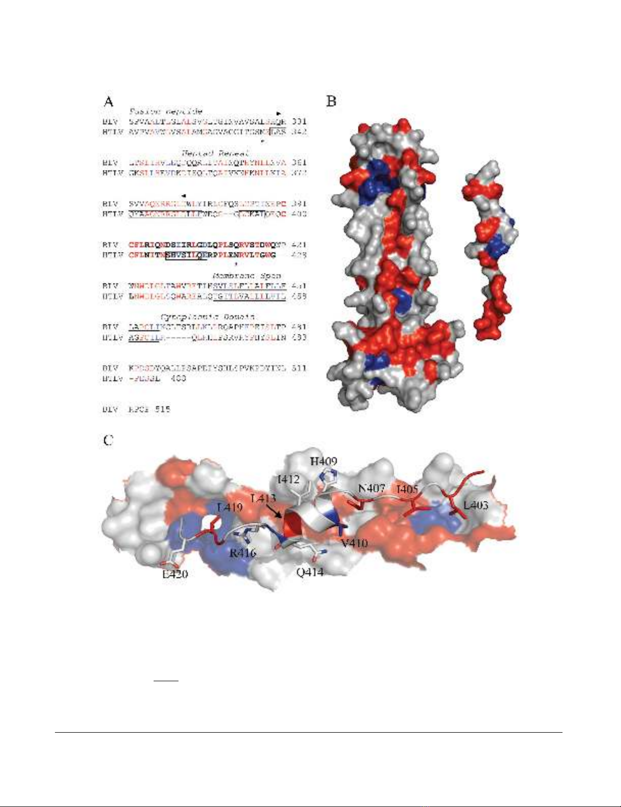
Analysis of the conserved regions of BLV and HTLV-1 TMFigure 1
Analysis of the conserved regions of BLV and HTLV-1 TM. (A) Alignment of the BLV and HTLV-1 TM sequences, the
predicted coiled coil of BLV TM is indicated between the arrow heads; the LHR is in bold; the helical regions of the HTLV-1
TM are boxed; the limits of the HTLV-1 crystal structure are marked by asters; and the membrane spanning region is under-
lined. (B) The HTLV-1 core coiled-coil and, on the right, the leash and α-helical region that is mimicked by the HTLV-1 inhibi-
tory peptide (from PDB 1MG1). The face of the peptide that interacts with the coiled coil is shown. For the sequence
alignment and structural renderings, residues identical between BLV and HTLV-1 are shown in red, conservative substitutions
are blue, and non-conserved are rendered white. Amino acid coordinates refer to the full-length envelope precursor. (C)
Detail of the predicted interaction of the HTLV-1 LHR-mimetic peptide (ribbon structure) with the surface of the coiled coil
(space filling form) based on the structure of Kobe et al. [16]; shading as above.


%20--%3e%3cdefs%3e%3cstyle%3e%20.st0%20{%20fill:%20%23fff;%20}%20.st1%20{%20fill:%20%237800fa;%20}%20%3c/style%3e%3c/defs%3e%3cpath%20class='st1'%20d='M117.78,12.18H43.11c2.9,3.47,4.65,7.94,4.65,12.82,0,5.6-2.3,10.66-6.01,14.29h76.02l7.22-13.56-7.22-13.56Z'/%3e%3cg%3e%3cpath%20class='st0'%20d='M53.58,26.17h-.59v-1.46h.59v-4.96h2.83c1.78,0,2.67.94,2.67,2.82v5.76c0,1.87-.89,2.81-2.67,2.81h-2.83v-4.96ZM55.36,21.37v3.34h1.1v1.46h-1.1v3.34h1.01c.61,0,.91-.37.91-1.1v-5.93c0-.74-.3-1.1-.91-1.1h-1.01Z'/%3e%3cpath%20class='st0'%20d='M65.99,31.14h-1.8l-.31-2.07h-2.19l-.31,2.07h-1.64l1.82-11.39h2.62l1.82,11.39ZM65.28,18.04c-.25.46-.51.77-.75.94-.21.15-.47.22-.79.22-.26,0-.57-.07-.92-.22l-.38-.15c-.14-.05-.26-.07-.37-.07-.3,0-.53.18-.71.54l-.91-.68c.25-.46.51-.77.75-.94.21-.14.48-.21.79-.21.26,0,.57.07.92.21l.38.15c.14.05.26.07.37.07.3,0,.53-.18.71-.54l.91.68ZM61.91,27.52h1.73l-.87-5.76-.87,5.76Z'/%3e%3cpath%20class='st0'%20d='M74.53,26.89v1.52c0,1.91-.89,2.86-2.67,2.86s-2.67-.95-2.67-2.86v-5.93c0-1.91.89-2.86,2.67-2.86s2.67.95,2.67,2.86v1.11h-1.69v-1.22c0-.75-.31-1.12-.93-1.12s-.93.37-.93,1.12v6.15c0,.74.31,1.11.93,1.11s.93-.37.93-1.11v-1.63h1.69Z'/%3e%3cpath%20class='st0'%20d='M81.4,31.14h-1.8l-.31-2.07h-2.19l-.31,2.07h-1.64l1.82-11.39h2.62l1.82,11.39ZM75.9,19.2l1.52-1.91h1.71l1.51,1.91h-1.61l-.76-.95-.75.95h-1.61ZM77.32,27.52h1.73l-.87-5.76-.87,5.76ZM83.1,15.99l-1.76,1.91h-1.26l1.17-1.91h1.86Z'/%3e%3cpath%20class='st0'%20d='M84.86,19.75c1.78,0,2.67.94,2.67,2.82v1.48c0,1.87-.89,2.81-2.67,2.81h-.85v4.28h-1.79v-11.39h2.64ZM84.01,21.37v3.86h.85c.58,0,.87-.36.87-1.08v-1.71c0-.71-.29-1.07-.87-1.07h-.85Z'/%3e%3cpath%20class='st0'%20d='M93.51,19.75c1.78,0,2.67.94,2.67,2.82v1.48c0,1.87-.89,2.81-2.67,2.81h-.85v4.28h-1.79v-11.39h2.64ZM92.66,21.37v3.86h.85c.58,0,.87-.36.87-1.08v-1.71c0-.71-.29-1.07-.87-1.07h-.85Z'/%3e%3cpath%20class='st0'%20d='M98.8,31.14h-1.79v-11.39h1.79v4.88h2.03v-4.88h1.83v11.39h-1.83v-4.88h-2.03v4.88Z'/%3e%3cpath%20class='st0'%20d='M105.36,24.55h2.46v1.62h-2.46v3.34h3.09v1.63h-4.88v-11.39h4.88v1.63h-3.09v3.18ZM108.17,17.29l-1.76,1.91h-1.26l1.17-1.91h1.86Z'/%3e%3cpath%20class='st0'%20d='M112.2,19.75c1.78,0,2.67.94,2.67,2.82v1.48c0,1.87-.89,2.81-2.67,2.81h-.85v4.28h-1.79v-11.39h2.64ZM111.35,21.37v3.86h.85c.58,0,.87-.36.87-1.08v-1.71c0-.71-.29-1.07-.87-1.07h-.85Z'/%3e%3c/g%3e%3ccircle%20class='st1'%20cx='25'%20cy='25'%20r='20'/%3e%3cpath%20class='st0'%20d='M32.78,19.27c2.92,0,4.43,2.55,5.28,5.33l.71,2.17c.14.38-.33.75-.71.75h-5.61c.19-.33.24-.71.09-1.08l-.75-2.45c-.43-1.32-.99-2.64-1.79-3.77.75-.57,1.65-.94,2.78-.94h0ZM25,18.38c3.25,0,4.9,2.78,5.89,5.89l.76,2.45c.14.42-.33.8-.8.8h-11.69c-.42,0-.94-.38-.8-.8l.75-2.45c.99-3.11,2.64-5.89,5.89-5.89h0ZM25,11.35c1.74,0,3.11,1.37,3.11,3.11s-1.37,3.11-3.11,3.11-3.11-1.41-3.11-3.11,1.41-3.11,3.11-3.11h0ZM17.27,19.27c1.08,0,1.98.38,2.73.94-.8,1.13-1.37,2.45-1.74,3.77l-.8,2.45c-.14.38-.05.75.09,1.08h-5.56c-.42,0-.9-.38-.75-.75l.71-2.17c.9-2.78,2.41-5.33,5.33-5.33h0ZM17.27,12.91c1.51,0,2.78,1.27,2.78,2.83s-1.27,2.83-2.78,2.83-2.83-1.27-2.83-2.83,1.27-2.83,2.83-2.83h0ZM32.78,12.91c1.56,0,2.78,1.27,2.78,2.83s-1.23,2.83-2.78,2.83-2.83-1.27-2.83-2.83,1.27-2.83,2.83-2.83h0ZM27.07,28.56v.09c0,.57-.24,1.08-.61,1.46h0v.05c-.38.33-.9.57-1.46.57s-1.08-.24-1.46-.61h0c-.38-.38-.61-.9-.61-1.46v-.09h1.41v.09c0,.19.05.38.19.47v.05c.09.09.28.19.47.19s.38-.09.47-.19v-.05c.14-.09.24-.28.24-.47t-.05-.09h1.41ZM30.99,28.56v.09c0,1.65-.66,3.16-1.74,4.24-1.08,1.08-2.59,1.79-4.24,1.79s-3.16-.71-4.24-1.79l-.05-.05c-1.04-1.08-1.7-2.55-1.7-4.2v-.09h1.41v.09c0,1.27.47,2.4,1.27,3.25h.05c.85.85,1.98,1.37,3.25,1.37s2.4-.52,3.25-1.37c.85-.8,1.37-1.98,1.37-3.25v-.09h1.37ZM34.99,28.56v.09c0,2.78-1.13,5.28-2.92,7.07-1.79,1.79-4.29,2.92-7.07,2.92s-5.23-1.13-7.07-2.92c-1.79-1.79-2.92-4.29-2.92-7.07v-.09h1.41v.09c0,2.4.94,4.53,2.5,6.08,1.56,1.56,3.72,2.5,6.08,2.5s4.52-.94,6.08-2.5c1.56-1.56,2.5-3.68,2.5-6.08v-.09h1.41Z'/%3e%3c/svg%3e)