
Bệnh viện Trung ương Huế
40 Journal of Clinical Medicine - Hue Central Hospital - Volume 17, number 2 - 2025
Study of periostin levels in patients before and after acute myocardial infarction
Received: 27/01/2025. Revised: 01/03/2025. Accepted: 19/3/2025.
Corresponding author: Doan Chi Thang. Email: thangdoanchi1981@gmail.com. Phone: 0905469595
DOI: 10.38103/jcmhch.17.2.6 Original research
STUDY OF PERIOSTIN LEVELS IN PATIENTS BEFORE AND AFTER ACUTE
MYOCARDIAL INFARCTION
Doan Chi Thang1, Tran Khoi Nguyen1, Nguyen Trung Tin2, Huynh Van Minh2, Hoang Anh Tien2
1Cardiology Department, Hue Central Hospital, Vietnam
2Department of Internal Medicine, Hue University of Medicine and Pharmacy, Vietnam
ABSTRACT
Background: There is a clear correlation between inflammatory outcome signs and adverse outcomes in patients
after acute myocardial infarction. Periostin - an inflammatory biomarker in recent times promises to be an effective and
necessary factor in predicting disease progression. This study described characteristics of serum periostin levels in
patients with acute MI and some follow up results of this biomaker.
Methods: Study design: Analytical cross-sectional study. Non-probability, purposive sampling. The research
subjects were divided into 2 groups: the group of patients diagnosed with acute MI (including 153 patients) and the
remaining group was the control group (including 153 healthy people) . All patient groups and the control group were
hospitalized and treated from September 2019 to March 2023.
Results: There was no difference in age, BMI and sex between the patient group and the control group (p < 0.05).
The serum periostin level in acute MI patients was the highest (149.37ng/ml, IQR: 120.69 - 208.18), then the level at 3
months post-MI (77.69 ng/ml, IQR: 61.63 - 101.05), the control group’s periostin level was the lowest (63.04 ng/ml, IQR:
40.96 - 80.98), the difference was statistically significant (p < 0.05). Median periostin level in Killip I group (132.28 ng/ml)
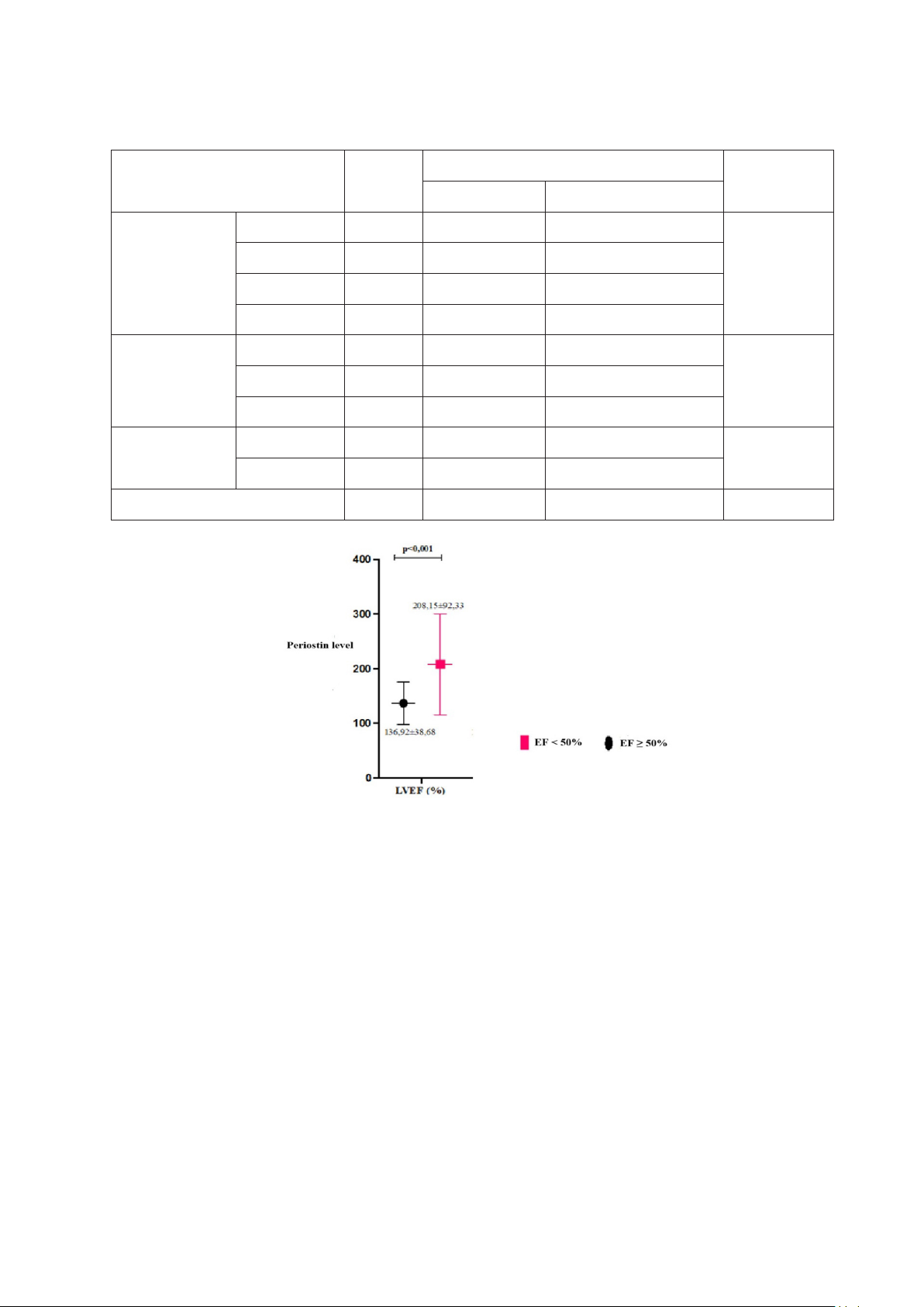
was lower than in the remaining group (187.84 ng/ml) (p < 0.05). Mean serum periostin level in the LVEF < 50% group
(208.15 ± 92.33 ng/ml) was significantly greater than that in the LVEF ≥ 50% group (136.92 ± 38.68 ng/ml), (p < 0.05).
Conclusion: Serum periostin levels increase in acute myocardial infarction and decrease gradually post myocardial
infarction. There is a difference in admission periostin levels between preserverd and reduced EF groups.
Keywords: Periostin, myocardial infarction.
I. BACKGROUND
Cardiovascular diseases, of which myocardial
infarction (MI) is the main cause of morbidity and
mortality in Western countries, are now rapidly
becoming more common in developing and
underdeveloped countries [1]. Up to 3 million people
worldwide suffer from MI and 1 million people
die from this disease every year [2]. According
to the World Health Organization in 2016, it is
estimated that 31% of deaths in Vietnam are due to
cardiovascular disease, more than half of which are
coronary artery disease [3]. Research on biomarkers
in patients with MI has made great progress in recent
years and a solution to this problem is expected to
be found in the near future, possibly in the form
of multi-marker assessment [4]. There are many
biomarkers studied in patients with myocardial
infarction such as troponin, myoglobin, CK-MB,
h-FAPB, IMA, etc. However, in general, their role
is still limited because they are mainly studied in
the acute phase. There is a clear correlation between
inflammatory outcome signs and adverse outcomes
in patients after acute myocardial infarction. In that
general trend, periostin - an inflammatory biomarker
in recent times promises to be an effective and
necessary factor in accurately predicting disease
progression, helping to choose treatment and is a
target in preventive care of coronary artery disease

Bệnh viện Trung ương Huế
Journal of Clinical Medicine - Hue Central Hospital - Volume 17, number 2 - 2025 41
Study of periostin levels in patients before and after acute myocardial infarction
[5]. In the world, the role of periostin on the heart is
increasingly studied in developed countries [6, 7].
In Vietnam, there is currently no research on the role
of periostin in acute MI. Therefore, we conducted a
study on the characteristics of serum periostin levels
in patients with acute MI.
II. MATERIALS AND METHODS
2.1. Materials
The research subjects were divided into 2
groups: the group of patients diagnosed with acute
MI and the remaining group was the control group.
All patient groups and the control group were
hospitalized and treated from September 2019 to
March 2023.
Disease group: Including 153 patients diagnosed
with acute MI treated at Hue Central Hospital and
Vinh Long Hospital, including 55 patients with
STEMI and 98 patients with NSTEMI.
Exclusion criteria for the disease group:
moderate to severe valvular heart disease,
concurrent inflammation, malignancy, hypertrophic
cardiomyopathy, dilated cardiomyopathy, blood
creatinine ≥ 4 mg/dL (353.6 µmol/L), disagreeing
to participate in the study.
Control group: Including 153 healthy subjects
who did not have acute MI (clinically no chest
pain, hs-Troponin T within normal limits), who had
regular health check-ups at Vinh Long hospital.
2.2. Methods
Study design: Analytical cross-sectional study.
Non-probability, purposive sampling.
All patients will be clinically examined and
assigned routine investigations during the patient’s
hospital stay, except for serum periostin levels
which is performed 5-7 days after MI (levels at first
time). Patients will be periodically re-examined after
discharge. After 3 months of MI, an echocardiogram
(assessing LVEF parameters) and a second periostin
blood test will be performed (levels at second time).
Serum periostin concentration testing is performed by
enzyme-linked immunosorbent assay, which is ELISA
(Enzyme-Linked ImmunoSorbent Assay) and serum
periostin concentration quantification is performed
at the Department of Immunology, University of
Medicine and Pharmacy, Hue University with the
Human Periostin Kit from MyBioSource.com.
Data processing: Data were processed using
SPSS version 26 (IBM SPSS Statistics for Windows,
Version 26.0), Medcalc version 22.017 and Excel
2013 software.
III. RESULTS
The baseline characteristics are listed in Table 1.
There was no difference in age, BMI and sex between
the patient group and the control group, p > 0.05. The
serum periostin level in acute MI patients was the
highest, the control group’s periostin level was the
lowest, p < 0.001 (Table 2). Periostin level in Killip
I group was lower than in the remaining group, the
difference was statistically significant with p < 0.001
(Table 3). Mean serum periostin level in the LVEF <
50% group was significantly greater than that in the
LVEF ≥ 50% group, p < 0.05 (Figure 1).
Table 1: Common characteristics between the disease group and the control group
Characteristics
Disease group Control group
p
nX ± SD
(min-max) nX ± SD
(min-max)
BMI (kg/m2)
Male 92 23.95 ± 3.44
(17,10 - 35,38) 92 23.60 ± 2.16
(18,49 - 28,76) 0.642
Female 61 24.99 ± 4.18
(16.89 - 36.21) 61 25.43 ± 3.03
(15.77 - 37.46) 0.327
Total 153 24.36 ± 3.77
(16.89 - 36.21) 153 24.33 ± 2.70
(18.49 - 30.82) 0.848

Bệnh viện Trung ương Huế
42 Journal of Clinical Medicine - Hue Central Hospital - Volume 17, number 2 - 2025
Study of periostin levels in patients before and after acute myocardial infarction
Characteristics
Disease group Control group
p
nX ± SD
(min-max) nX ± SD
(min-max)
Age (years)
Male 92 67.70 ± 12.70
(38.00 - 96.00) 92 69.10 ± 11.94
(47.00 - 88.00) 0.417
Female 61 74.18 ± 10.67
(44.00 - 96.00) 61 72.13 ± 12.10
(50.00 - 90.00) 0.330
Total 153 70.29 ± 12.32
(38.00 - 96.00) 153 70.31 ± 12.08
(47.00 - 90.00) 0.986
Age groups n % n %
0.178
< 60 29 18.95 37 24.18
60 - 69 52 33.99 35 22.88
70 - 79 32 20.92 34 22.22
≥ 80 40 26.14 47 30.72
Gender
(%)
Male 92 60.13 92 60.13
1.000
Female 61 39.87 61 39.87
Table 2: Periostin level between groups
Periostin (ng/mL) n Median IQR p
Disease group at
1st time
Male 92 142.41 117.17 - 203.56
0.236*
Female 61 156.00 124.04 - 218.01
Disease group at
2nd time
Male 92 73.61 59.07 - 97.20
0.158
Female 61 78.61 66.74 - 112.63
Comtrol group
Male 92 65.76 36.14 - 80.71
0.579
Female 61 60.66 42.33 - 81.09
Comparing
1st time 149.37 120.69 - 208.18
p(a-b) < 0.001
2nd time 77.69 61.63 - 101.05
Control 63.04 40.96 - 80.98 p(a-c) < 0.001
p(b-c) < 0.001
a: periostin concentration in the first patient group, b: periostin concentration in the second patient group,
c: periostin concentration in the control group
Bệnh viện Trung ương Huế
Journal of Clinical Medicine - Hue Central Hospital - Volume 17, number 2 - 2025 43
Study of periostin levels in patients before and after acute myocardial infarction
Table 3: Periostin levels at admission and general characteristics
General characteristics n
Periostin (ng/mL)
p
Median IQR
Age
< 60 29 151.20 116.30 - 210.89
0.082
60 - 69 52 132.28 113.93 - 179.29
70 - 79 32 175.49 124.82 - 223.07
≥ 80 40 159.84 126.06 - 205.64
BMI (kg/m2)
< 23 61 138.61 116.94 - 201.40
0.36923 - 24.9 27 179.29 127.22 - 234.66
≥ 25 65 147.78 120.93 - 207.52
Killip class I 93 132.28 114.56 - 180.94 < 0.001
II - IV 60 187.84 132.85 - 235.16
Total 153 149.37 120.69 - 208.18
Figure 1: Mean periostin level at admission between EF groups on echocardiography 3 months after MI
IV. DISCUSSION
Regarding the general characteristics of the study
subjects, in our study there was no difference in the
ratio of gender, BMI and age between the two groups
of patients and the control group (Table 1), which is
similar to the subjects of studies related to periostin
after acute MI in the world [8]. There is a change in
serum periostin concentration over time after acute
MI, from 2016 to present, it has been shown that
although periostin concentration has increased from
the first day of injury, it only peaks after 5 - 7 days
of illness and then gradually decreases [9]. That is
the basis for us to choose the time for the first serum
periostin test on the 5 - 7th day of MI, when the
periostin concentration reaches its maximum value.
In our study, the serum periostin concentration in
patients with acute MI at the initial test was the
highest, with a median of 149,37 ng/mL (Table 2).
The second test 3 months after acute MI, the median
serum periostin concentration decreased to 77.69 ng/
mL, while the periostin concentration in the control
group was the lowest, with a median of 63.04 ng/
mL, compared with the reference value of normal
serum periostin concentration of < 100 ng/mL
according to Mybiosource.com - the manufacturer
of the serum periostin test kit. The median periostin

Bệnh viện Trung ương Huế
44 Journal of Clinical Medicine - Hue Central Hospital - Volume 17, number 2 - 2025
Study of periostin levels in patients before and after acute myocardial infarction
concentration in our control group was 63.04 ng/
mL, not much different from the results of study
of Evan Tan examining periostin levelsin Chinese
adults with a median value of 57.0 ng/mL [10]. The
study results were consistent with most previous
literature [9], except Chi-Wen Cheng showed that
periostin concentration decreased early after AMI,
which is not similar to the natural progression of the
disease and was not expected by the author himself
[11]. However, the sample size in Chi-Wen Cheng’s
study was small (45 MI patients), so it is only for
reference. Under normal conditions, serum periostin
levels is very low, but when there is damage such
as acute MI, periostin levels will increase rapidly.
Research of Lin Ling et al(2014) recorded the
median periostin concentration in acute MI patients
as 188 ng/mL [12]. Periostin concentration peaked
on days 5 to 7 after a acute MI and then gradually
decreased over time [13]. Xinwei He et al studied
patients with large-vessel atherosclerotic stroke,
tested blood periostin levelsat 3 different times on
days 1, 6, and 28 of stroke and found that periostin
levelsbegan to increase from the first day of the
disease, reached the highest concentration on day 6,
and remained higher than normal after stroke for at
least 28 days [14].
According to Caswell-Smith, periostin levels
did not differ by age or common comorbidities
other than respiratory diseases [15]. We noted no
difference in serum periostin levels by age group
(Table 3). In addition, there was no difference in
periostin levels by BMI classification. This suggests
that serum periostin levels are independent of age
and body mass index. Meanwhile, periostin levels in
the Killip I classification group were lower than in
the Killip II - IV classification group, suggesting that
serum periostin levels are more likely to correlate
with disease severity. Regarding the difference of
serum periostin in echocardiographic parameters
after 3 months of MI: after the patient has an acute
MI, periostin stimulates excessive myocardial
remodeling, leading to a state of fibrotic scar tissue
that does not function to replace the damaged
myocardial area, which will reduce heart function,
reduce LVEF, reduce cardiac contractility and lead
to expansion of the size of the heart chamber, shown
by an increase in the diameter of the heart chamber
on echocardiography. The more periostin is
produced, the more obvious the remodeling process
becomes. The study results showed that the serum
periostin concentration in the LVEF ≥ 50% group
was much lower than that in the remaining group
(Figure 1). This contributes to the demonstration
that periostin is associated with cardiac remodeling
after acute MI.
V. CONCLUSION
Serum periostin levels increase in acute
myocardial infarction and decrease gradually post
myocardial infarction. There is a difference in
admission periostin levels between preserverd and
reduced EF groups.
Ethics approval
This study was conducted in accordance with the
Declaration of Helsinki and approved by the Ethics
Committee of Hue Central hospital.
Competing interests
The authors declare that they have no competing
interests.
REFERENCE
1. Weil BR, Neelamegham S. Selectins and Immune Cells in
Acute Myocardial Infarction and Post-infarction Ventricular
Remodeling: Pathophysiology and Novel Treatments. Front
Immunol. 2019; 10: 300.
2. Mechanic OJ, Gavin M, Grossman SA. Acute Myocardial
Infarction. StatPearls 2023; Available from: https://www.
ncbi.nlm.nih.gov/books/NBK459269/
3. Pham Manh Hung, Clinical cardiology. 2019, Hanoi,
Vietnam: Medical Publishing House.
4. Tilea I, Varga A, Serban RC. Past, Present, and Future of
Blood Biomarkers for the Diagnosis of Acute Myocardial
Infarction-Promises and Challenges. Diagnostics (Basel).
2021; 11(5): 881.
5. Seropian IM, Sonnino C, Van Tassell BW, Biasucci LM,
Abbate A. Inflammatory markers in ST-elevation acute
myocardial infarction. Eur Heart J Acute Cardiovasc Care.
2016; 5(4): 382-395.
6. Iekushi K, Taniyama Y, Azuma J, Katsuragi N, Dosaka N,
Sanada F. Novel mechanisms of valsartan on the treatment
of acute myocardial infarction through inhibition of the
antiadhesion molecule periostin. Hypertension. 2007;
49(6): 1409-1414.


![Bài giảng Cập nhật vấn đề hồi sức bệnh tay chân miệng nặng [mới nhất]](https://cdn.tailieu.vn/images/document/thumbnail/2025/20250920/hmn03091998@gmail.com/135x160/23301758514697.jpg)