
Lin et al. Journal of Biomedical Science 2010, 17:44
http://www.jbiomedsci.com/content/17/1/44
Open Access
RESEARCH
© 2010 Lin et al; licensee BioMed Central Ltd. This is an Open Access article distributed under the terms of the Creative Commons At-
tribution License (http://creativecommons.org/licenses/by/2.0), which permits unrestricted use, distribution, and reproduction in any
medium, provided the original work is properly cited.
Research
Sonoporation-mediated gene transfer into adult
rat dorsal root ganglion cells
Chung-Ren Lin*
1,3
, Kuan-Hung Chen
1
, Chien-Hui Yang
1
, Jiin-Tsuey Cheng
4
, Shyr-Ming Sheen-Chen
2
, Chih-
Hsien Wu
1,4
, Wei-Dih Sy
1
and Yi-Shen Chen
1
Abstract
Background: Gene transfer into many cell types has been successfully used to develop alternative and adjunct
approaches to conventional medical treatment. However, effective transfection of postmitotic neurons remains a
challenge. The aim of this study was to develop a method for gene transfer into rat primary dorsal root ganglion
neurons using sonoporation.
Methods: Dissociated cells from adult rat dorsal root ganglion (DRG) cells were sonicated for 1-8 s at 2.5-10 W to
determine the optimal ultrasound duration and power for gene transfection and cell survival. Transfection efficiency
was compared between sonoporation, liposome and lentiviral vector gene transfer techniques.
Results: The optimum ultrasound intensity was 5 W for 2 s and yielded an efficiency of gene transfection of 31% and a
survival rate of 35%.
Conclusions: Sonoporation can be optimized to minimize cell death and yield a high percentage of transfected
neurons and that this technique can be easily applied to primary cultures of rat dorsal root ganglion neurons.
Background
Methods for altering gene expression are widely used to
elucidate molecular mechanisms involved in cellular
physiopathology. Gene modification also has potential as
a therapeutic modality for treating many diseases. Many
gene transfer methods have been developed in the past
two decades, including calcium phosphate coprecipita-
tion, microinjection, recombinant viruses, liposome-
mediated gene transfer, lipids that do not form liposomes,
high molecular weight cationic polymers, particle bom-
bardment (biollistics) and electroporation.
Ultrasound-mediated gene transfer has recently
emerged as a promising technique with a broad range of
potential applications. Ultrasound can be used to modify
the permeability of the cell membrane to facilitate the
uptake of RNA [1-5] and DNA into the cell [6-11]. Low-
frequency ultrasound increases membrane permeability
to many drugs, including high molecular weight proteins
[12]. The degree of macromolecule uptake is correlated
with the acoustic energy and frequency of the stimulus
[13]. Miller et al. [14] demonstrated that the uptake of
fluorescent dextran by Chinese hamster ovary cells is
similar at ultrasound frequencies of 1.0 MHz and 3.3
MHz, but is greatly reduced at 5.3 MHz and 7.15 MHz.
Huber and Pfisterer reported that focused ultrasound
enhanced the transfer of DNA plasmids into several cell
lines in vitro and into a Dunning prostate tumor after
direct DNA injection in vivo. They showed that the pres-
sure amplitude and duration of sonication affect transfec-
tion ratio and cell survival [15].
Transfection of postmitotic neurons has been a major
challenge in the past. With few exceptions, neuronal
transfections have been unreliable, cytotoxic, labor-
intensive and inefficient [16]. Although sonoporation-
mediated gene transfer into cell lines [8,17,18], silkworm
larvae [19], the ovary and uterus [20], muscle cells [21],
the salivary gland [22], the joint synovium [4] and the
chicken embryo [23] has been described, there are few
reports on sonoporation-mediated gene transfer into
neuronal cells. Shimamura et al. used ultrasound and
microbubble-mediated cavitation to transfect rat brain
cells [24]. They successfully transfected meningeal and
* Correspondence: chungren@ntu.edu.tw
1 Department of Anesthesiology, Chang Gung Memorial Hospital-Kaohsiung
Medical Center, Chang Gung University College of Medicine, Kaohsiung,
T
aiwan
Full list of author information is available at the end of the article

Lin et al. Journal of Biomedical Science 2010, 17:44
http://www.jbiomedsci.com/content/17/1/44
Page 2 of 6
glial cells, but failed to transfect neurons. Manome et al.
reported that they transfected neuronal cells in brain
slices, but in very small amounts [7]. Fischer et al. used
sonoporation to transfect plasmids into the retinal neu-
rons, dorsal forebrain and optic tectum of the chicken,
into the cerebellar neurons of the rat and into the hip-
pocampal neurons of the mouse. Although the ability of
ultrasound to enhance gene transfer has been demon-
strated in numerous studies, its efficacy for gene transfer
into the dorsal root ganglion (DRG) has not been deter-
mined.
This study was designed to investigate the feasibility of
gene transfer into the DRG using a standard laboratory
sonicator and to determine the optimal ultrasound dura-
tion and power for gene transfection and DRG survival.
We also compared transfection (transduction) efficiency
between the sonoporation, liposome and lentiviral vector
gene transfer techniques.
Materials and methods
Animals
The animals were used in accordance with the guidelines
of the Chang Gung University.
Tissue culture
DRG cells were prepared as previously described [25].
DRGs were dissected in sterile Hanks' buffered saline
solution (HBSS) containing 3% D-glucose and 0.01 M
HEPES buffer (HBSS+). DRG cells were dissociated by
mild trituration using a Pasteur pipette after incubation
for 10 min at 37°C in Ca2+/Mg2+-free HBSS containing
0.05% trypsin. The cell suspensions were initially plated
into noncoated plastic tissue culture flasks and left for 3 h
at room temperature to minimize the number of fibro-
blasts in the cultures. The supernatant, which contained
DRG neurons and satellite glial cells, was then aspirated.
Cell density was determined using a hemacytometer.
Between 100,000 and 200,000 cells were plated onto 12
mm glass cover slips that were coated sequentially with
poly-D-lysine and laminin (Sigma) diluted 1:100 in HBSS.
Cell cultures were maintained at 37°C under a 5% CO
2
atmosphere in culture medium (Neurobasal Media,
Gibco) containing N-2 supplement (Gibco), 100 U/ml
penicillin, 100 mg/ml streptomycin and 2% fetal bovine
serum (Gibco BRL).
Virus preparation
Enhanced green fluorescence protein (E-GFP)-containing
lentiviral vectors were obtained from the National RNAi
Core Facility, Academia Sinica, Taiwan. Lentiviruses were
prepared according to a standard protocol. Titers were
assayed using 3T3 cells and serial dilutions of the vector
preparations. The titers of these vector stocks were also
estimated by measuring the level of viral p24gag antigen
using a human immunodeficiency virus-1 p24 antigen
assay kit (Beckman Coulter, Fullerton, CA).
Liposome-mediated gene transfer
Lipofectamine 2000 (Invitrogen) was mixed with plasmid
E-GFP DNA (pE-GFP C1) in the ratio of 3 μl per 2 μg,
respectively, in 50 μl of HBSS+. The vector is described
elsewhere [26]. The optimum ratio of lipofectamine to
pE-GFP C1 was determined according to the manufac-
turer's instructions. These reagents were incubated at
room temperature for 15 min and were then added to a
solution containing freshly dissected DRG cells. The
medium was replaced with Lipofectamine 2000 after 12
h.
Sonoporation
We used a Sonics and Materials VC130 sonicator (New-
town, CT), which produces continuous wave ultrasound
at 20 kHz and has an adjustable output range of 2.5-130
W and a 12 mm diameter probe tip. pE-GFP C1 was
diluted in HBSS+, added to the wells at concentrations of
0.5-20 μg/ml and incubated for 5-10 min at 37°C. The
sonicator probe was sterilized by sonication in 70% etha-
nol followed by sonication in sterile water. It was then
placed into the wells, each of which contained 1000 μl of
medium, and activated for 1-8 s, which delivered 2.5-10
W of energy. Cells were cultured for 1-14 d after sonopo-
ration and were then fixed and processed for immunocy-
tochemical labeling using standard methods [27].
Cell survival assay
Cell survival after sonoporation was assessed using the
Live/Dead assay (Molecular Probes) according to the
manufacturer's specifications. DRG cells were main-
tained in vitro for 48 h after sonication. The medium was
then aspirated and cells were rinsed twice with HBSS+.
After adding 200 μl of HBSS+ containing 1 μM ethidium
homodimer and 1 μM calcein AM, the cells were incu-
bated for 30 min at 37°C, rinsed twice in HBSS+ and
counted using a fluorescence microscope.
Fixation and immunocytochemistry
Cells were fixed and immunolabeled as described previ-
ously [27]. The primary antibodies were mouse anti-β-
tubulin isotype III (1:1000; Sigma) and rabbit anti-E-GFP
(1:1000; Sigma). The secondary antibodies were goat
anti-rabbit Alexa568, goat anti-mouse Alexa568 and goat
anti-rat Alexa488 (Molecular Probes Inc., Eugene, OR)
and were diluted 1:1000 in PBS containing 0.2% Triton X-
100. Cells were stained using 1 μg/ml of DAPI (Sigma) in
PBS and then mounted on cover slips using Fluoro-
mount-G.
Lin et al. Journal of Biomedical Science 2010, 17:44
http://www.jbiomedsci.com/content/17/1/44
Page 3 of 6
Statistical analysis
Means for experimental variables were compared using
ANOVA and Student's t-test post hoc. Means and stan-
dard errors for cell numbers were calculated by counting
cells in at least five view fields from at least three cover
slips per treatment.
Results
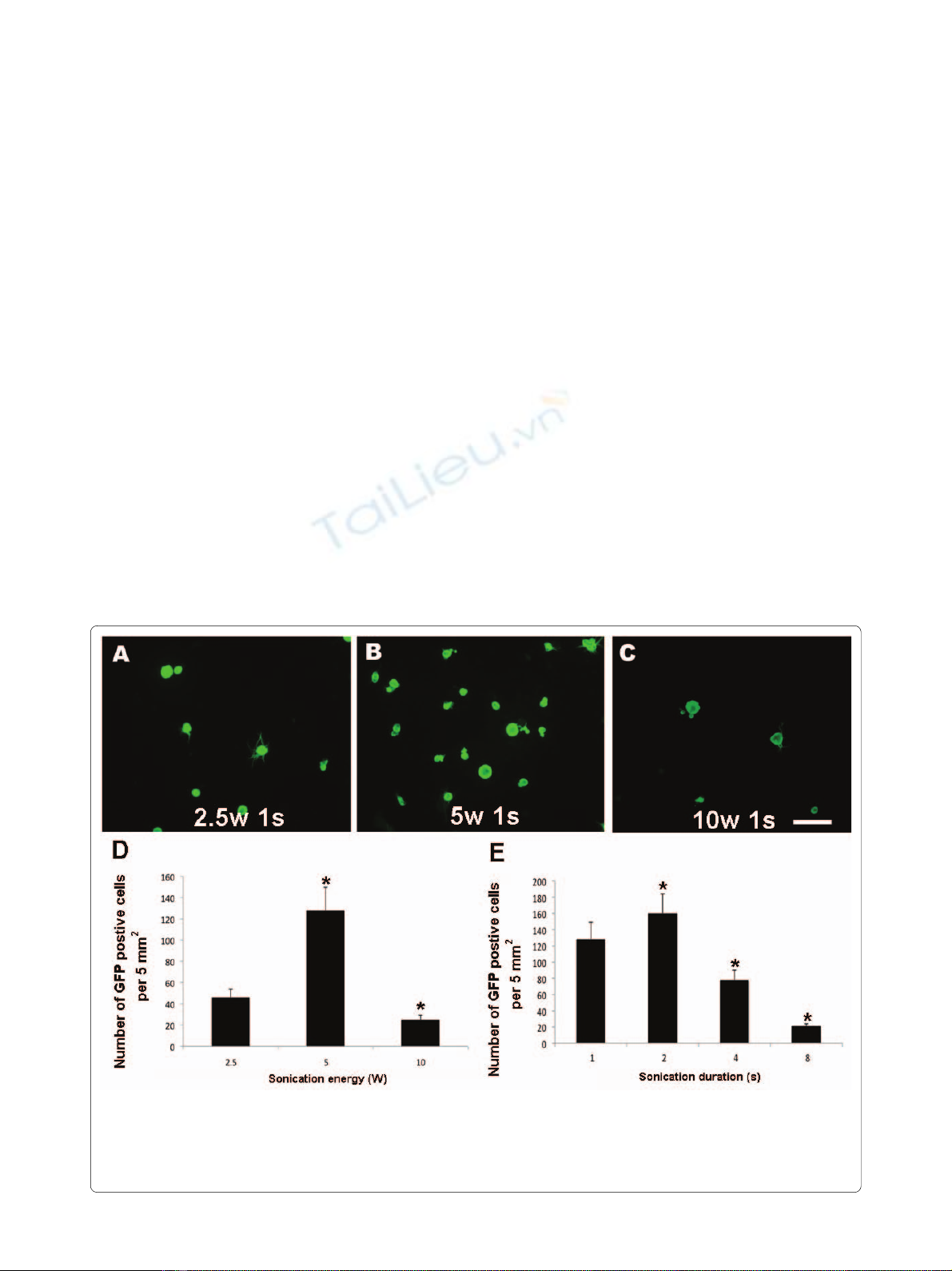
The optimal power for sonoporation of DRG cells is 5 W
We conducted a series of experiments using cultures of
DRG cells to determine the optimal energy output for
sonoporation. Dissociated cells from adult rat DRGs were
suspended at a density of 10,000 cells per cm3. After addi-
tion of 10 μg/ml of pE-GFP C1, the suspensions were son-
icated for 1 s at 2.5-10 W. The cells were maintained in
vitro for 48 h after sonoporation. GFP expression showed
that a few cells were transfected by treatment for 1 s at 2.5
W (Figure 1A). A twofold increase in output energy from
2.5 W to 5 W increased the number of GFP-positive cells
by threefold (Figure 1B). However, the number of GFP-
positive cells decreased when the output energy was
increased further to 10 W (Figure 1C), probably because
of decreased cell survival (Figure 2). The optimal output
energy for sonoporation for 2 s was 5 W according to the
number of GFP-positive cells (Figure 1D).
The optimal duration of sonoporation of DRG cells is 2 s
To determine the optimal duration of sonoporation, we
conducted another series of experiments using DRG cells.
Dissociated cells from adult rat DRGs were suspended at
a concentration of 10,000 cells per cm3 and 10 μg/ml of
pE-GFP C1 was added to the medium. The suspensions
were then sonicated at 5 W for 1-8 s. The cells were main-
tained in vitro for 48 h after sonoporation. The number of
GFP-positive cells increased up to a sonoporation dura-
tion of 2 s (Figure 1E).
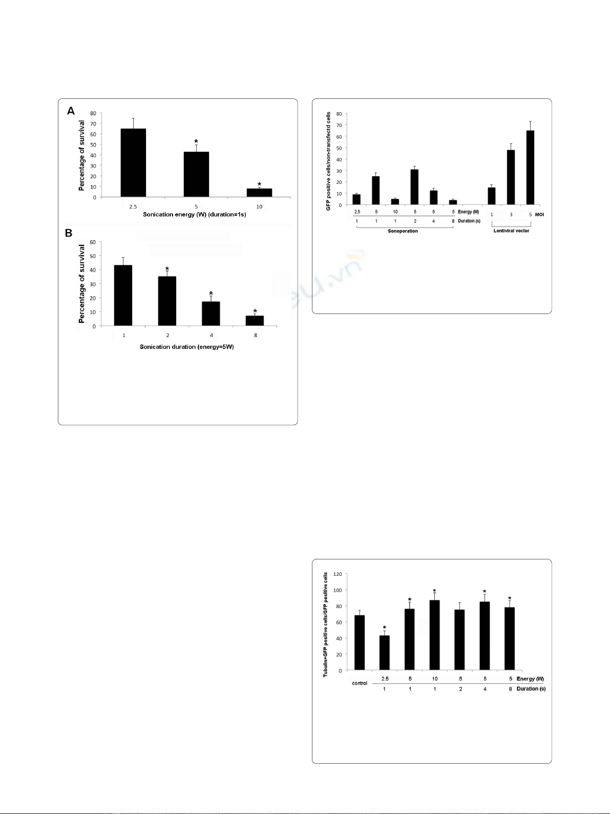
The survival rate of DRG cells after sonoporation for 2 s at 5
W is 35%
The number of dead cells after sonoporation was
assessed according to the accumulation of ethidium
homodimer and the number of live cells was assessed
according to the accumulation of calcein AM. The num-
ber of dead cells after sonication for 1 s at 2.5 W was 35%
of that of the unsonicated control. Sonication for 1 s at 5
W or 10 W increased the number of dead cells to 43% of
that of the control. At 5 W, sonication for 2 s, 4 s and 8 s
Figure 1 Optimal conditions for DRG transfection via sonoporation. Dissociated cells from adult rat DRGs were suspended at a density of 10,000
cells per cm3 in a solution containing 10 μg/ml of pE-GFP C1 and were sonicated for 1 s at 2.5-10 W using a 12 mm diameter probe tip. The cells were
maintained in vitro for 48 h after sonoporation. The calibration bar in Panel c represents 100 μm and applies to Panels a-c. Panel d shows the number
of GFP-expressing cells per 5 mm2 after sonication at various energy levels. Panel e shows the number of GFP-expressing cells per 5 mm2 after soni-
cation for various sonication durations at 5 W.
Lin et al. Journal of Biomedical Science 2010, 17:44
http://www.jbiomedsci.com/content/17/1/44
Page 4 of 6
increased the number of dead cells to 65%, 87% and 93%,
respectively, of that of the control (Figure 2).
Comparison of transfection (transduction) efficiency
between the sonoporation, liposome and lentiviral vector
methods
Cells used for these experiments were obtained from the
pooled dissociated adult rat preparations before they
were allocated to the free-floating and adherent-cell
treatments. Less than 1% of cells transfected with lipo-
somes were GFP-positive (data not shown). Lentiviral
vectors added at MOIs of 1, 3 and 5 transduced 10%, 48%
and 65%, respectively, of the DRG cells (Figure 3).
Although the efficiency of the lentivirus method was
much higher than that of the sonoporation method, prep-
aration of the viral vector was much more labor-intensive
than the sonoporation method.
Percentage of neuronal cells transfected using
sonoporation
We used immunocytochemistry against β-III tubulin to
identify neuronal cells in primary DRG cultures incu-
bated with 10 ng/ml of NGF for 48 h. The percentage of
β-III tubulin-positive cells in the control culture was 68.0
± 6.2%. After sonoporation for 1 s at energy outputs of 2.5
W, 5 W and 10 W, cells immunoreactive for β-III tubulin
constituted 43%, 76% and 87%, respectively, of GFP-
transfected cells. At an energy output of 5 W and sonopo-
ration for 1 s, 2 s, 4 s and 8 s, cells immunoreactive for
β-III tubulin constituted 76%, 75%, 85% and 78%, respec-
tively, of GFP-transfected cells (Figure 4). These findings
show that sonoporation-mediated gene transfer is effec-
tive in DRG cells.
Discussion
Transfection of postmitotic neurons is labor-intensive,
inefficient, unreliable and may have cytotoxic effects. The
inability to express foreign proteins in postmitotic neu-
rons has hampered neuroscience research [16]. Our
results show that sonoporation is a feasible in vitro
method for gene transfer into cultured DRG cells from
adult rats. These results were achieved using a standard
laboratory sonicator of the type used to disrupt cells and
homogenize solutions. Sonoporation for 2 s at 5 W
Figure 3 Comparison of gene transfection efficiency between the
sonoporation and lentiviral vector methods. The histogram shows
the relative number of transfected GFP-positive cells per 5 mm2 after
sonoporation (left side of the panel) and gene transfer using the lenti-
viral vector (right side of the panel). All DRG cells were derived from the
same pool of cells.
Figure 4 β-tubulin III- and GFP-immunoreactive DRG cells. After
addition of 10 μg/ml of pE-GFP C1, the cells were sonicated for 2 s at 5
W using a 12 mm probe. The cells were processed for immunocy-
tochemistry 48 h after sonoporation and labeled with antibodies
against GFP and β-tubulin III (a marker of neuronal cells).
Figure 2 Cell survival for various sonoporation durations and en-
ergy levels. Survival was defined as the number of calcein-positive
cells 2 d after sonoporation and expressed as a percentage of the num-
ber of calcein-positive cells in the unsonoporated control. Results are
expressed as the mean ± SD. *p < 0.05.

Lin et al. Journal of Biomedical Science 2010, 17:44
http://www.jbiomedsci.com/content/17/1/44
Page 5 of 6
resulted in optimum transfection efficiency (31%) and a
cell survival rate equivalent to 35% of that of the control.
Virus-based methods are the most successful of effect-
ing gene transfer to neuronal cells [16,28]. The increasing
use of viral vectors to transfer DNA into neurons has
arisen because of their high infection efficiencies com-
pared with nonviral methods. However, preparation of
recombinant viruses is expensive and labor-intensive.
Other limitations of this method are its potential toxicity
for neurons, a DNA expression cassette of limited size
and the production of severe immune reactions in vivo.
These approaches also constitute a potential health haz-
ard for laboratory personnel [29-32]. Furthermore, seri-
ous concerns about the insertional mutagenesis have
arisen, especially concerning the use of viral vectors when
clinical trials are involved [11,33]. By contrast, nonviral
methods such as naked plasmid DNA injection, elec-
troporation, and sonoporation should have a higher
potential for clinical application, even although their effi-
cacy for gene delivery is lower [34].
Liposome-mediated gene transfer involves the fusion of
synthetic lipids into the plasmid membrane, which may
affect cell membrane proteins. Therefore, sonoporation is
preferable to liposome-mediated gene transfer for study-
ing expression of transmembrane proteins. Furthermore,
the efficacy of liposomal transfection is less than 1%.
Although gene transfer via electroporation in vivo is
effective using DNA injection followed by the application
of electric fields, the tissue damage caused by the electric
pulse is problematic for cell survival. Ultrasound, on the
other hand, makes the cell membrane porous and
enhances the intracellular delivery of naked DNA in vitro.
The membrane damage induced by ultrasound is tran-
sient and the holes (or pores) can reseal and allow sur-
vival of the cells. During sonoporation, large molecules in
the medium can leak into the cells and remain trapped
there after the membrane reseals [17,35]. Sonoporation
has opened tremendous opportunities for targeted gene
transfer. Conceptually, gene vectors mixed with ultra-
sound contrast agents could be injected into animal cells
and targeted gene transfer could be achieved by selective
application to a predefined area. Indeed, promising
results have now been reported in animal models [36]. By
using this approach, the risk of systemic exposure (a
major drawback of current clinical gene transfer proto-
cols) could be reduced substantially reduced.
Sonoporation of freshly dissected DRG cells was highly
selective for neuronal cells. Neuronal cells constituted a
much higher percentage of the total number of trans-
fected DRG cells with the sonoporation method than
with the lentivirus method. The percentage of sonopo-
rated neuronal cells depended on energy level; under
optimal sonoporation conditions, 75% of sonoporated
DRG cells were neuronal cells. The mechanism underly-
ing the preferred transfection of neuronal cells by sonop-
oration remains unknown. Sonoporation facilitates the
entry of macromolecules into cells via microbubble-
mediated cavitation and transient disruption of the
plasma membrane [37,38]. As the average diameter of
neuronal cells is larger than that of glial cells, it is possible
that the effects of ultrasound frequency and intensity
depend on cell diameter.
Sonoporation is an alternative method for transferring
naked plasmid DNA into neuronal cells and may avoid
side effects associated with other methods. We found that
sonoporated cells maintained transgene expression for at
least 2 weeks after treatment. In addition, sonoporation
did not harm normal cellular functions such as the out-
growth of dendrites and axons, and all of these cell types
had well-developed dendritic and axonal arbors. Neu-
ronal cells with elaborate neurites can be transfected via
sonoporation without physical disruption [39]. Thus,
sonoporation does not result in genomic instability or
other forms of permanent cellular damage that would
limit sonoporation to short-term applications.
However, our study had several limitations. We have
not evaluated a comprehensive comparison of ultrasound
modalities and contrast agents. Indeed, the number of
possible combinations of ultrasound field characteristics
(e.g., continuous versus pulsed wave, frequency or duty
cycle) and contrast microbubbles is enormous. Therefore
the optimal conditions for gene transfer by sonoporation
require further investigation.
Conclusions
This study demonstrates that sonoporation enables deliv-
ery of plasmids into dorsal root ganglion cells in vitro.
Sonoporation is a simple and economic method for
studying physiological pathways in DRG cells in vitro.
Sonoporation of dorsal root ganglion cells allows the
preferential transfection of neuronal cells. Therefore, we
propose that sonoporation could be applied to the intact
nervous system to transfer foreign DNA for physiological
research.
Competing interests
The authors declare that they have no competing interests.
Authors' contributions
CR carried out the all the animal studies, participated in design of the study
and coordination and drafted the manuscript. KH carried out the immunohis-
tochemistry. JT participated in participated in the design of the study and per-
formed the statistical analysis. SH and CH carried out the cell counting. WD and
YS carried out the virus preparation. All authors read and approved the final
manuscript.
Acknowledgements
This work was supported in part by grant No. 870641, 860231, and 860232
from Chang Gung Memorial Hospital Research, Kaohsiung, Taiwan, and by
grant No. 95-2745-B-182A-004, 96-2628-B-182A-005-MY3, and 98-2314-B-
182A-035-MY2 from the Taiwan National Science Council Research, Taipei,
Taiwan.


%20--%3e%3cdefs%3e%3cstyle%3e%20.st0%20{%20fill:%20%23fff;%20}%20.st1%20{%20fill:%20%237800fa;%20}%20%3c/style%3e%3c/defs%3e%3cpath%20class='st1'%20d='M117.78,12.18H43.11c2.9,3.47,4.65,7.94,4.65,12.82,0,5.6-2.3,10.66-6.01,14.29h76.02l7.22-13.56-7.22-13.56Z'/%3e%3cg%3e%3cpath%20class='st0'%20d='M53.58,26.17h-.59v-1.46h.59v-4.96h2.83c1.78,0,2.67.94,2.67,2.82v5.76c0,1.87-.89,2.81-2.67,2.81h-2.83v-4.96ZM55.36,21.37v3.34h1.1v1.46h-1.1v3.34h1.01c.61,0,.91-.37.91-1.1v-5.93c0-.74-.3-1.1-.91-1.1h-1.01Z'/%3e%3cpath%20class='st0'%20d='M65.99,31.14h-1.8l-.31-2.07h-2.19l-.31,2.07h-1.64l1.82-11.39h2.62l1.82,11.39ZM65.28,18.04c-.25.46-.51.77-.75.94-.21.15-.47.22-.79.22-.26,0-.57-.07-.92-.22l-.38-.15c-.14-.05-.26-.07-.37-.07-.3,0-.53.18-.71.54l-.91-.68c.25-.46.51-.77.75-.94.21-.14.48-.21.79-.21.26,0,.57.07.92.21l.38.15c.14.05.26.07.37.07.3,0,.53-.18.71-.54l.91.68ZM61.91,27.52h1.73l-.87-5.76-.87,5.76Z'/%3e%3cpath%20class='st0'%20d='M74.53,26.89v1.52c0,1.91-.89,2.86-2.67,2.86s-2.67-.95-2.67-2.86v-5.93c0-1.91.89-2.86,2.67-2.86s2.67.95,2.67,2.86v1.11h-1.69v-1.22c0-.75-.31-1.12-.93-1.12s-.93.37-.93,1.12v6.15c0,.74.31,1.11.93,1.11s.93-.37.93-1.11v-1.63h1.69Z'/%3e%3cpath%20class='st0'%20d='M81.4,31.14h-1.8l-.31-2.07h-2.19l-.31,2.07h-1.64l1.82-11.39h2.62l1.82,11.39ZM75.9,19.2l1.52-1.91h1.71l1.51,1.91h-1.61l-.76-.95-.75.95h-1.61ZM77.32,27.52h1.73l-.87-5.76-.87,5.76ZM83.1,15.99l-1.76,1.91h-1.26l1.17-1.91h1.86Z'/%3e%3cpath%20class='st0'%20d='M84.86,19.75c1.78,0,2.67.94,2.67,2.82v1.48c0,1.87-.89,2.81-2.67,2.81h-.85v4.28h-1.79v-11.39h2.64ZM84.01,21.37v3.86h.85c.58,0,.87-.36.87-1.08v-1.71c0-.71-.29-1.07-.87-1.07h-.85Z'/%3e%3cpath%20class='st0'%20d='M93.51,19.75c1.78,0,2.67.94,2.67,2.82v1.48c0,1.87-.89,2.81-2.67,2.81h-.85v4.28h-1.79v-11.39h2.64ZM92.66,21.37v3.86h.85c.58,0,.87-.36.87-1.08v-1.71c0-.71-.29-1.07-.87-1.07h-.85Z'/%3e%3cpath%20class='st0'%20d='M98.8,31.14h-1.79v-11.39h1.79v4.88h2.03v-4.88h1.83v11.39h-1.83v-4.88h-2.03v4.88Z'/%3e%3cpath%20class='st0'%20d='M105.36,24.55h2.46v1.62h-2.46v3.34h3.09v1.63h-4.88v-11.39h4.88v1.63h-3.09v3.18ZM108.17,17.29l-1.76,1.91h-1.26l1.17-1.91h1.86Z'/%3e%3cpath%20class='st0'%20d='M112.2,19.75c1.78,0,2.67.94,2.67,2.82v1.48c0,1.87-.89,2.81-2.67,2.81h-.85v4.28h-1.79v-11.39h2.64ZM111.35,21.37v3.86h.85c.58,0,.87-.36.87-1.08v-1.71c0-.71-.29-1.07-.87-1.07h-.85Z'/%3e%3c/g%3e%3ccircle%20class='st1'%20cx='25'%20cy='25'%20r='20'/%3e%3cpath%20class='st0'%20d='M32.78,19.27c2.92,0,4.43,2.55,5.28,5.33l.71,2.17c.14.38-.33.75-.71.75h-5.61c.19-.33.24-.71.09-1.08l-.75-2.45c-.43-1.32-.99-2.64-1.79-3.77.75-.57,1.65-.94,2.78-.94h0ZM25,18.38c3.25,0,4.9,2.78,5.89,5.89l.76,2.45c.14.42-.33.8-.8.8h-11.69c-.42,0-.94-.38-.8-.8l.75-2.45c.99-3.11,2.64-5.89,5.89-5.89h0ZM25,11.35c1.74,0,3.11,1.37,3.11,3.11s-1.37,3.11-3.11,3.11-3.11-1.41-3.11-3.11,1.41-3.11,3.11-3.11h0ZM17.27,19.27c1.08,0,1.98.38,2.73.94-.8,1.13-1.37,2.45-1.74,3.77l-.8,2.45c-.14.38-.05.75.09,1.08h-5.56c-.42,0-.9-.38-.75-.75l.71-2.17c.9-2.78,2.41-5.33,5.33-5.33h0ZM17.27,12.91c1.51,0,2.78,1.27,2.78,2.83s-1.27,2.83-2.78,2.83-2.83-1.27-2.83-2.83,1.27-2.83,2.83-2.83h0ZM32.78,12.91c1.56,0,2.78,1.27,2.78,2.83s-1.23,2.83-2.78,2.83-2.83-1.27-2.83-2.83,1.27-2.83,2.83-2.83h0ZM27.07,28.56v.09c0,.57-.24,1.08-.61,1.46h0v.05c-.38.33-.9.57-1.46.57s-1.08-.24-1.46-.61h0c-.38-.38-.61-.9-.61-1.46v-.09h1.41v.09c0,.19.05.38.19.47v.05c.09.09.28.19.47.19s.38-.09.47-.19v-.05c.14-.09.24-.28.24-.47t-.05-.09h1.41ZM30.99,28.56v.09c0,1.65-.66,3.16-1.74,4.24-1.08,1.08-2.59,1.79-4.24,1.79s-3.16-.71-4.24-1.79l-.05-.05c-1.04-1.08-1.7-2.55-1.7-4.2v-.09h1.41v.09c0,1.27.47,2.4,1.27,3.25h.05c.85.85,1.98,1.37,3.25,1.37s2.4-.52,3.25-1.37c.85-.8,1.37-1.98,1.37-3.25v-.09h1.37ZM34.99,28.56v.09c0,2.78-1.13,5.28-2.92,7.07-1.79,1.79-4.29,2.92-7.07,2.92s-5.23-1.13-7.07-2.92c-1.79-1.79-2.92-4.29-2.92-7.07v-.09h1.41v.09c0,2.4.94,4.53,2.5,6.08,1.56,1.56,3.72,2.5,6.08,2.5s4.52-.94,6.08-2.5c1.56-1.56,2.5-3.68,2.5-6.08v-.09h1.41Z'/%3e%3c/svg%3e)