
Open Access
Available online http://ccforum.com/content/10/3/R87
Page 1 of 9
(page number not for citation purposes)
Vol 10 No 3
Research
Effects of thoraco-pelvic supports during prone position in
patients with acute lung injury/acute respiratory distress
syndrome: a physiological study
Davide Chiumello1, Massimo Cressoni2, Milena Racagni2, Laura Landi2, Gianluigi Li Bassi2,
Federico Polli2, Eleonora Carlesso2 and Luciano Gattinoni1,2
1Dipartimento di Anestesia e Rianimazione, Fondazione IRCCS – 'Ospedale Maggiore Policlinico, Mangiagalli, Regina Elena', Via F. Sforza 35, 20122
Milan, Italy
2Istituto di Anestesia e Rianimazione Università degli Studi di Milano, Via F. Sforza 35, 20122 Milan, Italy
Corresponding author: Luciano Gattinoni, gattinon@policlinico.mi.it
Received: 2 Feb 2006 Revisions requested: 23 Feb 2006 Revisions received: 2 Apr 2006 Accepted: 2 May 2006 Published: 8 Jun 2006
Critical Care 2006, 10:R87 (doi:10.1186/cc4933)
This article is online at: http://ccforum.com/content/10/3/R87
© 2006 Chiumello et al.; licensee BioMed Central Ltd.
This is an open access article distributed under the terms of the Creative Commons Attribution License (http://creativecommons.org/licenses/by/2.0),
which permits unrestricted use, distribution, and reproduction in any medium, provided the original work is properly cited.
Abstract
Introduction This study sought to assess whether the use of
thoraco-pelvic supports during prone positioning in patients
with acute lung injury/acute respiratory distress syndrome (ALI/
ARDS) improves, deteriorates or leaves unmodified gas
exchange, hemodynamics and respiratory mechanics.
Methods We studied 11 patients with ALI/ARDS, sedated and
paralyzed, mechanically ventilated in volume control ventilation.
Prone positioning with or without thoraco-pelvic supports was
applied in a random sequence and maintained for a 1-hour
period without changing the ventilation setting. In four healthy
subjects the pressures between the body and the contact
surface were measured with and without thoraco-pelvic
supports. Oxygenation variables (arterial and central venous),
physiologic dead space, end-expiratory lung volume (helium
dilution technique) and respiratory mechanics (partitioned
between lung and chest wall) were measured after 60 minutes
in each condition.
Results With thoraco-pelvic supports, the contact pressures
almost doubled in comparison with those measured without
supports (19.1 ± 15.2 versus 10.8 ± 7.0 cmH2O, p ≤ 0.05;
means ± SD). The oxygenation-related variables were not
different in the prone position, with or without thoraco-pelvic
supports; neither were the CO2-related variables. The lung
volumes were similar in the prone position with and without
thoraco-pelvic supports. The use of thoraco-pelvic supports,
however, did lead to a significant decrease in chest wall
compliance from 158.1 ± 77.8 to 102.5 ± 38.0 ml/cmH2O and
a significantly increased pleural pressure from 4.3 ± 1.9 to 6.1
± 1.8 cmH2O, in comparison with the prone position without
supports. Moreover, when thoraco-pelvic supports were added,
heart rate increased significantly from 82.1 ± 17.9 to 86.7 ±
16.7 beats/minute and stroke volume index decreased
significantly from 37.8 ± 6.8 to 34.9 ± 5.4 ml/m2. The increase
in pleural pressure change was associated with a significant
increase in heart rate (p = 0.0003) and decrease in stroke
volume index (p = 0.0241).
Conclusion The application of thoraco-pelvic supports
decreases chest wall compliance, increases pleural pressure
and slightly deteriorates hemodynamics without any advantage
in gas exchange. Consequently, we stopped their use in clinical
practice.
Introduction
Prone positioning is used and recommended as a rescue
maneuver to improve arterial oxygenation in adult patients with
acute lung injury (ALI), acute respiratory distress syndrome
(ARDS) [1,2] or chronic obstructive pulmonary disease [3],
although its benefits with regard to outcome are not proven
[4,5].
Improved oxygenation implies, by definition, improvement of
the ventilation/perfusion ratio. This can be achieved through
different mechanisms, not mutually exclusive, each
ALI = acute lung injury; ARDS = acute respiratory distress syndrome; BSA = body surface area; EELV = end-expiratory lung volume; PEEP = positive
end-expiratory pressure.

Critical Care Vol 10 No 3 Chiumello et al.
Page 2 of 9
(page number not for citation purposes)
documented in the literature: (1) a more uniform distribution of
alveolar inflation/ventilation, due to the lower gradient of
transpulmonary pressure resulting from the changes in chest
wall mechanics, with perfusion being less affected [6-9]; (2) a
greater recruitment of the dorsal lung regions in comparison
with the derecruitment of the ventral lung regions when chang-
ing from the supine to the prone position [10]; (3) an overall
increase in end-expiratory lung volume (EELV) as a result of
the more favorable position of the diaphragm [11].
Douglas and colleagues [12] used supports under the ribcage
and the pelvis of patients with respiratory failure, to prevent
their abdomen from bearing the entire weight of the torso.
Indeed, some authors have advocated the use of thoraco-pel-
vic supports to avoid an increase in intra-abdominal pressure,
which could limit diaphragm excursion and, consequently,
alveolar ventilation in the most dependent lung regions
[13,14]. A survey study, in 29 intensive care units, found that
thoraco-pelvic supports were routinely applied in 18 of them
[15].
However, the use of thoraco-pelvic supports in the prone posi-
tion has potential drawbacks, such as the possibility of devel-
oping pressure sores at the contact surfaces [16]. Because
the effectiveness of this intervention is debated, in the present
study we set out to investigate whether the use of thoraco-pel-
vic supports on patients with ALI/ARDS improves, worsens, or
has no effect on respiratory mechanics, gas exchange, and
hemodynamics.
Materials and methods
Study population
Eleven consecutive intubated patients with ALI/ARDS,
defined in accordance with standard criteria [17], were
included in the study. None of them had a history of chronic
obstructive pulmonary disease, heart failure or severe head
trauma. Their main clinical characteristics are summarized in
Table 1. After completing the study and analyzing the data we
realized the possible importance of the contact pressures. We
therefore measured the contact pressures directly in four
healthy volunteers with or without the thoraco-pelvic supports.
The study was approved by the Institutional Review Board of
our hospital. Informed consent, because the patients were
incompetent, was obtained in accordance with Italian national
regulations (waived consent).
Study design
The patients were first studied in the supine position (1-hour
baseline). Subsequently, they were studied in the prone posi-
tion for 2 hours, for 1 hour with supports and for 1 hour with-
out, in a randomized manner (see flow diagram in Figure 1) for
a total duration of 3 hours study time.
The patients were lying on air-cushioned beds (Total Care®;
Hill Rom Services Inc., Batesville, IN, USA). In the supine posi-
tion and in the prone position without supports, the body of
each patient was in direct contact with the mattress. In prone
position with supports, a roll was placed under the cranial part
of the ribcage and a pillow under the pelvic region, so that
most of the body weight rested on them. The thoraco-pelvic
supports were placed so as to allow free abdominal move-
ments (see Figure 2 and Table 2).
The patients were studied while sedated with fentanyl (1.5 to
5.5 µg/kg per hour) and midazolam (4 to 8 mg/hour), para-
lyzed with pancuronium bromide (0.05 to 0.1 mg/kg per hour)
and ventilated in volume-control mode with a Servo Ventilator
300 C (Siemens, Solna, Sweden). Mechanical ventilation was
set by the attending physician on a clinical basis and remained
unchanged throughout the study periods. The baseline mean
tidal volume was 565.3 ± 160.5 ml (7.2 ± 1.4 ml/kgIBW, where
IBW stands for ideal body weight; means ± SD), respiratory
rate was 17.1 ± 3.5 breaths/minute, inspiratory oxygen frac-
tion was 0.43 ± 0.04, positive end-expiratory pressure (PEEP)
was 10.8 ± 1.8 cmH2O, and plateau pressure was 22.4 ± 4.3
cmH2O.
Fluids, drug infusions and ventilator settings remained
unchanged throughout the whole study period.
Measurements
Contact pressures
The pressures between the air-cushioned beds or thoraco-pel-
vic supports and the body (namely, the contact pressures)
were measured in four healthy volunteers (age 28.7 ± 4.9
years, weight 66.2 ± 11.8 kg, body mass index 22.1 ± 2.0 kg/
m2), in the same three conditions and body positions in which
the patients were studied. A plastic bag with a volume of 250
ml containing 100 ml of water and equipped with a pressure
transducer (Transpec IV L974; Abbott Ireland, Sligo, Ireland)
was used. The zero of the pressure transducer was at the level
of the plastic bag. In the supine position, pressure transducers
were placed under the shoulders, the lumbar spine, and the
sacrum. In the prone position, with and without the thoraco-
pelvic supports, pressure transducers were placed in the cor-
responding positions, under the upper chest, the mesogas-
trium, and the pelvic region (Figure 2).
Gas exchanges and hemodynamics
All variables were recorded at the end of each study period.
Blood gas tensions in the arterial and central venous blood
were analysed with a blood gas analyzer (IL-1312 Blood Gas
Manager; Instrumentation Laboratory, Milan, Italy). Minute met-
abolic carbon dioxide production, partial pressure of CO2 in
mixed expired air, and end-tidal concentration of carbon diox-
ide were measured with a respiratory function monitor
(CO2SMO™; Novametrix Medical Systems Inc., Wallingford,
CT, USA). The venous admixture (estimated from the central

Available online http://ccforum.com/content/10/3/R87
Page 3 of 9
(page number not for citation purposes)
venous blood values), the physiological dead space, and the
alveolar dead space were computed from standard formulae.
Blood pressures (central and arterial) were measured with dis-
posable pressure transducers (Transpec IV L974) positioned
at the mid-axillary line. Cardiac output was measured with the
thermo-dilution method, using a Swan–Ganz Oximetry Pace-
port Thermo-dilution Catheter (Edwards Lifesciences, Irvine,
CA, USA) in five patients, and by pulse contour analysis
(PiCCO System™ version 4.1; Pulsion Medical System,
Munich, Germany) in four. In the five patients with a Swan–
Ganz catheter, pulmonary artery and wedge pressures were
also recorded. The stroke volume index was computed as the
stroke volume divided by the body surface area (BSA). The
BSA was obtained with the formula BSA [m2] = 0.20247 ×
height [m]0.725 × weight [kg]0.425 [18].
End-expiratory lung volume and respiratory mechanics
EELVs at PEEP were measured with a simplified closed-circuit
helium-dilution method, during an end-expiratory pause [19].
An anesthesia bag, filled with 1.5 liters of a known gas mixture
(13% helium in oxygen) was connected to the airway opening
previously clamped at end-expiration to maintain the PEEP
level. Ten manual breaths were subsequently performed. The
helium concentration in the bag was then measured with a
helium analyzer (PK Morgan Ltd, Chatham, UK) and EELV was
computed from the formula EELV = (Vi × [He]i/[He]f) - Vi,
where Vi is the initial gas volume in the anesthesia bag and
[He]i and [He]f are the initial and final concentrations of helium
in the bag, respectively.
Airway pressures were measured proximally to the endotra-
cheal tube with a dedicated pressure transducer (MPX 2010
DP; Motorola, Phoenix, AZ, USA). Mean airway pressures
were calculated as the area under the airway pressure–time
trace, divided by the duration of each breath. Esophageal and
gastric pressures were measured with two radio-opaque bal-
loons inflated with 0.5 to 1.0 ml of air (SmartCath; Bicore,
Irvine, CA, USA) connected to a pressure transducer (Bentley
Trantec; Bentley Laboratories, Irvine, USA). The esophageal
and gastric balloons were both positioned in the stomach with
the use of an endotracheal tube inserted through the mouth as
a guide through the pharynx. The esophageal balloon was then
retracted until it reached the upper third of the esophagus. In
addition, to ensure the correct position of the catheters, an
inspiratory occlusion was made, so that a check for concord-
ant changes in airway, esophageal, and gastric pressures
could be made.
Respiratory flow rates were measured with a heated pneumo-
tachograph (Fleisch no. 2; Fleisch, Lausanne, Switzerland)
inserted between the proximal tip of the endotracheal tube and
the Y-piece of the breathing circuit. Flow and pressure signals
were recorded on a personal computer for subsequent analy-
sis with dedicated software (Colligo; Elekton, Milan, Italy).
Tidal volumes were obtained by mathematical integration of
the measured flow signal. The static compliance of each com-
ponent of the respiratory system – respiratory system, chest
wall, and lung – was calculated as a chord compliance, using
standard formulae, with the rapid occlusion method [20]. The
end-inspiratory pause button of the ventilator was actioned
until airway, esophageal, and gastric pressures decreased
from their maximum value to an apparent plateau. Similarly,
Table 1
Patients' main characteristics
Patient Sex (M/F) Age (years) Measured
weight (kg)
BMI (kg/m2) PEEP (cmH2O) PaO2/FiO2 (Torr) Diagnosis Days of ALI/
ARDS
Outcome
1 M 73 75 23,2 9.4 180 Sepsis (from peritonitis) 2 S
2 F 55 55 19,9 10.9 245 Sepsis (from peritonitis) 9 S
3 M 76 85 23,3 8.3 138 Community-acquired pneumonia 4 S
4 M 43 90 23,3 10.6 225 Pneumonia (ab ingestis) – sepsis 2 S
5 M 80 70 23,0 11.3 210 Nosocomial pneumonia 13 S
6 M 48 85 24,2 12.8 265 Polytrauma 8 S
7 M 44 80 24,7 9.3 178 Nosocomial pneumonia 6 S
8 M 38 92 23,3 8.8 225 Pneumonia (ab ingestis)7S
9 M 77 55 22,1 14.0 204 Idiopathic pneumonia in bone
marrow transplantation
1D
10 M 27 80 23,3 12.7 237 Nosocomial pneumonia 4 S
11 M 59 93 23,3 10.3 160 Sepsis 2 S
Overall 10 M, 1 F 56.4 ± 18.0 78.2 ± 13.4 23.1 ± 1.2 10.8 ± 1.81 206.2 ± 38.7 – 5.2 ± 3.7 1D, 10S
BMI, Body mass index; PEEP, positive end-expiratory pressure; PaO2/FiO2, ratio of arterial oxygen tension to fraction of inspired oxygen; ALI, acute
lung injury; ARDS, acute respiratory distress syndrome; S, survived; D, died. Overall results are means ± SD.
Critical Care Vol 10 No 3 Chiumello et al.
Page 4 of 9
(page number not for citation purposes)
end-expiratory airway, esophageal, and gastric pressures were
recorded after an end-expiratory hold maneuver.
Transpulmonary pressure was computed as the difference
between airway pressure and esophageal pressure, and the
transdiaphragmatic pressure as the difference between
esophageal pressure and gastric pressure. Pleural pressure
change, gastric pressure change, and transpulmonary pres-
sure change were calculated as the differences between end-
inspiratory and end-expiratory esophageal pressure, gastric
pressure, and transpulmonary pressure, respectively.
Intra-abdominal pressure was estimated by measuring the
bladder pressure by the method of Cheatham and Safcsak
[21].
Statistical analysis
Data are shown as means ± SD. All data were analyzed with
SAS software (version 8.2; SAS Institute, Cary, NC, USA).
The study design included a baseline condition (supine) and
two treatments (prone without supports and prone with sup-
ports). The treatments were administered to each patient in a
randomized order, in accordance with a crossover design.
The effect of the two treatments and of the sequence of their
administration was evaluated with an analysis of variance for
repeated measures, performed with the SAS MIXED proce-
dure. In addition, each study treatment (prone with and without
supports) was compared with baseline (supine) by using
paired t tests.
To explore the possible association between pleural pressure
change and several tested variables, we used the SAS MIXED
procedure, building a mixed-effect linear model, in which each
patient was treated as a random coefficient. This procedure
yielded the parameters of a global regression model, as well
as an indication (p value) of the significance of the association
itself.
Results
Contact pressures
Contact pressures recorded in four healthy subjects in the
supine and in the prone position with and without supports are
summarized in Figure 2. As shown, in shifting the subjects
from the supine to the prone position without thoraco-pelvic
supports, the contact pressures at thorax and sacrum/pubis
did not change significantly, whereas pressures recorded at
the abdominal wall surface increased (11.0 ± 1.8 versus 5.8
± 2.9 cmH2O for the supine position). After application of the
thoraco-pelvic supports the contact pressures at thorax and
Figure 1
Flow chart of the study protocolFlow chart of the study protocol.
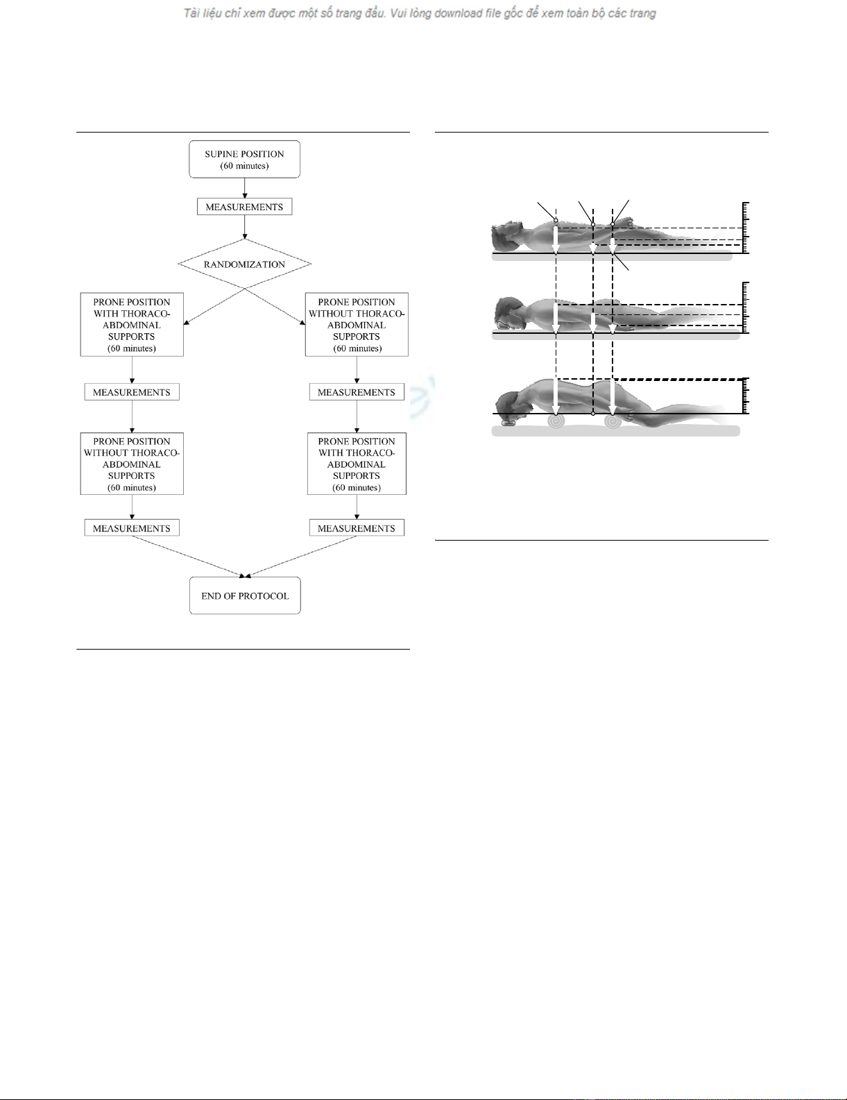
Figure 2
Patients' positions and contact pressuresPatients' positions and contact pressures. Patients' positions used in
the study: supine (top), prone without supports (center) and prone with
thoraco-pelvic supports (bottom). The mean contact pressures (meas-
ured with pressure transducers in four healthy volunteers) are also indi-
cated by white arrows. Table 2 shows detailed contact pressures at
different sites and global values.
30
0
Supine
15 8
5
navel pubes
inter-nipple
line
sacrum
Prone
without
support
Contact Pressure (cmH O)
2
30
17 11 4.5
Prone
with
support
0
30
2829 0
0
15
8
5
17
11
4.5
29
28

Available online http://ccforum.com/content/10/3/R87
Page 5 of 9
(page number not for citation purposes)
pubis increased significantly compared with those in the prone
position without supports (29.0 ± 6.5 versus 17.0 ± 7.4
cmH2O and 28.3 ± 8.9 versus 4.5 ± 4.2 cmH2O, respectively)
whereas the contact pressure at the abdominal wall surface
was zero because the abdomen remained suspended.
End-expiratory lung volume and respiratory mechanics
The EELVs and the mechanics of the respiratory system, par-
titioned into the chest wall and lung components, are summa-
rized in Table 3. Shifting the patients from the supine to the
prone position, without supports, led to a decreasing trend of
chest wall compliance and to a significant increase in lung
compliance. Adding the thoraco-pelvic supports in the prone
position led to a further significant decrease in chest wall com-
pliance and a significant increase in pleural pressure. We
found no sequence effect (that is, prone after supine or supine
after prone; see Figure 1) on lung volumes and respiratory
mechanics variables.
Gas exchange
Table 4 summarizes the gas exchange variables in the supine
and in the prone position with and without thoraco-pelvic sup-
ports. As shown, the oxygenation-related variables in the arte-
rial and central venous blood improved significantly in shifting
the patients from supine to prone without thoraco-pelvic sup-
ports. The application of thoraco-pelvic supports did not lead
to any further significant change. No significant differences
were observed in CO2-related variables between the supine
and the prone position with or without thoraco-pelvic sup-
ports. We found no sequence effect on gas exchange
variables.
Hemodynamics
The application of thoraco-pelvic supports caused a signifi-
cant increase in heart rate and a decrease in stroke volume
index and in pulmonary artery pressures, in comparison with
the prone position without supports. The other hemodynamic
variables (notably cardiac index and systemic vascular resist-
Table 2
Detailed contact pressures at different sites and global values
Position Units Supine Prone without supports Prone with supports
Thorax cmH2O 15.4 ± 4.1 17.0 ± 7.4 29.0 ± 6.5a,b
Abdomen cmH2O 5.8 ± 2.9 11.0 ± 1.8a0.0 ± 0.0a,b
Sacrum/pubis cmH2O 8.0 ± 5.7 4.5 ± 4.2 28.3 ± 8.9a,b
Global cmH2O 9.7 ± 5.8 10.8 ± 7.0 19.1 ± 15.2a,b
Results are means ± SD. ap ≤ 0.05 compared with supine; bp ≤ 0.05 compared with prone without supports.
Table 3
Lung volumes and respiratory mechanics
Variable Units Supine Prone without support Prone with support
Tidal volume (VT) ml 565.3 ± 160.5 577.6 ± 185.3 593.4 ± 200.7
Tidal volume per kg IBW (VT/kgIBW) ml/kg 7.2 ± 1.4 7.4 ± 1.6 7.6 ± 1.8
EELV l 1.12 ± 0.49 1.00 ± 0.26 1.07 ± 0.31
Mean airway pressure cmH2O 15.1 ± 2.1 15.6 ± 2.3 15.7 ± 2.1a
Plateau pressure (Pplat)cmH
2O 22.4 ± 4.3 22.1 ± 3.8 23.6 ± 4.5a,b
Respiratory system compliance ml/cmH2O 52.1 ± 17.6 52.9 ± 18.8 49.3 ± 18.1b
Lung compliance ml/cmH2O 71.5 ± 23.8 93.5 ± 47.3a102.0 ± 47.0a
Chest wall compliance ml/cmH2O 235.2 ± 152.5 158.1 ± 77.8 102.5 ± 38.0a,b
Transpulmonary pressure changeccmH2O 8.4 ± 2.2 7.1 ± 2.2a6.6 ± 2.3a
Pleural pressure changeccmH2O 3.2 ± 1.9 4.3 ± 1.9 6.1 ± 1.8a,b
Gastric pressure cmH2O 13.4 ± 4.0 14.3 ± 3.5 13.2 ± 4.3
Gastric pressure changeccmH2O 2.6 ± 0.8 3.4 ± 1.1a4.5 ± 1.9a,b
Transdiaphragmatic pressure cmH2O 0.6 ± 2.0 0.9 ± 1.6 1.6 ± 1.7
Bladder pressure cmH2O 12.0 ± 2.8 14.5 ± 3.4a14.5 ± 3.7a
Results are means ± SD. IBW, ideal body weight; EELV, end-expiratory lung volume. ap ≤ 0.05 compared with supine; bp ≤ 0.05 compared with
prone without supports; cdifference between end-inspiration and end-expiration


![PET/CT trong ung thư phổi: Báo cáo [Năm]](https://cdn.tailieu.vn/images/document/thumbnail/2024/20240705/sanhobien01/135x160/8121720150427.jpg)


%20--%3e%3cdefs%3e%3cstyle%3e%20.st0%20{%20fill:%20%23fff;%20}%20.st1%20{%20fill:%20%237800fa;%20}%20%3c/style%3e%3c/defs%3e%3cpath%20class='st1'%20d='M117.78,12.18H43.11c2.9,3.47,4.65,7.94,4.65,12.82,0,5.6-2.3,10.66-6.01,14.29h76.02l7.22-13.56-7.22-13.56Z'/%3e%3cg%3e%3cpath%20class='st0'%20d='M53.58,26.17h-.59v-1.46h.59v-4.96h2.83c1.78,0,2.67.94,2.67,2.82v5.76c0,1.87-.89,2.81-2.67,2.81h-2.83v-4.96ZM55.36,21.37v3.34h1.1v1.46h-1.1v3.34h1.01c.61,0,.91-.37.91-1.1v-5.93c0-.74-.3-1.1-.91-1.1h-1.01Z'/%3e%3cpath%20class='st0'%20d='M65.99,31.14h-1.8l-.31-2.07h-2.19l-.31,2.07h-1.64l1.82-11.39h2.62l1.82,11.39ZM65.28,18.04c-.25.46-.51.77-.75.94-.21.15-.47.22-.79.22-.26,0-.57-.07-.92-.22l-.38-.15c-.14-.05-.26-.07-.37-.07-.3,0-.53.18-.71.54l-.91-.68c.25-.46.51-.77.75-.94.21-.14.48-.21.79-.21.26,0,.57.07.92.21l.38.15c.14.05.26.07.37.07.3,0,.53-.18.71-.54l.91.68ZM61.91,27.52h1.73l-.87-5.76-.87,5.76Z'/%3e%3cpath%20class='st0'%20d='M74.53,26.89v1.52c0,1.91-.89,2.86-2.67,2.86s-2.67-.95-2.67-2.86v-5.93c0-1.91.89-2.86,2.67-2.86s2.67.95,2.67,2.86v1.11h-1.69v-1.22c0-.75-.31-1.12-.93-1.12s-.93.37-.93,1.12v6.15c0,.74.31,1.11.93,1.11s.93-.37.93-1.11v-1.63h1.69Z'/%3e%3cpath%20class='st0'%20d='M81.4,31.14h-1.8l-.31-2.07h-2.19l-.31,2.07h-1.64l1.82-11.39h2.62l1.82,11.39ZM75.9,19.2l1.52-1.91h1.71l1.51,1.91h-1.61l-.76-.95-.75.95h-1.61ZM77.32,27.52h1.73l-.87-5.76-.87,5.76ZM83.1,15.99l-1.76,1.91h-1.26l1.17-1.91h1.86Z'/%3e%3cpath%20class='st0'%20d='M84.86,19.75c1.78,0,2.67.94,2.67,2.82v1.48c0,1.87-.89,2.81-2.67,2.81h-.85v4.28h-1.79v-11.39h2.64ZM84.01,21.37v3.86h.85c.58,0,.87-.36.87-1.08v-1.71c0-.71-.29-1.07-.87-1.07h-.85Z'/%3e%3cpath%20class='st0'%20d='M93.51,19.75c1.78,0,2.67.94,2.67,2.82v1.48c0,1.87-.89,2.81-2.67,2.81h-.85v4.28h-1.79v-11.39h2.64ZM92.66,21.37v3.86h.85c.58,0,.87-.36.87-1.08v-1.71c0-.71-.29-1.07-.87-1.07h-.85Z'/%3e%3cpath%20class='st0'%20d='M98.8,31.14h-1.79v-11.39h1.79v4.88h2.03v-4.88h1.83v11.39h-1.83v-4.88h-2.03v4.88Z'/%3e%3cpath%20class='st0'%20d='M105.36,24.55h2.46v1.62h-2.46v3.34h3.09v1.63h-4.88v-11.39h4.88v1.63h-3.09v3.18ZM108.17,17.29l-1.76,1.91h-1.26l1.17-1.91h1.86Z'/%3e%3cpath%20class='st0'%20d='M112.2,19.75c1.78,0,2.67.94,2.67,2.82v1.48c0,1.87-.89,2.81-2.67,2.81h-.85v4.28h-1.79v-11.39h2.64ZM111.35,21.37v3.86h.85c.58,0,.87-.36.87-1.08v-1.71c0-.71-.29-1.07-.87-1.07h-.85Z'/%3e%3c/g%3e%3ccircle%20class='st1'%20cx='25'%20cy='25'%20r='20'/%3e%3cpath%20class='st0'%20d='M32.78,19.27c2.92,0,4.43,2.55,5.28,5.33l.71,2.17c.14.38-.33.75-.71.75h-5.61c.19-.33.24-.71.09-1.08l-.75-2.45c-.43-1.32-.99-2.64-1.79-3.77.75-.57,1.65-.94,2.78-.94h0ZM25,18.38c3.25,0,4.9,2.78,5.89,5.89l.76,2.45c.14.42-.33.8-.8.8h-11.69c-.42,0-.94-.38-.8-.8l.75-2.45c.99-3.11,2.64-5.89,5.89-5.89h0ZM25,11.35c1.74,0,3.11,1.37,3.11,3.11s-1.37,3.11-3.11,3.11-3.11-1.41-3.11-3.11,1.41-3.11,3.11-3.11h0ZM17.27,19.27c1.08,0,1.98.38,2.73.94-.8,1.13-1.37,2.45-1.74,3.77l-.8,2.45c-.14.38-.05.75.09,1.08h-5.56c-.42,0-.9-.38-.75-.75l.71-2.17c.9-2.78,2.41-5.33,5.33-5.33h0ZM17.27,12.91c1.51,0,2.78,1.27,2.78,2.83s-1.27,2.83-2.78,2.83-2.83-1.27-2.83-2.83,1.27-2.83,2.83-2.83h0ZM32.78,12.91c1.56,0,2.78,1.27,2.78,2.83s-1.23,2.83-2.78,2.83-2.83-1.27-2.83-2.83,1.27-2.83,2.83-2.83h0ZM27.07,28.56v.09c0,.57-.24,1.08-.61,1.46h0v.05c-.38.33-.9.57-1.46.57s-1.08-.24-1.46-.61h0c-.38-.38-.61-.9-.61-1.46v-.09h1.41v.09c0,.19.05.38.19.47v.05c.09.09.28.19.47.19s.38-.09.47-.19v-.05c.14-.09.24-.28.24-.47t-.05-.09h1.41ZM30.99,28.56v.09c0,1.65-.66,3.16-1.74,4.24-1.08,1.08-2.59,1.79-4.24,1.79s-3.16-.71-4.24-1.79l-.05-.05c-1.04-1.08-1.7-2.55-1.7-4.2v-.09h1.41v.09c0,1.27.47,2.4,1.27,3.25h.05c.85.85,1.98,1.37,3.25,1.37s2.4-.52,3.25-1.37c.85-.8,1.37-1.98,1.37-3.25v-.09h1.37ZM34.99,28.56v.09c0,2.78-1.13,5.28-2.92,7.07-1.79,1.79-4.29,2.92-7.07,2.92s-5.23-1.13-7.07-2.92c-1.79-1.79-2.92-4.29-2.92-7.07v-.09h1.41v.09c0,2.4.94,4.53,2.5,6.08,1.56,1.56,3.72,2.5,6.08,2.5s4.52-.94,6.08-2.5c1.56-1.56,2.5-3.68,2.5-6.08v-.09h1.41Z'/%3e%3c/svg%3e)