
JOURNAL OF 108 - CLINICAL MEDICINE AND PHARMACY Vol. 19 - Dec./2024 DOI: https://doi.org/10.52389/ydls.v19ita.2526
122
The evaluation of ultrasound-guided core biopsy in
detection of abnormal cervical lymph nodes
Vu Thi Hoa, Le Huy Thai*, Vu Thi Thu Lan,
Le Thi Loan, Nguyen Quynh Tu, Phan Thi Nga,
Nguyen Thi Thanh Tam, Tran Thi Thanh Nga,
Nguyen Thi Giang and Ngo Tien Quan
108 Mi
litary Central Hospital
Summary
Objective: To assess the value of routine ultrasound (US) imaging and histopathological results of
ultrasound-guided core needle biopsy (US-CNB) of abnormal cervical lymphadenopathy. Subject and
method: From September 2022 to August 2023, a total 112 patients with clinical suspected cervical
lymph nodes (CLNs) and/or have suspected signs on US (width ≥ 5mm, round in shape and absent hilus
of CLNs) underwent US-CNB at 108 Military Central Hospital. Result: Among 112 patients, there were 56
metastatic lymph nodes, 10 lymphomas, 10 tuberculous and 32 nonspecific inflammatory lymph nodes.
Level IV nodes included benign and malignant lesions was predominant. In the group of malignant
CLNs: Irregular margin, absence of hilum and hypoechogenicity were found in 65.7%, 70% and 94.3%
respectively, these proportions were significantly greater than that of benign group, with p<0.05.
Comparison of US and histopathology of CLNs diagnosis: The sensitivity, specificity, positive predictive
value, negative predictive value were 88.6%, 66.7%, 81.6%, 77.8%, respectively, when there were ≥ 2
suspected signs. Conclusion: Ultrasound is often considered as the first imaging diagnostic and valuable
tool for detecting suspicious CLNs due to its convenience, non-invasiveness and cost-effectiveness, to
helps reduce unnecessary interventions for benign lymph nodes. US-CNB is a minimally invasive
technique that allows accurate diagnosis of the lymph node's histopathology.
Keywords: Ultrasound, core needle biopsy, lymph node.
I. Background
Metastatic cervical lymphadenopathy is quite
common in patients with head and neck cancers or
cancers outside this region. For patients with
squamous cell carcinoma of the head and neck, the
presence of metastatic CLNs reduces the 5-year
survival rate to 50% and contralateral CLNs metastasis
decreases the 5-year survival rate to 25%1. Therefore,
evaluating metastatic CLNs plays a crucial role in
cancer patients, aiding in prognosis and optimal
treatment. Treatment and prognosis depend on the
Received: 19 September 2023, Accepted: 16 January 2024
*Corresponding author: thailehuymch@gmail.com -
108 Millitary Central Hospital
histopathology and stage of the cancer. Additionally,
cervical lymphadenopathy is also a common site for
lymphoma, tuberculosis and other benign CLNs
disorders such as Kikuchi's disease, Kimura's disease
and Rosai-Dorfman disease1.
Historically, the primary method for diagnosing
and evaluating CLNs relied on clinical examination,
which often led to the oversight of small or deep-
seated nodes. US, utilizing high-frequency probes,
has emerged as a non-invasive and flexible
diagnostic approach. Through US, we can assess the
size, structure, interrelationships between nodes
and adjacent structures, vascularization and capsule
disruption... Therefore, US not only plays a crucial
role in distinguishing between benign and
malignant but also monitors the effect of radio-

JOURNAL OF 108 - CLINICAL MEDICINE AND PHARMACY Vol. 19 - Dec./2024 DOI: https://doi.org/10.52389/ydls.v19ita.2526
123
chemotherapy of metastatic nodes as well as for the
detection of recurrent lymph node metastases in the
neck. The recent reports have demonstrated that US
and US-CNB of CLNs are simple and safe procedures
for detecting and diagnosing abnormal CLNs2. In
Viet Nam, there are a number of studies of thyroid
cancer but there is a few of reports study about
other malignant CLNs. So we performed this study
was to assess the value of routine ultrasound
imaging and histopathological results of ultrasound-
guided core neede biopsy in the diagnosis of
abnormal cervical lymphadenopathy.
II. SUBJECT AND METHOD
2.1. Subject
A total 112 patients with abnormal CLNs on
clinic and on US examinations, were performed US-
CNB. Clinical suspicions include stiff, irregular
margin and unmovable lymph nodes. According to
Vassallo P et al., ultrasound signs of suspected CLNs
include: Width ≥ 5mm, a round shape (width/length
ratio ≥ 0.5) and the absence of a hilum structure of
the lymph node3.
2.2. Method
The design of our study was a cross-sectional
descriptive study. Before the biopsy, we performed
US of CLNs by the GE Voluson S8 machines with a 5-
12MHz linear transducer. Using US to evaluate the
location, size, echogenicity, calcification, necrosis,
hilum and vascularization of CLNs. US-CNB was
performed by interventional radiologists free-
handed with a 13cm-long manual 14-18G needle
(US Biopsy, Franklin, IN, Japan). US was used to point
the biopsy location, carefully identifying the needle
path, avoiding major blood vessels and nerve
structures. The skin was disinfected. The physician
washed their hands, put on gloves and prepared a
sterile drape with a hole for the biopsy site, covering
the probe with a sterile endoscopy nylon. Local
anaesthesia was administered by using 1%
lidocaine. The biopsy needle was passed through
the lymph node's capsule. Then, the inner cutting
needle was withdrawn while simultaneously
advancing the cutting needle to obtain the tissue
sample. At least 3-6 tissue specimens were placed in
a specimen container. Potential bleeding was
stopped by compression. The puncture site was
disinfected and pressure dressing was applied. To
assess any procedure-related complications, US
imaging was performed again 30-45 minutes after
the biopsy. Furthermore, patients were encouraged
to communicate with the clinical physician or
directly contact the biopsy-performing physician
regarding any signs of pain or swelling at the
intervention site4.
2.3. Statistical analysis
The data were collected and processed using
SPSS 20.0 software. Statistical analysis was
performed using Chi-square Tests and Fisher’s Exact
Test. The significance level was set at p<0.05. Base
on three characteristics (margin, echogenicity and
absent of hilum of lymph node) and histopathology
report, we calculated the sensitivity, specificity,
positive predictive value and negative predictive
value of ultrasound.
2.4. Ethical standards
Consent was obtained from all participants
through written informed consent after providing
detailed explanations before the biopsy.
III. RESULT
3.1. Patient’s general characteristics
Table 1. Patient’s general characteristics
Characteristics Value
Age 52.9 ± 16.2 Min 12
Max 87
Gender Male 75 (67%)
Female 37 (33%)
In our study, the patient’s average age was 52.9
± 16.2 years old. Majority of patients were male with
the rate of 67%.
JOURNAL OF 108 - CLINICAL MEDICINE AND PHARMACY Vol. 19 - Dec./2024 DOI: https://doi.org/10.52389/ydls.v19ita.2526
124
3.2. Ultrasound image characteristics of cervical lymph node
Table 2. Correlation of characteristics of cervical lymph node ultrasound
with histopathological results
Pathology
Characteristics Malignant CLNs Benign CLNs Sum p
Width <8mm 8 (11.4%) 10 (23.8%) 18 (16.1%) 0.11
≥ 8mm 62 (88.6%) 32 (76.2%) 94 (83.9%)
Shape Width/Long < 0.5 15 (21.4%) 13 (31%) 28 (25%) 0.27
Width/Long ≥ 0.5 55 (78.6%) 29 (69%) 84 (75%)
Margin Regular 24 (34.3%) 38 (90.5%) 77 (68.8%) 0.00
Irregular 46 (65.7%) 4 (9.5%) 35 (31.2%)
Echogenicity
Hypoechoic 49 (70%) 25 (59.5%) 39 (34.8%)
0.00 Hyperechoic 14 (20%) 1 (2.4%) 50 (44.6%)
Heterogeneous echo 7 (10%) 16 (38.1%) 23 (20.5%)
Calcification No 67 (95.7%) 42 (100%) 109 (7.3%) 0.29
Yes 3 (4.3%) 0 (0%) 3 (2.7%)
Necrosis No 63 (90%) 32 (76.2%) 95 (84.8%) 0.06
Yes 7 (10%) 10 (23.8%) 17 (15.2%)
Hilum Absent 66 (94.3%) 26 (61.9%) 92 (82.1%) 0.00
Present 4 (5.7%) 16 (38.1%) 20 (17.9%)
Vascularization
No angiogenesis 50 (71.4%) 33 (78.6%) 83 (74.1%)
0.23
Central 3 (4.3%) 4 (9.5%) 7 (6.2%)
Peripheral 8 (11.4%) 1 (2.4%) 9 (8%)
Central and peripheral 9 (12.9%) 4 (9.5%) 13 (11.6%)
p<0.05 is considered significant. Chi-square and Fisher Exact tests were used for analysis.
In group of malignant CLNs, irregular margin, absence of hilum and hypoechogenicity was found in
65.7%, 70% and 94.3% respectively, these proportions were greater than rates of benign group, the variance
had statistical significance with p<0.05. The differences in nodal size, shape, calcification, necrosis and
vascularization characteristics between benign and malignant CLNs were not statistically significant.
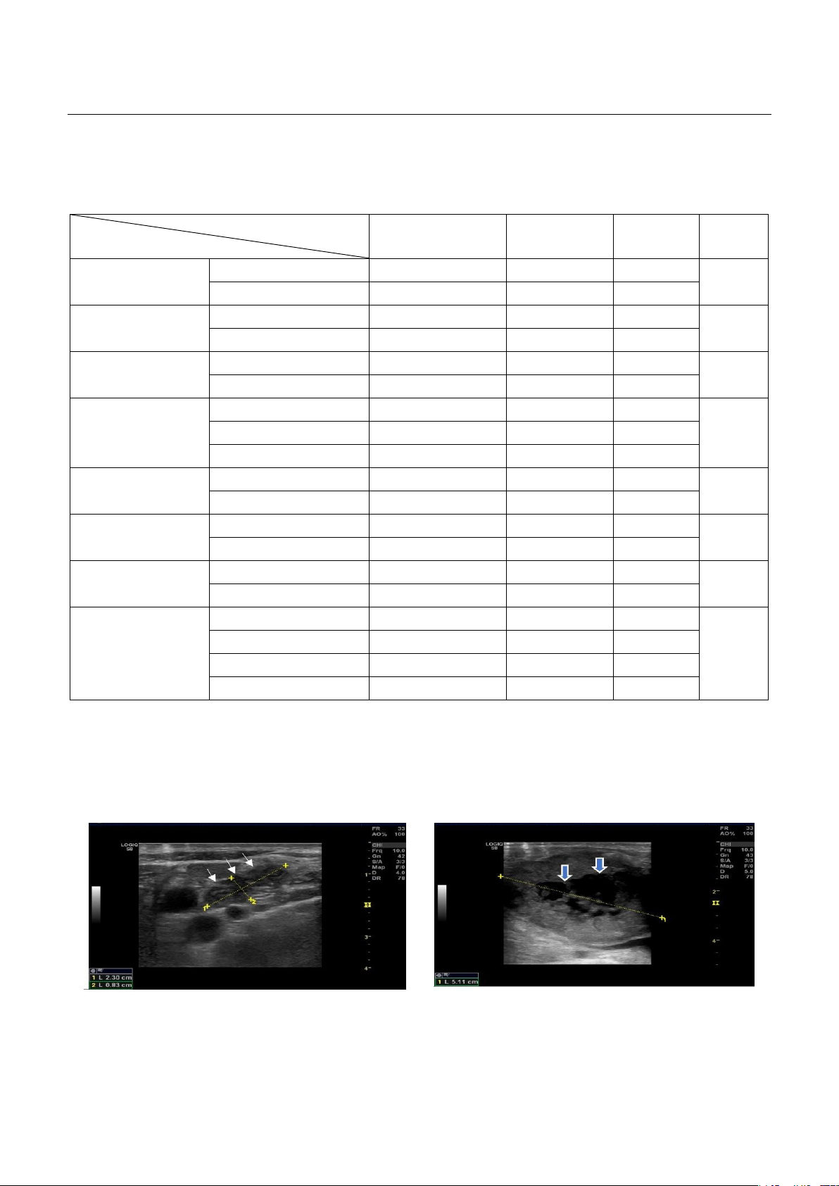
Figure 1. A hypoechoic mestastatic lymph node with
oval in shape, absence of echogenic hilus, irregular
margin (arrows).
Figure 2. A hypoechoic mestastatic lymph node with
round in shape, absence of echogenic hilus, irregular
margin and necrosis (arrows)
JOURNAL OF 108 - CLINICAL MEDICINE AND PHARMACY Vol. 19 - Dec./2024 DOI: https://doi.org/10.52389/ydls.v19ita.2526
125
3.3. Histopathological characteristics and ultrasound-guided cervical lymph node biopsy results
Complications of the ultrasound-guided cervical lymph node biopsy: There were no cases of bleeding,
death, nerve damage or infection along the biopsy tract in our study.
Table 3. Histopathological results of patients
Patients (n) Percentage (%) Sum (%)
Malignant lymph node Metastase 56 50 62.5
Lymphoma 14 12.5
Benign lymph node Tuberculosis 10 8.9 37.5
Inflammatory 32 28.6
In our study, metastatic CLNs were predominant, accounting for 50%, while the minority were cases of
tuberculous lymph node accounting for 8.9%.
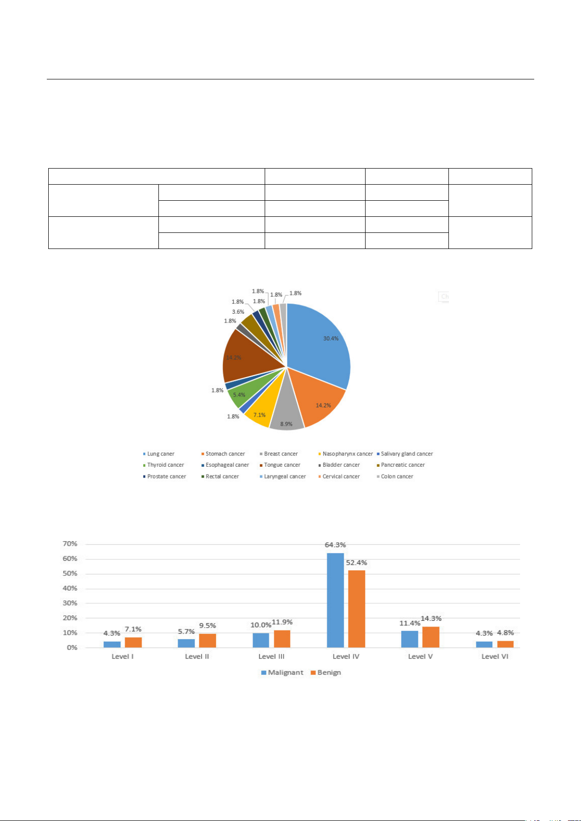
Chart 1. Distribution of metastatic cancers in cervical lymph nodes
Among the 112 patients included in the study, metastases originating from lung cancer were
predominant, constituting 30.4% of cases.
Chart 2. Distribution of cervical lymph node levels according to Robbins
Cervical lymph node level IV were predominant for both malignant and benign lymph nodes with the
rate of 64.3% and 52.4% respectively.

JOURNAL OF 108 - CLINICAL MEDICINE AND PHARMACY Vol. 19 - Dec./2024 DOI: https://doi.org/10.52389/ydls.v19ita.2526
126
3.4. The value of ultrasound in diagnosing abnormal cervical lymph nodes
Table 4. Sensitivity (Se), specificity (Sp), positive predictive value (PPV),
and negative predictive value (NPV) of ultrasound images
Value
Characteristic Se (%) Sp (%) PPV (%) NPV (%)
≥ 1 sign 100% 7.1% 64.2% 100%
≥ 2 signs 88.6% 66.7% 81.6% 77.8%
≥ 3 signs 41.4% 95.2% 93.5% 49.4%
To achieve both high sensitivity and high
specificity, it was advisable to choose ≥ 2 suspicious
signs.
IV. DISCUSSION
Regarding the high proportion of CLNs group IV
it can be explained by the predominance of
malignant CLNs, accounting for 50% in our study.
These metastatic CLNs often involve various types of
cancers. According to Ellison's research, CLNs level
IV play a significant role in the lymphatic drainage of
the chest, lungs, and esophagus...5. They are
commonly affected by both malignant and benign
CLNs. Among the metastatic group, lung cancer is
the most common because it is the second most
common cancer in Vietnam, following liver cancer,
according to Globocan 2018 statistics. Regarding the
nodal size and shape, although larger CLNs tend to
have a higher likelihood of malignancy, reactive and
tuberculous CLNs can also be large and appear
round in shape. Moreover, even early-stage
metastatic CLNs can be very small. It is similar to the
findings of Ying M et al6. Therefore, nodal size plays
a more crucial role in monitoring lymphadenopathy
than differing benign or malignant nature.
According to Vassallo P et al3, eccentric cortical
hypertrophy had been a useful sign for identifying
CLNs with malignant potential before changing
shape. This is due to early-stage cancer cells tend to
develop in a specific region of CLNs, causing an
eccentric shift in the axis, while inflammatory CLNs
tend to spread diffusely. The presence of the hilum
is often associated with benign CLNs; however,
early-stage metastatic lymph node can also be
present of hilum. In our study, presence of hilum
was observed in 5.7% of malignant CLNs, which is
consistent with the findings of other authors 4-
51%3,6-8. In addition, tuberculous CLNs are typically
associated with absent of hilum on ultrasound1, 9.
Malignant CLNs at an early stage, before capsule
invasion or rupture, often have more distinct and
well-defined borders similar to benign nodes.
Because cancer cells infiltrate and disrupt the
internal lymph node structure, causing a loss of
echogenicity within the lymph node. When cancer
cells invade and rupture the capsule, the margin
may appear irregular. However, the nature of CLNs
margin is not considered a definitive criterion for
distinguishing between benign and malignant CLNs,
according to Ahuja AT, Ying M1, 6.
Regarding calcification, our study encountered
very few cases, primarily due to the low incidence of
metastatic CLNs from thyroid cancer (only 3 cases).
In contrast, metastatic CLNs from other cancers
often exhibited coarser calcifications when there
was a history of recurrent bleeding in CLNs. This can
be attributed to the relatively low demand for
biopsy in cases of thyroid cancer metastasis, as most
clinicians rely on fine-needle aspiration combined
with evidence of malignant changes in the thyroid
gland for diagnosis. Moreover, the assessment of
neovascularization in our study did not yield
statistically significant results. This could be because
malignant CLNs tend to generate new blood vessels
as cancer cells produce angiogenic factors, leading
to increased vascularity within CLNs. However, acute
inflammatory lymph nodes can also exhibit
numerous vascular signals. These values are crucial
in assessing the diagnostic accuracy of US in
detecting abnormal CLNs, helping clinicians make
informed decisions regarding further evaluation and
intervention. Therefore, using two out of the three


![Bài giảng Cập nhật vấn đề hồi sức bệnh tay chân miệng nặng [mới nhất]](https://cdn.tailieu.vn/images/document/thumbnail/2025/20250920/hmn03091998@gmail.com/135x160/23301758514697.jpg)