5
Chapter 2 - SUBJECTS AND METHODS
2.1. Studying subjects
Severe cerebral infarction patients above the cerebellum tent were
treated at Strokecenter-Central MilitaryHospital108from 9/2013 – 6/2017.
2.1.1. Criteria for selecting a patient
The patients was diagnosed as stroke according to the World Health
Organization (1989) stroke definition, arriving at the hospital 72 hours
prior to the onset of c erebral infarction. Images of hemispherical infarction
on CT/MRI/ Severe nerve damage with NIHSS≥15 score (if the patient was
hospitalized prematurely, the damage on the first CT was unknown, the
patient would be diagnosed for a second time on CT. Patients were divided
into 2 groups: mechanical ventilation group and non mechanical ventilation
group.
2.1.2. Exclusion criteria
History of stroke with mRS score> 2 points, patients with severe
medical conditions such as liver failure, severe kidney failure, cancer,
COPD,…
2.2. Research methods:
2.2.1. Study design:Progressive, description, follow-up study
2.2.2.Sample size:
Sample size is determined by formula:
p (1-p) n = Sample size to study
n = Z
2
(1-α/2)
-------- Z
2
(1-α/2)
: At the probability level 95% (Z =1,96)
d
2
d: The desired accuracy (d = 0,05)
p: Estimated ratio, the rate ofcerebral
infarctionpatients requiring mechanical
ventilationin previous studies, p= 0,11.
→ Based on the above formula, the estimated patient sample sizeis 150.
In the period of taking data from 9/2013 – 6/2017, we collected 166
patients including 84 ventilated severe cerebral infarction patients and 82
severe cerebral infarction patients without mechanical ventilation.
2.2.3. Research variables
6
Clinical variables: gender, age, medical history, time of admission, pulse,
blood pressure, temperature, level of consciousness at admission on
Glasgow scale, NIHSS score, degree of paralysis, language disorder,
sensory disorders, urinaryincontinence, pupil abnormalities, light reflexes,
head-eye deviation, progression of symptoms, related mechanical
ventilation complications.
Subclinical variables: hematology, biochemistry, coagulation, arterial blood
gases.
Imaging variables: CT, CTA, DSA: parenchymal, artery damage, midline shift.
Variables of treatment outcome: death, live, mRS at discharge, 1 year.
2.2.4. Research contents
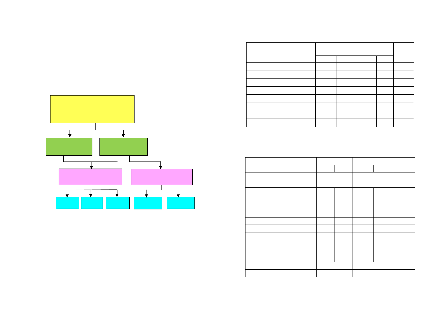
Patients were divided into 2 groups: MVgroup and non MVgroup. The
patients were divided into two groups, the MVgroup and the non MV group.
MVis indicated when at least one of the following criteria: Glasgow ≤ 8, loss
of reflexes protects airway causing mucus congestion, patients with
consciousness disorders, stimulation must use safety drugs strong spirit causes
respiratory depression, patients with respiratory failure, circulatory failure.
Describe the clinical and paraclinical features with analysis and comparison
between two groups of MV and non MV group to highlight c linical and
subclinical characteristics of patients requiring MV.
Identify factors related to MV, factors related to prognosis of death at
hospital discharge and mRS 0-3 at 1 year. The supposedly relevant
variables are included in univariate analysis and logistic multivariate
regressions to find meaningful prognostic factors.
2.3. Data analysis
Data processing using SPSS 16.0 software.
Description of clinical, subclinical, imaging features: neurological signs
on onset, on admission and during hospitalization, intubation designation,
subclinical characteristics, imaging, complications during MV, treatments
and outcome.