
Open Access
Available online http://ccforum.com/content/10/6/R157
Page 1 of 7
(page number not for citation purposes)
Vol 10 No 6
Research
Activated protein C improves intestinal microcirculation in
experimental endotoxaemia in the rat
Christian Lehmann1, Konrad Meissner2, Andreas Knöck1, Stephan Diedrich1, Dragan Pavlovic1,
Matthias Gründling1, Taras Usichenko1, Michael Wendt1 and Jürgen Birnbaum3
1Klinik und Poliklinik für Anästhesiologie und Intensivmedizin, Ernst Moritz Arndt University, Fr.-Loeffler-Str. 23a, D-17475 Greifswald, Germany
2Washington University Medical Center, Department of Anesthesiology, 660 S. Euclid Ave., St. Louis, MO 63110, USA
3Charité – Universitätsmedizin Berlin, Kliniken für Anästhesiologie und operative Intensivmedizin, Campus Charité Mitte, Charitéplatz 1, D-10117
Berlin, Germany
Corresponding author: Christian Lehmann, christian.lehmann@uni-greifswald.de
Received: 7 Aug 2006 Revisions requested: 8 Sep 2006 Revisions received: 21 Sep 2006 Accepted: 13 Nov 2006 Published: 13 Nov 2006
Critical Care 2006, 10:R157 (doi:10.1186/cc5093)
This article is online at: http://ccforum.com/content/10/6/R157
© 2006 Lehmann et al; licensee BioMed Central Ltd.
This is an open access article distributed under the terms of the Creative Commons Attribution License (http://creativecommons.org/licenses/by/2.0),
which permits unrestricted use, distribution, and reproduction in any medium, provided the original work is properly cited.
Abstract
Introduction Successful treatment of severe sepsis and septic
shock remains a major challenge in critical care medicine. The
recently introduced recombinant human activated protein C
(APC) remarkably improved the outcome of septic patients. The
influence of APC on intestinal circulation is still poorly
understood. Therefore, the present study aimed to investigate
the effects of APC on intestinal microcirculation during
experimental endotoxaemia in rats by using intravital
microscopy.
Methods A total of 44 male Lewis rats were randomly assigned
to receive intravenous injections of 15 mg/kg
lipopolysaccharide alone (LPS) (n = 11) or LPS followed by
subsequent injection of 2 mg/kg recombinant human APC (LPS
+ APC) (n = 11), whereas control animals received either APC
(n = 11) or saline (n = 11). Animals underwent observations of
functional capillary density and leucocyte adherence on venular
endothelium in the microcirculation of the intestinal wall by
means of intravital fluorescence microscopy. Indicators of
macrocirculation as well as plasma levels of tumour necrosis
factor-α, interleukin (IL)-1β, IL-6, and IL-10 were measured.
Results Although APC administration of both LPS-treated and
control rats did not change macrocirculation or release of
inflammatory cytokines, it increased mucosal and muscular
functional capillary density (p < 0.001 and p < 0.05,
respectively) and reduced the number of firmly adhering
leucocytes in intestinal submucosal V1 and V3 venules (p <
0.01) in LPS + APC-treated compared with LPS-treated
animals, which did not receive APC. No remarkable differences
that could be attributed to APC treatment were observed
between the two control groups.
Conclusion APC administration during experimental
endotoxaemia improved intestinal microcirculation by protecting
functional capillary density as a measure of microvascular
perfusion and exerted anti-inflammatory effects by reducing
leucocyte adherence to the endothelium in submucosal venules.
Therefore, beneficial effects of APC in septic patients might be
due, in part, to improved intestinal microcirculation.
Introduction
Sepsis, severe sepsis, and septic shock represent progressive
stages of the same illness, in which a systemic response to an
infection mediated by endogenous mediators leads to a gen-
eralised inflammatory reaction in organs remote from the initial
insult and eventually to organ dysfunction and/or failure [1].
Impairment of gut perfusion is regarded as one important
mechanism in the development of sepsis. The splanchnic per-
fusion is reduced early in the course of any circulatory shock
[2]. The mucosa of the gut suffers most as it experiences a
high oxygen demand even in steady state [2]. Intestinal
mucosal hypoperfusion with subsequent ischaemia during
endotoxaemia might cause a breakdown of the gut barrier
function with translocation of bacteria and their toxins into the
systemic circulation, thus maintaining a 'gut-derived' septic
APC = activated protein C; FCD = functional capillary density; FITC = fluorescein isothiocyanate; HR = heart rate; IL = interleukin; i.v. = intravenous;
IVM = intravital fluorescence microscopy; LPS = lipopolysaccharide; MAP = mean arterial pressure; TNF-α = tumour necrosis factor-α; V1 = grade
I venule; V3 = grade III venule.

Critical Care Vol 10 No 6 Lehmann et al.
Page 2 of 7
(page number not for citation purposes)
state [3]. Gut mucosal hypoperfusion plays a major role in the
pathogenesis of ongoing sepsis and multiple organ dysfunc-
tion syndrome [4] because subsequent ischaemia leads to
translocation of endotoxin [5,6] and induces both vasocon-
striction and hypoperfusion of small intestinal microcirculation
[7]. A number of animal experiments using several different
agents have aimed to improve microcirculation, particularly of
the intestine, in septic conditions [4,6,8,9]. There is an
increasing body of evidence that activated protein C (APC)
exerts beneficial effects in the microcirculation. Human
plasma-derived and human cell-produced recombinant protein
C inhibits E-selectin-mediated cell adhesion to the vascular
endothelium [10]. APC also attenuated endotoxin-derived pul-
monary vascular injury in rats by inhibiting activated leucocytes
[11]. Recently, published studies investigated the effects of
APC on microcirculation during experimental endotoxaemia by
intravital fluorescence microscopy (IVM) [12,13]. They were
able to show that APC diminishes endotoxin-derived reduction
of functional capillary density (FCD) as well as leucocyte
adherence to the endothelium in dorsal skinfold chamber
preparations and in the mesentery, but they did not investigate
the intestinal wall. With respect to the role of the intestinal
microcirculation in sepsis [14], the aim of our study was to
evaluate the effects of APC administration during experimental
endotoxaemia in the terminal ileum wall of the rat by using IVM.
Materials and methods
Animals
After approval by the local standing committee on animal
experiments, a total of 44 male Lewis rats were used in the
experiments (body weight 250 ± 50 g; Department of Labora-
tory Animal Science, Karlsburg, Ernst Moritz Arndt University,
Greifswald, Germany). All experimental procedures were per-
formed according to German animal safety legislations. Ani-
mals were kept under 12-hour light/dark rhythmic conditions
(temperature 22°C, humidity 55% to 60%). Standard diet and
water were available ad libitum. After the experiment, all ani-
mals were euthanised by overdose of intravenous (i.v.)
pentobarbital.
Anaesthesia and preparation
Anaesthesia was induced via intraperitoneal administration of
60 mg/kg pentobarbital. Maintaining of anaesthesia was
achieved with repeated i.v. injections of 5 mg/kg pentobarbital
(Fagron GmbH & Co. KG (previously Synopharm GmbH & Co.
KG) Barsbüttel, Germany). With the animals positioned in a
supine position, polyethylene catheters (PE 50, internal diam-
eter 0.58 mm, external diameter 0.96 mm; Portex, brand of
Smiths Medical, Hythe, Kent, UK) were introduced into the left
external jugular vein and common carotid artery. A continuous
monitoring of arterial blood pressure and heart rate (HR) was
thereby undertaken (Hewlett-Packard monitor, Model 66S;
Hewlett-Packard, Saronno, Italy). All animals received a tra-
cheostomy to permit access to the airway. The animals spon-
taneously breathed room air. A specially tempered microscopy
bench served to maintain a continuous body temperature of
37°C ± 0.5°C. Subsequent to shaving and disinfection,
median laparotomy was performed from the xyphoid process
to the symphysis.
General protocol
The experiment started after a 15-minute equilibration period
following preparation. Animals were randomly assigned to one
of four groups (n = 11, respectively). In 22 animals, endotox-
aemia was induced by administration of 15 mg/kg lipopolysac-
charide (LPS) from Escherichia coli, serotype O111:B4
(Sigma-Aldrich, Steinheim, Germany). The 22 control animals
were given an equivalent amount of saline. Eleven animals out
of each group received 2 mg/kg APC (Drotrecogin alpha [acti-
vated], Xigris®; Lilly Deutschland GmbH, Bad Homburg, Ger-
many) immediately after endotoxin or saline administration,
respectively.
Intravital fluorescence microscopy
IVM was performed 2 hours after the onset of the experiment.
The examination was directed upon an isolated segment
(approximately 5 cm) of the terminal ileum proximal to the ileo-
caecal valve, held in place by a supporting device. A coverslip
served as a transparent cover. By means of this method,
approximately 1 cm2 of gut surface could be evaluated by
microscopy. Areas of the intestine not being examined were
covered with gauze and continuously superfused with isotonic
saline kept at 37°C to avoid dehydration and exposure to
ambient air. IVM was performed using the epifluorescent
microscope Axiotech Vario (Carl Zeiss, Jena, Germany), light
source HBO 50 (Carl Zeiss), oculars ×10 (Carl Zeiss), lens
×20/0.5 Achroplan (Carl Zeiss), filter type no. 20 (Carl Zeiss)
for examinations with Rhodamine 6G solution (Sigma-Aldrich),
filter type no. 10 (Carl Zeiss) for examinations with fluorescein
isothiocyanate (FITC)-albumin, a black-and-white CCD
(charge-coupled device) video camera (BC-12; AVT-Horn,
Aalen, Germany), an S-VHS video tape recorder (Panasonic
NV-SV120EG-S; Matsushita Audio Video GmbH, Lüneburg,
Germany), and a black-and-white monitor (PM-159; Ikegami
Electronics [Europe] GmbH, Neuss, Germany). Within the
described configurations, a total magnification of ×500 at the
14-inch monitor was achieved. Initially, staining of the leuco-
cytes was performed through the i.v. injection of 200 μl of
0.05% Rhodamine 6G solution. The microscope was then set
to focus on the submucosa of the prepared intestinal section.
Five visual fields containing non-branching, grade I stretching
venules (V1) over a length of at least 300 μm, as well as
another five visual fields revealing similar grade III venules (V3),
were observed and recorded for 30 seconds. Two hundred
microlitres of 5% FITC-albumin solution (Sigma-Aldrich) dis-
solved in normal saline was subsequently given to facilitate a
better evaluation of the capillary flow bed through the resultant
amplified contrast of the plasma. After focus setting, five video
sequences (30 seconds each) of random fields of the capillar-
ies within the longitudinal musculature as well as five fields of

Available online http://ccforum.com/content/10/6/R157
Page 3 of 7
(page number not for citation purposes)
the capillaries within the circular muscle were recorded. Then,
a section of the intestinal lumen (2 cm, antimesenteric) was
opened using a microcautery knife (Geiger Model-100; Gei-
ger Medical Technologies, Inc., Council Bluffs, IA, USA) to
facilitate the examination of the mucosa. Sections filled with
chymus were preferred to avoid heat damage of the opposing
mesenteric wall. After flushing with isotonic saline kept at body
temperature, the intestine was once again lifted and held by
the supporting device. Sections of the mucosa directly border-
ing the mesentery were examined to circumvent possible influ-
ences from microcauterisation. Again, five video sequences
(30 seconds each) of randomly chosen mucosa sections were
recorded. Evaluation of all the video sequences took place off-
line on a video monitor. Leucocyte adherence (the number of
leucocytes that during an observation period stayed immobile
for at least 30 seconds on an oblique, cylindrical endothelial
surface; units, n/mm2) and FCD (the length of capillaries with
observable erythrocyte perfusion in relation to a predeter-
mined rectangular field; units, cm/cm2 or cm-1) were deter-
mined according to Schmid-Schoenbein et al. [15].
Laboratory analysis
Blood samples (0.55 ml) were taken at the beginning and the
end of the experiments for arterial blood gas and haematocrit
analysis (ABL 330; Radiometer, Hamburg, Germany). Moreo-
ver, 280 μl of plasma was fractionated and stored at -70°C for
cytokine analysis (tumour necrosis factor-α [TNF-α], inter-
leukin [IL]-1β, IL-6, and IL-10) using Rat-Quantikine ELISA
[enzyme-linked immunosorbent assay] kits (R&D Systems
GmbH, Wiesbaden-Nordenstadt, Germany) according to the
manufacturer's instructions.
Statistical analysis
Data analysis was performed with a statistical software pack-
age (SigmaStat; Jandel Scientific, Erkrath, Germany). All data
were expressed as group means ± standard deviation and
analysed using a one-way analysis of variance followed by the
Newman-Keuls multiple comparison test. Mean arterial pres-
sure (MAP) and HR were analysed by a two-way analysis of
variance (repeated measures in the factor of time) followed by
the Newman-Keuls multiple comparison test. A p value of less
than 0.05 was considered significant.
Results
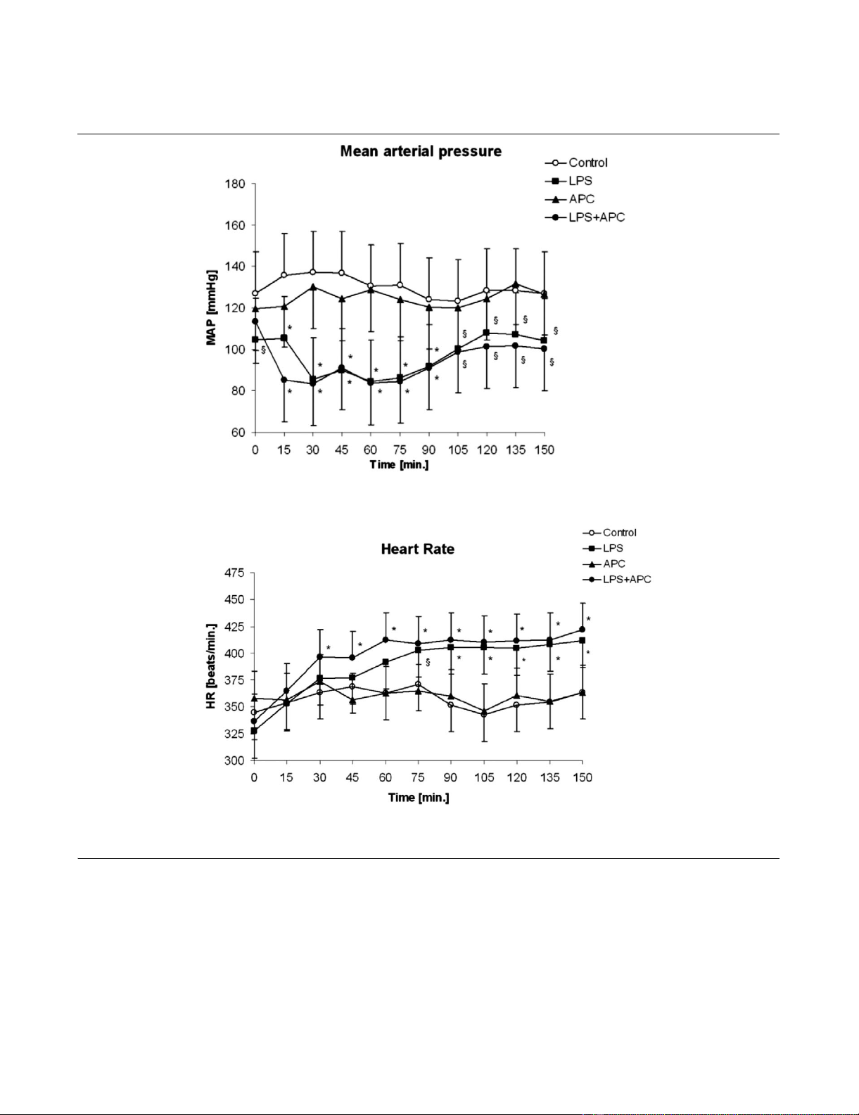
Haemodynamic changes in the macrocirculation
MAP and HR remained stable in the non-LPS control groups
(Figure 1). Endotoxin challenge resulted in a significantly
decreased MAP after 30 minutes (Figure 1a). MAP was stabi-
lised in both endotoxaemic groups two hours after LPS admin-
istration. LPS groups with and without APC treatment did not
differ in MAP or HR two hours after endotoxin challenge. HR
of the endotoxaemic groups was still significantly increased
compared with the control groups at this time point (Figure
1b).
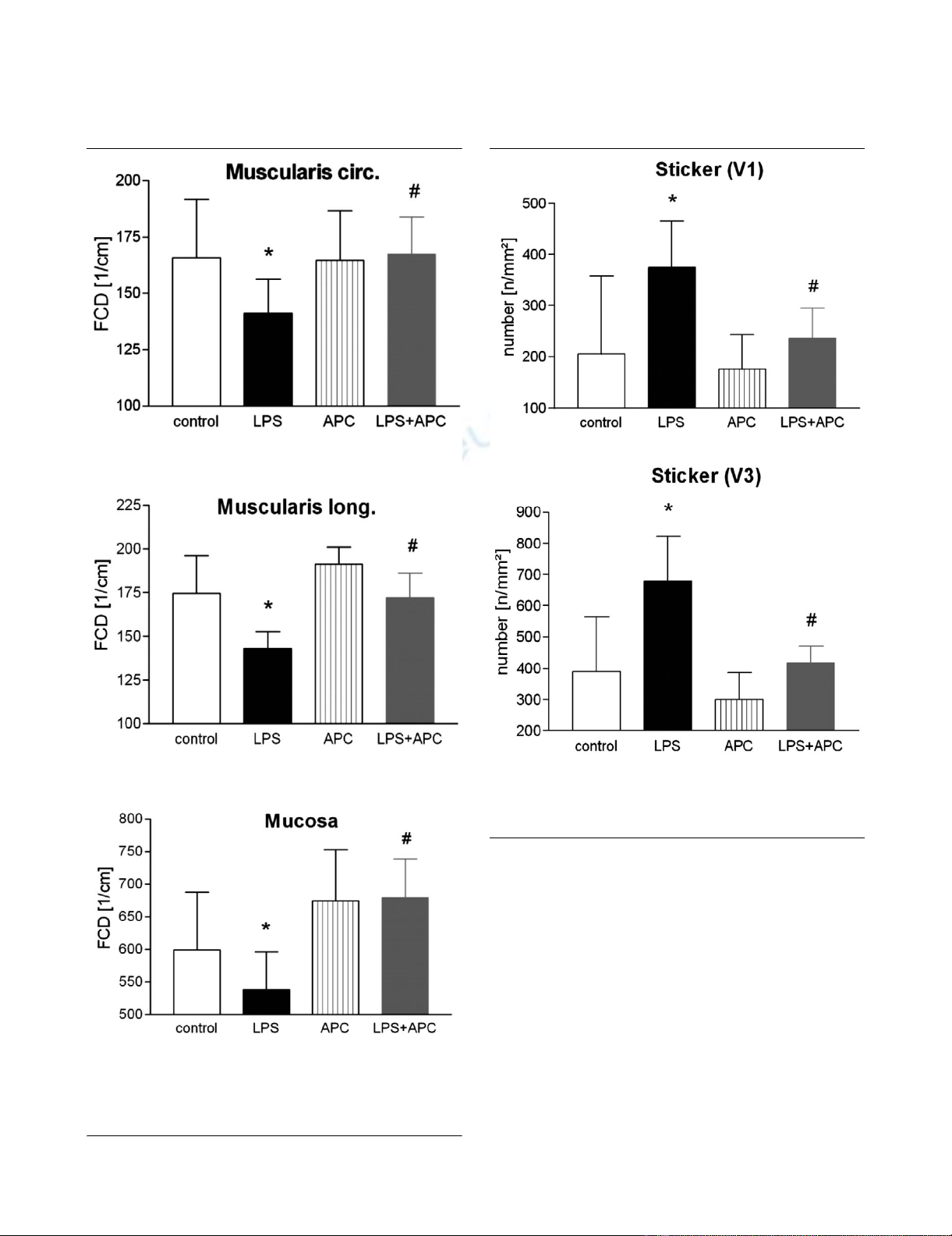
Functional capillary density
Changes in the FCD could be attributed to the treatment reg-
imens of the study. Two hours after endotoxin challenge, a sig-
nificant reduction of the FCD in both the circular and the
longitudinal muscular layers of LPS-treated animals were
observed. APC administration prevented the LPS-induced
decrease of mucosal and both muscular FCDs (all p < 0.001;
Figure 2).
Leucocyte adherence
Figure 3 shows the number of firmly adherent leucocytes two
hours after endotoxin challenge. In the untreated LPS group,
we saw an increase in the number of sticking leucocytes in the
postcapillary venules (+45% versus control group; p < 0.01).
In the collecting venules (V1), we saw similar effects as in the
V3 venule subpopulation (+43% versus control group; p <
0.001). In the V3 venules of the APC-treated animals, the
increase was significantly attenuated (-40% versus LPS
group; p < 0.01). There was also an attenuation of the
increase in the number of sticking leucocytes in the V1 venules
(p < 0.01 versus LPS group).
Blood gas and IL analysis
Blood gas and haematocrit analysis did not differ between
APC-treated and control animals, which compares to the fact
that we did not observe bleeding complications. LPS signifi-
cantly increased inflammatory cytokines as well as IL-10 com-
pared with the control groups (Table 1). However, APC
treatment did not affect cytokine release.
Discussion
In the present study, we showed that APC administration
improved FCD as a measure of microvascular perfusion in the
intestinal wall during experimental endotoxaemia. Moreover,
APC treatment revealed anti-inflammatory effects by reducing
leucocyte adherence to the endothelium in the intestinal sub-
mucosal venules. To the best of our knowledge, these findings
have not been reported for the intestinal wall, which is one of
the key sites for the manifestation of bacterial sepsis and thus
for all treatment strategies for severe sepsis and septic shock
alike.
There are several biologic activities of APC, besides the inhi-
bition on coagulation, which may affect the microcirculation.
Important actions of APC are also the profibrinolytic effect by
inhibiting plasminogen activator-inhibitor [16] as well as anti-
inflammatory actions via limited leucocyte-endothelium inter-
action. It could be shown that APC significantly inhibited leu-
cocyte activation in renal ischaemia/reperfusion [17] as well
as in LPS-induced pulmonary injury [11]. Two recent studies
showed the beneficial effect of APC on microvascular per-
fusion and leucocyte-endothelium interaction in the dorsal
skinfold chamber preparation of hamsters and in rat mesentery
during experimental endotoxaemia [12,13].
Critical Care Vol 10 No 6 Lehmann et al.
Page 4 of 7
(page number not for citation purposes)
In clinical studies, APC treatment reduced the mortality in
patients with severe sepsis [18]. Furthermore, it is known that
acquired protein C deficiency leads to higher mortality of sep-
tic patients [19]. Despite difficulties in the bedside diagnosis,
an impaired microcirculation of various organs is frequently
assumed in the clinical course of sepsis. Intestinal microcircu-
latory blood flow especially is diminished, and subsequent
hypoxaemia impairs mucosal barrier function [2,20]. The rea-
sons for the impairment of capillary perfusion in sepsis are
manifold and not yet entirely understood [21]. One mechanism
under consideration is the increased leucocyte adhesion to
the endothelium, which can be visualised by IVM [12,13] and
was confirmed in our work regarding intestinal microcircula-
tion. Piper et al. [22] investigated leucocyte activation and flow
behaviour in the microcirculation of septic rat skeletal muscle.
As anticipated, leucocyte adhesion increased in the first 24
hours after sepsis. But interestingly, after 24 to 48 hours, they
found a decrease of leucocyte adhesion in postcapillary
venules in correlation to the reduction of circulating white
blood cell count. From these data, they concluded that
Figure 1
Haemodynamic dataHaemodynamic data. Mean arterial pressure (a) and heart rate (b). *p < 0.01 versus control; §p < 0.05 versus control. APC, activated protein C-
only group; LPS, lipopolysaccharide-only group; LPS + APC, activated protein C-treated endotoxaemic group; MAP, mean arterial pressure.
Available online http://ccforum.com/content/10/6/R157
Page 5 of 7
(page number not for citation purposes)
leucocyte adhesion is not responsible for the heterogeneity in
microcirculatory blood flow. Another possible cause of a
reduced blood flow in the microcirculation is the activation of
coagulation. Although APC has anticoagulatory effects, other
potent inhibitors of coagulation (such as antithrombin III) fail in
reducing mortality of septic patients compared with APC [23].
Taking into consideration the multifactorial actions of APC on
microvascular distress, which are closely linked to inflamma-
tion and coagulation [24], it becomes evident that intravital
microscopy of the intestinal wall, which is described in the
present study, might represent a potent tool for gaining more
insight into the actions of APC.
Several cytokines have been implicated in the development of
systemic inflammatory response syndrome and sepsis [25].
High levels of circulating TNF-α, IL-1β, IL-6, IL-8, and IL-10
Figure 2
Functional capillary density (FCD) in the circular (a) and longitudinal (b) muscularis layer and in the mucosal layer (c)Functional capillary density (FCD) in the circular (a) and longitudinal (b)
muscularis layer and in the mucosal layer (c). *p < 0.05 LPS versus
control; #p < 0.05 versus LPS. APC, activated protein C-only group;
LPS, lipopolysaccharide-only group; LPS + APC, activated protein C-
treated endotoxaemic group.
Figure 3
Number of closely adherent leucocytes (sticker) in V1 (a) and V3 (b) venulesNumber of closely adherent leucocytes (sticker) in V1 (a) and V3 (b)
venules. *p < 0.05 versus control; #p < 0.05 versus LPS. APC, acti-
vated protein C-only group; LPS, lipopolysaccharide-only group; LPS +
APC, activated protein C-treated endotoxaemic group.


%20--%3e%3cdefs%3e%3cstyle%3e%20.st0%20{%20fill:%20%23fff;%20}%20.st1%20{%20fill:%20%237800fa;%20}%20%3c/style%3e%3c/defs%3e%3cpath%20class='st1'%20d='M117.78,12.18H43.11c2.9,3.47,4.65,7.94,4.65,12.82,0,5.6-2.3,10.66-6.01,14.29h76.02l7.22-13.56-7.22-13.56Z'/%3e%3cg%3e%3cpath%20class='st0'%20d='M53.58,26.17h-.59v-1.46h.59v-4.96h2.83c1.78,0,2.67.94,2.67,2.82v5.76c0,1.87-.89,2.81-2.67,2.81h-2.83v-4.96ZM55.36,21.37v3.34h1.1v1.46h-1.1v3.34h1.01c.61,0,.91-.37.91-1.1v-5.93c0-.74-.3-1.1-.91-1.1h-1.01Z'/%3e%3cpath%20class='st0'%20d='M65.99,31.14h-1.8l-.31-2.07h-2.19l-.31,2.07h-1.64l1.82-11.39h2.62l1.82,11.39ZM65.28,18.04c-.25.46-.51.77-.75.94-.21.15-.47.22-.79.22-.26,0-.57-.07-.92-.22l-.38-.15c-.14-.05-.26-.07-.37-.07-.3,0-.53.18-.71.54l-.91-.68c.25-.46.51-.77.75-.94.21-.14.48-.21.79-.21.26,0,.57.07.92.21l.38.15c.14.05.26.07.37.07.3,0,.53-.18.71-.54l.91.68ZM61.91,27.52h1.73l-.87-5.76-.87,5.76Z'/%3e%3cpath%20class='st0'%20d='M74.53,26.89v1.52c0,1.91-.89,2.86-2.67,2.86s-2.67-.95-2.67-2.86v-5.93c0-1.91.89-2.86,2.67-2.86s2.67.95,2.67,2.86v1.11h-1.69v-1.22c0-.75-.31-1.12-.93-1.12s-.93.37-.93,1.12v6.15c0,.74.31,1.11.93,1.11s.93-.37.93-1.11v-1.63h1.69Z'/%3e%3cpath%20class='st0'%20d='M81.4,31.14h-1.8l-.31-2.07h-2.19l-.31,2.07h-1.64l1.82-11.39h2.62l1.82,11.39ZM75.9,19.2l1.52-1.91h1.71l1.51,1.91h-1.61l-.76-.95-.75.95h-1.61ZM77.32,27.52h1.73l-.87-5.76-.87,5.76ZM83.1,15.99l-1.76,1.91h-1.26l1.17-1.91h1.86Z'/%3e%3cpath%20class='st0'%20d='M84.86,19.75c1.78,0,2.67.94,2.67,2.82v1.48c0,1.87-.89,2.81-2.67,2.81h-.85v4.28h-1.79v-11.39h2.64ZM84.01,21.37v3.86h.85c.58,0,.87-.36.87-1.08v-1.71c0-.71-.29-1.07-.87-1.07h-.85Z'/%3e%3cpath%20class='st0'%20d='M93.51,19.75c1.78,0,2.67.94,2.67,2.82v1.48c0,1.87-.89,2.81-2.67,2.81h-.85v4.28h-1.79v-11.39h2.64ZM92.66,21.37v3.86h.85c.58,0,.87-.36.87-1.08v-1.71c0-.71-.29-1.07-.87-1.07h-.85Z'/%3e%3cpath%20class='st0'%20d='M98.8,31.14h-1.79v-11.39h1.79v4.88h2.03v-4.88h1.83v11.39h-1.83v-4.88h-2.03v4.88Z'/%3e%3cpath%20class='st0'%20d='M105.36,24.55h2.46v1.62h-2.46v3.34h3.09v1.63h-4.88v-11.39h4.88v1.63h-3.09v3.18ZM108.17,17.29l-1.76,1.91h-1.26l1.17-1.91h1.86Z'/%3e%3cpath%20class='st0'%20d='M112.2,19.75c1.78,0,2.67.94,2.67,2.82v1.48c0,1.87-.89,2.81-2.67,2.81h-.85v4.28h-1.79v-11.39h2.64ZM111.35,21.37v3.86h.85c.58,0,.87-.36.87-1.08v-1.71c0-.71-.29-1.07-.87-1.07h-.85Z'/%3e%3c/g%3e%3ccircle%20class='st1'%20cx='25'%20cy='25'%20r='20'/%3e%3cpath%20class='st0'%20d='M32.78,19.27c2.92,0,4.43,2.55,5.28,5.33l.71,2.17c.14.38-.33.75-.71.75h-5.61c.19-.33.24-.71.09-1.08l-.75-2.45c-.43-1.32-.99-2.64-1.79-3.77.75-.57,1.65-.94,2.78-.94h0ZM25,18.38c3.25,0,4.9,2.78,5.89,5.89l.76,2.45c.14.42-.33.8-.8.8h-11.69c-.42,0-.94-.38-.8-.8l.75-2.45c.99-3.11,2.64-5.89,5.89-5.89h0ZM25,11.35c1.74,0,3.11,1.37,3.11,3.11s-1.37,3.11-3.11,3.11-3.11-1.41-3.11-3.11,1.41-3.11,3.11-3.11h0ZM17.27,19.27c1.08,0,1.98.38,2.73.94-.8,1.13-1.37,2.45-1.74,3.77l-.8,2.45c-.14.38-.05.75.09,1.08h-5.56c-.42,0-.9-.38-.75-.75l.71-2.17c.9-2.78,2.41-5.33,5.33-5.33h0ZM17.27,12.91c1.51,0,2.78,1.27,2.78,2.83s-1.27,2.83-2.78,2.83-2.83-1.27-2.83-2.83,1.27-2.83,2.83-2.83h0ZM32.78,12.91c1.56,0,2.78,1.27,2.78,2.83s-1.23,2.83-2.78,2.83-2.83-1.27-2.83-2.83,1.27-2.83,2.83-2.83h0ZM27.07,28.56v.09c0,.57-.24,1.08-.61,1.46h0v.05c-.38.33-.9.57-1.46.57s-1.08-.24-1.46-.61h0c-.38-.38-.61-.9-.61-1.46v-.09h1.41v.09c0,.19.05.38.19.47v.05c.09.09.28.19.47.19s.38-.09.47-.19v-.05c.14-.09.24-.28.24-.47t-.05-.09h1.41ZM30.99,28.56v.09c0,1.65-.66,3.16-1.74,4.24-1.08,1.08-2.59,1.79-4.24,1.79s-3.16-.71-4.24-1.79l-.05-.05c-1.04-1.08-1.7-2.55-1.7-4.2v-.09h1.41v.09c0,1.27.47,2.4,1.27,3.25h.05c.85.85,1.98,1.37,3.25,1.37s2.4-.52,3.25-1.37c.85-.8,1.37-1.98,1.37-3.25v-.09h1.37ZM34.99,28.56v.09c0,2.78-1.13,5.28-2.92,7.07-1.79,1.79-4.29,2.92-7.07,2.92s-5.23-1.13-7.07-2.92c-1.79-1.79-2.92-4.29-2.92-7.07v-.09h1.41v.09c0,2.4.94,4.53,2.5,6.08,1.56,1.56,3.72,2.5,6.08,2.5s4.52-.94,6.08-2.5c1.56-1.56,2.5-3.68,2.5-6.08v-.09h1.41Z'/%3e%3c/svg%3e)