
JNER
JOURNAL OF NEUROENGINEERING
AND REHABILITATION
Raspopovic et al. Journal of NeuroEngineering and Rehabilitation 2010, 7:17
http://www.jneuroengrehab.com/content/7/1/17
Open Access
RESEARCH
BioMed Central
© 2010 Raspopovic et al; licensee BioMed Central Ltd. This is an Open Access article distributed under the terms of the Creative Com-
mons Attribution License (http://creativecommons.org/licenses/by/2.0), which permits unrestricted use, distribution, and reproduc-
tion in any medium, provided the original work is properly cited.
Research
On the identification of sensory information from
mixed nerves by using single-channel cuff
electrodes
Stanisa Raspopovic
1
, Jacopo Carpaneto
1
, Esther Udina
2,3
, Xavier Navarro*
2,3
and Silvestro Micera*
1,4
Abstract
Background: Several groups have shown that the performance of motor neuroprostheses can be significantly
improved by detecting specific sensory events related to the ongoing motor task (e.g., the slippage of an object during
grasping). Algorithms have been developed to achieve this goal by processing electroneurographic (ENG) afferent
signals recorded by using single-channel cuff electrodes. However, no efforts have been made so far to understand the
number and type of detectable sensory events that can be differentiated from whole nerve recordings using this
approach.
Methods: To this aim, ENG afferent signals, evoked by different sensory stimuli were recorded using single-channel cuff
electrodes placed around the sciatic nerve of anesthetized rats. The ENG signals were digitally processed and several
features were extracted and used as inputs for the classification. The work was performed on integral datasets, without
eliminating any noisy parts, in order to be as close as possible to real application.
Results: The results obtained showed that single-channel cuff electrodes are able to provide information on two to
three different afferent (proprioceptive, mechanical and nociceptive) stimuli, with reasonably good discrimination
ability. The classification performances are affected by the SNR of the signal, which in turn is related to the diameter of
the fibers encoding a particular type of neurophysiological stimulus.
Conclusions: Our findings indicate that signals of acceptable SNR and corresponding to different physiological
modalities (e.g. mediated by different types of nerve fibers) may be distinguished.
Background
In the recent past, several groups have worked on the
development of neuroprostheses to restore sensory-
motor functions lost in patients affected by spinal cord
injury or stroke [1-3]. A number of these neuroprostheses
use functional electrical stimulation (FES) to elicit the
contraction of different muscles that are no longer con-
trolled by the central nervous system in order to obtain
functional movements. Although interesting results have
been achieved in the activation of lower extremity motion
and control of hand movements [4-7], various problems
still exist since, in most cases, FES is delivered in open
loop and does not take into account factors such as the
dynamic time-variant properties of the musculo-skeletal
system. This issue can be addressed by developing closed-
loop control algorithms based on the extraction of sen-
sory information, and its use for correcting deviations
caused by unexpected changes and non-linearities. Feed-
back information can be gathered by using implantable
[8,9] or external [10,11] artificial sensors or by processing
electroneurographic (ENG) signals recorded by means of
implanted interfaces with the peripheral nerves of the
subject [12]. In the latter case, the choice of the electrode
will make a difference on the type of processing available
based on the selectivity of the electrode and its place-
ment. For example, by using cuff electrodes only the
superposition of action potentials belonging to many dif-
ferent axons activated in the same nerve can be identified.
* Correspondence: micera@sssup.it, x.navarro@uab.cat
1 ARTS Lab, Scuola Superiore Sant'Anna, Piazza Martiri della Liberta' 33, Pisa,
Italy
2 Institute of Neurosciences and Dept. Cell Biology, Physiology and
Immunology, Universitat Autònoma de Barcelona (UAB), E-08193 Bellaterra,
Barcelona, Spain
Full list of author information is available at the end of the article
Raspopovic et al. Journal of NeuroEngineering and Rehabilitation 2010, 7:17
http://www.jneuroengrehab.com/content/7/1/17
Page 2 of 15
Thus, the contribution of single axons could be difficultly
extracted because of the low signal to noise ratio (SNR)
and of the possible overlapping between signal frequency
ranges (few hundred Hz to a few kHz) and noise [12].
In most cases the use of recorded neural activity has
been limited to sensory event onset detection for the
closed-loop control of FES systems [13-15] and for the
control of hand prostheses [16,17]. These limits can be
partly overcome by using multi-site cuff electrodes [18],
but it would still be important to enable strategies for dis-
criminating sensory information that can be extracted
from ENG signals recorded in a whole nerve using simple
cuff electrodes.
Cuff electrodes have been used for more than thirty
years [19] to stimulate peripheral nerves and also to
record electroneurographic (ENG) signals. Interestingly,
Haugland and coworkers [13-16] demonstrated that sen-
sory events, such as skin contacts or slip information,
could be recognized with respect to the background rest-
noise from cuff recorded neural signals in cats as well as
in humans. However, the main goal of these studies was
to identify the onset (and offset) of a specific neural activ-
ity, with the aim of triggering stimulation. The aim of our
work was to investigate the ability to discriminate differ-
ent types of sensory stimuli from the nerve signals
recorded by using a cuff electrode [20], and to propose an
optimal signal processing scheme. In particular, artificial
intelligence classifiers were used to discriminate different
features extracted from afferent signals, evoked by differ-
ent types of sensory stimuli and recorded with a cuff elec-
trode placed around the rat sciatic nerve. Our hypothesis
is that at least two stimuli can be discriminated with good
performance, and that classification performance
depends on the quality of neural signals recorded, which
in turn is related to the diameter of the fibers encoding a
particular type of neurophysiological stimulus.
For such purpose, particular attention must be devoted
to the selection of the features to be extracted. Whereas
several previous works have described the features to be
extracted from electromyographic (EMG) signals and
from intraneurally recorded ENG signals (e.g. using lon-
gitudinal intrafascicular electrodes and multielectrode
arrays), only a few studies have addressed this issue for
extraneurally recorded ENG. In fact, ENG signals
obtained by means of single-channel cuffs can be consid-
ered roughly in between cumulative EMG signals and
highly selective intraneural ENG signals.
In this paper, the features proposed in previous works
using single-channel cuff electrodes [21-24], as well as
those proposed in studies on EMG [25-27] signals were
analyzed in order to find the most informative feature
combination to feed into the classifiers. Finally, in order
to explore eventual presence of bursting nerve activity
(superposed to the background signal and not detectable
by visual perception) a wavelet denoising method, which
allowed the classification of spikes from neural signals
recorded using invasive intraneural electrodes [28,29],
was also tested.
Materials and methods
A. Experimental setup
Tripolar polyimide cuff electrodes (with three parallel
ring Pt electrodes), with an inner diameter of 1.2 mm and
a length of 12 mm were used. The fabrication process and
in vivo use have been described in detail previously [20].
The polyimide-based microstructure consists of a flat
rectangular piece (12 × 6.75 mm) - containing the elec-
trode contacts and rolled into a cylinder spiral shape -
and an interconnect ribbon (2 mm wide, 26 mm long)
with integrated contacts attached to a ceramic connector.
Experiments were performed in five Sprague-Dawley
rats. Under general anesthesia with ketamine/xylazine
(90/10 mg/kg i.p.), and with the aid of a dissecting micro-
scope and microsurgery tools, the sciatic nerve was
exposed at mid-thigh and carefully freed from surround-
ing tissues. The cuff was opened and placed around the
sciatic nerve avoiding compression and stretch. After
release, the spiral cuff was closed covering the whole
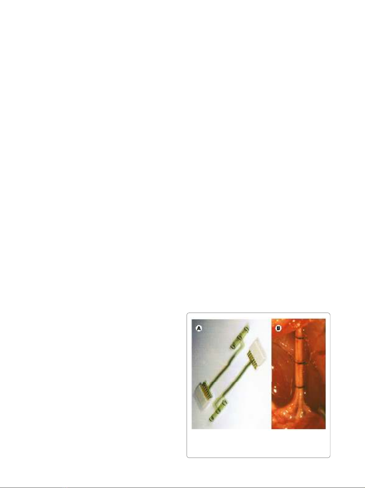
nerve perimeter (Figure 1).
Since the animals were under anesthesia during the
study, the problems related to the presence of movements
previously experienced [13,14] were mainly avoided.
Therefore, this represents an "optimal" condition for
detecting solely afferent activities, with minimal or absent
muscle artifacts.
All experiments were performed inside a Faraday cage,
in order to minimize the amount of electromagnetic
Figure 1 Polyimide tripolar cuff electrode used in the study. Cuff
electrode and connector (A), and its implantation around the sciatic
nerve of a rat before performing the experimental study (B).

Raspopovic et al. Journal of NeuroEngineering and Rehabilitation 2010, 7:17
http://www.jneuroengrehab.com/content/7/1/17
Page 3 of 15
noise interfering with the recordings. The experimental
procedures adhered to the recommendations of the Euro-
pean Union and the NIH Guide for Care and Use of Lab-
oratory Animals, and were approved by the Ethical
Committee of the Universitat Autònoma de Barcelona,
where the animal work was performed.
B. Stimuli application and signal recording
Different sensory stimuli were applied to discrete areas of
the hindpaw and the evoked neural activity was continu-
ously recorded. Three different types of stimuli were
sequentially applied, ten times each, to each animal: (1)
mechanical stimulus ("VF") of regulated intensity by
touching the plantar skin with a von Frey filament
(Stoelting Co, Illinois) (2) proprioceptive stimulus ("Pro-
prio") provoked by means of complete passive flexion of
the toes, and (3) nociceptive stimulus ("Nocio") provoked
by pinching the toes. These three types of stimuli were
selected because they elicit impulses conducted by three
different functional classes of afferent nerve fibers (Aβ
tactile mechanoreceptive, Aα proprioceptive, and Aδ/C
nociceptive, respectively).
Efforts were made to standardize the intensity of stim-
uli across trials: the same Von Frey filament was used in
all the tests, thus providing the same contact pressure;
passive flexion was produced by bending the toes from
the horizontal plane to about maximal flexion by means
of small wood sticks that were glued to the dorsum of the
nails, to avoid tactile stimulation; pinching the toe was
made using the same fine forceps (Dumont #5), aiming to
elicit pinching pain, with minimal touch.
Onset and duration of stimuli were identified by exper-
imenter's bottom pressure in synchrony with start and
end of stimulus application, while VF touch stimulation
was also recorded by means of a pressure sensor located
under the animal hindpaw, confirming good timing given
by means of bottom pressure. The duration of different
stimulus applications were not statistically different, and
had small standard deviation (touch stimulus (mean ±
standard deviation): 0.96 ± 0.11 sec; proprioceptive: 1.17
± 0.18 sec; nociceptive: 0.97 ± 0.25 sec).
Neural signals (Figure 2) were differentially amplified
(at 10,000X; Isolated Microamplifier, FHC Inc.), analogi-
cally filtered (band pass filter with cutoff frequencies of
10 Hz and 5 kHz), digitized at 20 kHz (PowerLab) and fed
into a PC running Chart v5.5 (AD Instruments). Datasets
consisted of ten applications of every type of stimuli dur-
ing the experiment. Noisy parts of the recordings - corre-
sponding to stochastic nerve and muscle discharges -
were not eliminated since they would be present also in
any real prosthetic applications. In this way, the experi-
ments should be able to indicate the real limits of this
approach.
C. Signal processing steps
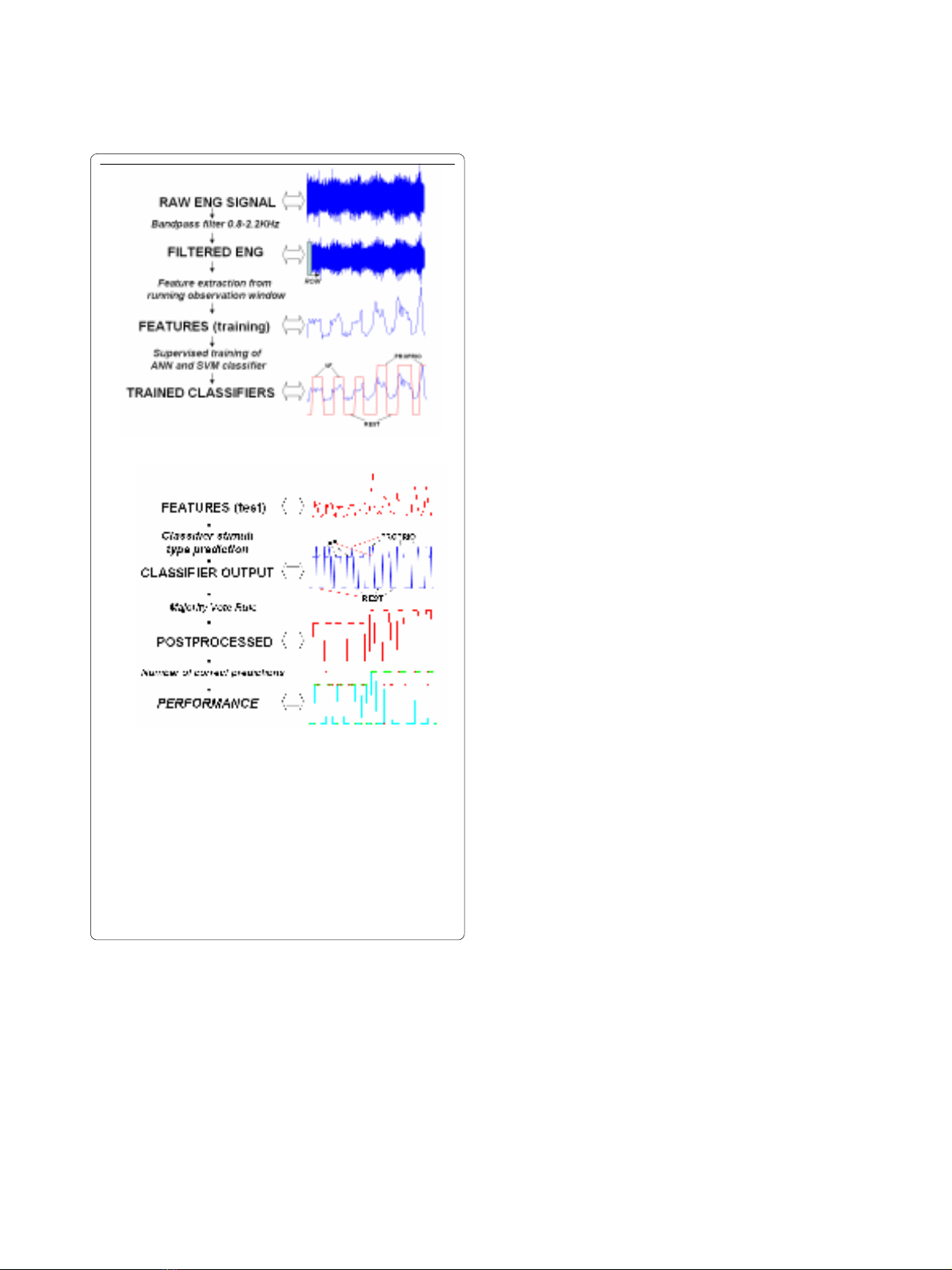
Figure 3 shows a block diagram of the proposed classifi-
cation scheme. Panel A describes the steps implemented
during the training phase. pre-processing, feature extrac-
tion, training of the classifiers using a supervised
approach. Panel B illustrates the steps performed during
the test phase: pre-processing, feature extraction (both
the same as during the training), identification of the
stimulus using the trained classifier, and a majority voting
technique. The different steps are described in detail in
this section.
Signal Pre-processing
Initially, a preliminary spectral analysis was performed in
order to impose correct filtering. Consistent with previ-
ous results [13], a neural signal peak between 1.0 and 2.0
kHz was observed for all the stimuli-evoked responses
analyzed. In a previous study [22], the nerve cuff signals
were found to be independently distributed Gaussian sig-
nals with zero mean and modulated in variance. Conse-
quently, the ENG signals recorded during the different
experimental conditions were digitally filtered using a
FIR bandpass filter with 0.8 KHz and 2.2 KHz cutoff fre-
quencies in order to reduce the presence of undesired sig-
nals (e.g. low frequency EMG signals and high frequency
amplifier noise). In fact, about 95% of the power spec-
trum of the EMG is accounted for by a band up to 400 Hz
- although there are some harmonics up to 800 Hz [25] -
while amplifier noise makes an important contribution
only at higher frequencies [21].
Length of running observation window and overlap
In this kind of signal processing paradigm, one of the
parameters to choose is the optimal length of the running
observation window (ROW), and possible overlap. In
EMG studies, the plateau in classification performance
for observation windows starts from 100 ms [30,31].
Since there are no indications in the literature either for
optimal window length with ENG signals or for overlap
(allowing a greater amount of samples for post-process-
ing rule [30,31]), the identification of these parameters
was analyzed first. Therefore, different observation win-
dow lengths were studied [25, 50, 75, 100, 125, 150, 200,
and 300 ms], and for the best performing lengths, differ-
ent overlaps [1/4, 1/2, 3/4] were tested.
Feature extraction. Several features were extracted
from the ENG signals (see Table 1 for mathematical defi-
nitions and references), in an attempt to enhance the
ENG signals conveying different sensory information
with respect to the resting-state ENG.
First, standard, time domain features used to process
EMG signals were estimated from the ROW: mean abso-
lute value (MAV), variance unbiased estimator (VAR),
and wave length (WL) [25].
Then, the features proposed in the few previous studies
on single-channel cuff ENG processing were tested. In
Raspopovic et al. Journal of NeuroEngineering and Rehabilitation 2010, 7:17
http://www.jneuroengrehab.com/content/7/1/17
Page 4 of 15
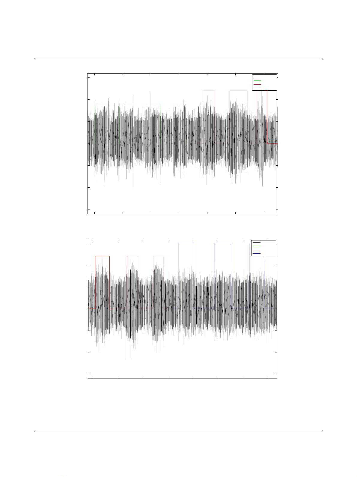
Figure 2 Examples of raw ENG recordings. In black is presented raw voltage; green labeled steps represents application of Touch stimulus; red la-
beled steps represent Proprioceptive stimulus application; in both cases the label with value 0 represents absence of stimulus (A). In black is presented
raw voltage; red labeled steps represents application of Proprioceptive stimulus; blue labeled steps represent Nociceptive stimulus application; in
both cases the label with value 0 represents absence of stimulus (B).
!&
(
)'%#%%#!'" &#%'%%"'&'!( %&'
)'
#(
%#$%#
# #
!&
(
)'%#%%#!'" &#%'%%"'&'!( %&'
)'
#(
%#$%#
##
Raspopovic et al. Journal of NeuroEngineering and Rehabilitation 2010, 7:17
http://www.jneuroengrehab.com/content/7/1/17
Page 5 of 15
[21] a higher order statistics approach was proposed,
which is able to separate the space of the noise with
respect to the space of the signal of interest. Briefly, this
means: a) constructing the Toeplitz matrix based on sec-
ond order estimation (autocorrelation) (HOS2) or third
order statistics (HOS3); b) transforming it into the eigen-
values matrix, by means of singular values decomposi-
tion, and c) taking the values higher than an empirical
threshold.
On another hand, [23,24] proposed to use the autocor-
relation function to distinguish different activities by ana-
lyzing whole nerve signals recorded with cuff electrodes,
based on the differences in fiber conduction velocity. Five
possible factors may be extracted from this feature
(ACORR): zero-cross time, time of minimum, minimum
value, time of maximum, and maximum value. We tested
these five parameters and found that the first minimum
value showed the greatest difference between noise and
elicited ENG activity.
Energy based on Discrete Fourier Transformation
(DFT) of the signal was used to understand whether our
ENG signals are more separable in the frequency domain
[25].
Features based on time-series analysis have already
shown to be useful in EMG signal processing, hence cep-
stral (CEPS) [26], and autoregressive (AR) [27] coeffi-
cients were included in the present study.
Finally, a wavelet-denoise with hard-thresholding and
Symmlet 7 mother wavelet (WDEN) was implemented
[28,29], in order to extract the bursting activity, possibly
superimposed to compound signals and not identifiable
visually. All these features were extracted from the ROW,
and were used as inputs to the classification systems.
Classification algorithms
The above features were normalized with respect to the
corresponding maximal values, and were used as inputs
to two non-linear classifiers applied in this study:
1. An artificial neural network (ANN) [32]: a feed-for-
ward neural classifier, trained by back-propagation rule,
comprising two hidden layers with 10 neurons was used.
Since there is no standard way to define the appropriate
topology of a neural network nor the number of neurons,
the parameters were determined by means of iterative
search. The numbers of hidden layers (from 1 to 3) and
neurons (from 1 to 11), and the optimal topology and
number were found with respect to the peak of classifica-
tion accuracy (this is not shown in the manuscript for the
sake of brevity). The optimal configuration used had two
hidden layers with 10 neurons each. The input layer was
composed of neurons corresponding to the number of
features used during simulations (from one to four), while
in the output layer there were four neurons, related to the
possible states-classes of the problem (rest, mechanical
stimulus, nociceptive stimulus and proprioceptive stimu-
lus).
2. Support vector machine (SVM) classifier [33] maps
input data into the feature space where they may become
linearly separable. Due to its superiority in terms of good
generalization derived from minimizing structure risk,
SVM has been applied successfully in bio-information
and pattern recognition [29,31,34]. The SVM network
was investigated using Gaussian Radial Basis function
(RBF) kernel, which yielded the best results during pre-
liminary investigations. A grid-search was employed as a
method of model selection to adjust SVM parameters, as
proposed in [31,34]. In this method, the performance of a
Figure 3 Block diagram of the proposed classification system for
ENG signals. Training is performed on the first and testing on the sec-
ond half of the data. (A) Training procedure consisting of: Filtering (in
order to eliminate the EMG low band, and amplifiers high band noise);
Feature extraction from running observation window (ROW); Training
of the classifier with stimuli type knowledge (VF, Proprioceptive, Rest as
labeled); recorded during the experimentation. (B) Test procedure: fil-
tering and feature extraction are first steps (both the same as during
the training); then the classifiers (trained in A) answer is post-processed
by means of majority vote rule. Evaluation is carried out by report be-
tween correctly classified instances and all samples in each test set.