J. Sci. Dev. 2011, 9 (Eng.Iss. 1): 84 - 90 HANOI UNIVERSITY OF AGRICULTURE
EFFECT OF MUCUNA PRURIENS ON CATECHOLAMINE BIOSYNTHETIC ENZYME GENE EXPRESSION IN RAT BRAIN
Ảnh hưởng của Mucuna pruriens đến sự biểu hiện gen tổng hợp catecholamine trên mô não chuột
Nguyen Thi Hiep1, Truong Thi Thanh Mai2, Bui Thai Hang1, Kim Sung Jun3
1College of Food Industry, Da Nang City, Vietnam 2College of Education – University of Danang, Da Nang City, Vietnam 3College of Science, Chosun University, Gwang ju, Korea Corresponding author email: bluebio203@gmail.com Received date: 13.04.2011 Accepted date: 05.05.2011
TÓM TẮT
Để xác định ảnh hưởng của Mucuna pruriens (MP) đến sự biểu hiện của một số gen tổng hợp catecholamine, nghiên cứu này xác định sự thay đổi của hàm lượng mRNA và protein của tyrosine hydroxylase (TH) và aromatic l- amino acid decarboxylase (AADC). TH là enzyme thứ nhất và AADC là enzyme thứ hai trong chu trình tổng hợp catecholamine. Hàm lượng mRNA của TH và AADC được xác định trong mô não của chuột bằng kỹ thuật RT- PCR. Hàm lượng protein được xác định bằng kỹ thuật Western blot. Chuột được nghiên cứu với liều lượng MP khác nhau (0, 100, 200, 300 và 400 mg/kg) lần lượt trong thời gian 2 giờ và 4 giờ. Kết quả chỉ ra rằng MP làm tăng cả hàm lượng mRNA và protein của TH và AADC ở liều lượng 400 mg/kg trong 2 giờ và ở liều lượng 200 mg/kg trong 4 giờ. Kết quả này cho biết, MP có ảnh hưởng tích cực đến sự biểu hiện của TH và AADC gen và mức độ ảnh hưởng đó phụ thuộc vào liều lượng cũng như thời gian sử dụng MP.
Từ khóa: AADC, biểu hiện gen, catecholamines, TH.
SUMMARY
To determine the effect of Mucuna pruriens (MP) on the catecholamine biosynthetic enzyme encoding genes, we determined changes in mRNA and protein levels of tyrosine hydroxylase (TH) and aromatic l- amino acid decarboxylase (AADC), the first rate – limiting and the second enzyme in the catecholamine biosynthetic pathway. TH and AADC mRNA levels were examined in rat brain tissue by RT- PCR. Protein levels were determined by Western blot analysis. Rats were treated with different doses of MP (0, 100, 200, 300 and 400 mg/kg) for 2 and 4 hrs.. The results showed that the MP significantly increased the levels of TH and AADC mRNA and protein at a dose of 400 mg/kg for 2 hrs and at a dose of 200 mg/kg for 4hrs, respectively. These results indicated that MP has significant effects on the expression of TH and AADC genes and the level of expression depends on the treatment dose and duration of administration.
Key words: AADC, catecholamines, gene expression, TH.
and epinephrine) called "catecholamines".
1. INTRODUCTION
Tyrosine hydroxylase (TH) is the first and rate-limiting enzyme the biosynthesis of in catecholamines, which is neurotransmitter involved in a variety of important physiological functions (Milsted et al., 2004). The regulation of TH protein level and activity represents a central means for controlling catecholamine synthesis (Kumer et al., 1996). Changes in TH expression generally reflect altered activity of dopaminergic neurons in brain. Parkinson’s disease (PD) is the most common neurodegenerative movement disorder and affects over six million people all over the world (Licker et al., 2009). This disease is a degenerative disease of the nervous system due to the progressive loss of nigrostriatal dopaminergic neurons and decrease in striatal dopamine. Dopamine is produced by dopaminergic neuronal cells and is one of three main neurotransmitters (dopamine, norepinephrine
84
Effect of Mucuna pruriens on catecholamine biosynthetic enzyme gene expression in rat brain
The fresh seeds of MP (from Gwang-ju city, South of Korea) were collected by removing peel and then ground to a paste by grinder. This powder was kept in deep freezer -700C and freezing-dry. A total of 100g of the dry powder was completely dissolved in 100ml of saline before use.
2.2. Animal experiments
regulating of
Male Sprague-Dawlay rats, 6 ~ 8 weeks of age and weights from 250~300g were used in all experiments. Animals were housed four per cage and under controlled environmental conditions (12 hrs light / dark cycle and room temperature 25 ± 2oC. Food and water were supplied all the time. Rats were treated by MP powder dissolved in saline with dose of 100, 200, 300 and 400 mg/kg each for 2 and 4 hrs (each group = 5 rats). The control groups received the same volume of saline instead of MP. The aromatic l- amino acid decarboxylase (AADC) is the second enzyme in the catecholamine biosynthesis. AADC is a non-specific enzyme mainly implicated in the synthesis of dopamine and serotonin through the decarboxylation of a substrates (L-DOPA), 5-hydroxytryptophan in neuronal and non-neuronal cells (Yuichi Okazaki and Yoshikazu Shizuri, 2001). Unlike other enzymes which are involved in catecholamine biosynthetic pathway, AADC is still generally considered not to be limiting catecholamine biosynthesis. in Nevertheless, some recent studies concerning the regulation of AADC in vivo and in vitro through phosphorylation indicate that this enzyme may play the role in regulation of catecholamine biosynthesis (Waymire and Haycock, 2002). The immobilization stress stimulation induced also increases in TH and AADC activity (Kubovcakova et al., 2004; Micutkova et al., 2003).
2.3. Total RNA isolation and relative quantification of mRNA levels by Reverse transcriptase polymerase chain reaction (RT – PCR)
four and
to
Seeds of Mucuna pruriens (MP) have been described as an useful therapeutic agent in various diseases of the human nervous and reproductive system including PD in the ancient Indian medical system (Manyam et al., 2004). MP contains L-dopa (5%), 5-indole compounds (tryptamine and 5- hydroxytriptamine), tetrahy- droisoquinolines alkaloidals (Eustace et al, 2006; Misra and Wagner, 2004). In addition, MP consists of co-enzyme Q-10 and nicotinamide adenine dinucleotide (NADH), which have neuroprotective activities (Manyam et al., 2004). Total RNA was isolated from frozen brain tissues by using the TrizolTM reagent (Invitrogen Co. USA). Reverse transcription was performed from 4 μg of total RNA samples using oligo (dT) primer and Moloney murine leukemia virus ribonuclease (M-MLV) (BioNEER co. Korea) according instructions. the manufacturer’s Quality of cDNA was verified by PCR amplification of β-actin. The cDNA was stored at - 20oC for further using. of The determining
enzyme and in Although many researches have focused on the therapeutic effects of natural plant extracts, however, until now, there are few researches on genetic analysis of catecholamine biosynthetic enzyme genes induced by MP. Therefore, in this study, we analyzed the effects of MP on the gene expression of TH and AADC, the first rate – limiting the second the catecholamine biosynthetic pathway,
2. MATERIALS AND METHODS
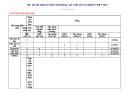
catecholamine synthesizing enzymes, TH, AADC as well as housekeeper β-actin gene expression was carried out by RT-PCR. Specific primers, annealing temperatures, number of cycles and size of each fragment for TH, AADC and β-actin mRNA are shown in table 1. PCR products were analyzed on 1.2% agarose gel in 0.5 X TBE (Tris-Boric acid- EDTA) buffers containing EtBr. Intensity of individual bands was evaluated by Gel Quant software (DNR Bio-Imaging Systems Ltd.). 2.1. MP sample preparation
Genes
Sequences of primers
Temp.of annealing
No. of cycles
Size of fragment
Gene Reference
TH
640C/45’’
35
380bp
M10244
AADC
600C/45’’
35
345 bp
U31884
30
155 bp
BC063166
β-actin
560C/45’’
5’ GCTGTCACGTCCCCAAGGTT 3’ 3’ TCAGACACCCGACGCACAGA 5’ 5’ CTTCAGATGGCAACTACTCC 3’ 3’ CTTCGGTTAGGTCAGTTCTC 5’ 5’ CCTCTATGCCAACACAGT 3’ 3’ AGCCACCAATCCACACAG 5’
Table 1. Oligonucleotide sequence of primers used in PCR for amplification
85
Nguyen Thi Hiep, Truong Thi Thanh Mai, Bui Thai Hang, Kim Sung Jun
Antibodies
Species
Dilution
Source
Table 2. Antibodies used for western blot assay
Primary antibodies
Affinity Bio Reagents
1:2000
Mouse (monoclonal IgG1)
Anti-TH (0MA-04051)
Abcam plc
1:1000
Rabbit polyclonal
Anti-AADC (ab3905)
Biomeda crop.
1:2000
Mouse (monoclonal IgG1)
Actin
Secondary antibodies
1:1000
Santa Cruz Biotechnology
Goat-anti-mouse IgG1
HRP conjugated(Sc-2054)
1:1000
Santa Cruz Biotechnology
Goat-anti-rabbit IgG1
HRP conjugated(Sc-2055)
2.4. Total protein isolation and western blot analysis
exposed to X-ray film (BioMax MS-1, Eastman Kodak). A digital image system was used to determine the density of the bands (Gel Quant, DNR Bio-Imaging Systems Ltd.).
2.5. Statistical analysis
Data were presented as mean ± S.E.M. Results were evaluated by Student’s test and by one- way analysis of variance (ANOVA). A value of P ≤ 0.05 was considered statistically significant.
3. RESULTS
Total protein was separated in the organic phase during the preparation of total RNA and subsequently precipitated with isopropanol, and then it was washed three times with 0.3 M guanidine hydrochloride. After washing with 70% ethanol, protein was dried and dissolved in 1% SDS. Protein concentration was determined by using a Bicinchoninic acid (BCA) protein assay kit (Cabres Co. USA). Bovine serum albumin (BSA) (Sigma-Aldrich Co. USA) was used as the standard.
3.1. Effects of MP on TH and AADC mRNA levels expression in rat brain tissue
increased
Total 10 μg of protein isolated from brain tissue was separated by sodium dodecyl sulphate- polyarylamide gel (SDS-PAGE; 5% stacking gel and 10.5% separating gel) and then transfered to a polyvinylidene fluoride (PVDF) membrane. Non- specific binding sites were blocked by immersing the membranes in 3% BSA in Tris- buffered saline Tween (TBST) for overnight at 4oC on a shaker. Levels of the TH and AADC immunoreactive protein were determined using TH and AADC primary antibody (Table 2). After membranes washed several times with washing buffer TBS-T, the membranes were incubated with primary anti- TH and AADC antibody for 2hrs at 4oC, followed by incubation with secondary antibodies (Table 2) for 2hrs at room temperature. The same amount of protein was loaded and reacted with anti-mouse actin antibody (Table 2) which was used for normalization of protein loading. All primary antibodies and secondary antibodies were diluted in TBS-T solution.
To reveal the reaction bands, the membrane was reacted with WEST-ZOL (plus) Western blot detection system (Intron Biotechnology, Inc.) and To determine the effects of MP on the catecholamine biosynthetic enzyme genes, we determined changes in mRNA level of TH and AADC, the first rate-limiting and the second enzyme in this pathway. TH and AADC mRNA levels were examined by RT-PCR. Rats were treated with different doses of MP (0, 100, 200, 300, and 400 mg/kg) each for 2 and 4 hrs. As shown in Fig.1 and Fig. 2, TH and AADC mRNA levels in dose-dependent manner. However, the AADC mRNA level was less than TH mRNA level. In particular, at 2 hrs TH mRNA increased by 2.2 ± 3 folds (P<0.001), while AADC mRNA level rose to 1.86 and 2 folds with doses 300 mg/kg, 400 mg/kg, respectively. At 4hrs, TH mRNA level increased sharply and was higher than AADC mRNA level, the highest expression (TH mRNA 7.4 times (P<0.0001) and AADC mRNA 1.3 times (P<0.05)) was observed with dose 200 mg/kg. The expression of TH and AADC mRNA levels depend on MP doses and duration of administration.
86
Effect of Mucuna pruriens on catecholamine biosynthetic enzyme gene expression in rat brain
Con 100 200 300 400 (mg/kg) Con 100 200 300 400 (mg/kg) Con 100 200 300 400 (mg/kg) Con 100 200 300 400 (mg/kg)
380 pb 380 pb 380 pb 380 pb
TH TH TH TH
345 pb 345 pb 345 pb 345 pb
AADC AADC AADC AADC
150 pb 150 pb 150 pb 150 pb
β-actin β-actin β-actin β-actin
TH TH TH TH AADC AADC AADC AADC
2.5 2.5 2.5 2.5
** ** **
** ** **
** ** **
** ** **
2.0 2.0 2.0 2.0
1.5 1.5 1.5 1.5
1.0 1.0 1.0 1.0
n n n n o o o o n i i i i o s s s s i s s s s s e e e e s r r r r e r p p p p p x x x x x e e e e e e e e e e n n n n n e e e e e g g g g g e e e e e v v v v v i i i i i t t t t t a a a a a l e l l l l e e e e R R R R R
0.5 0.5 0.5 0.5
0.0 0.0 0.0 0.0
(㎎/㎏) (㎎/㎏) (㎎/㎏) (㎎/㎏)
Con Con Con Con
100 100 100 100
200 200 200 200
300 300 300 300
400 400 400 400
Figure. 1. The effects of MP on the TH and AADC mRNA levels in rat brain tissue at after 2hrs treatment. Values (n=5/group) are presented as mean ± S.E and compared by one-way ANOVA and Tukey’s test: *P<0.05 and ** P< 0.001 versus control
Con 100 200 300 400 (mg/kg) Con 100 200 300 400 (mg/kg) Con 100 200 300 400 (mg/kg) Con 100 200 300 400 (mg/kg)
380 pb 380 pb 380 pb 380 pb
TH TH TH TH
345 pb 345 pb 345 pb 345 pb
AADC AADC AADC AADC
150 pb 150 pb 150 pb 150 pb
β-actin β-actin β-actin β-actin
TH TH TH TH AADC AADC AADC AADC
** ** **
8 8 8 8
** ** **
6 6 6 6
4 4 4 4
n n n n n o o o o o i i i i i s s s s s s s s s s e e e e e r r r r r p p p p p x x x x x e e e e e e e e e e n n n n n e e e e e g g g g g e e e e e v v v v v i i i i t t t t i t a a a a a l l l l l e e e e e R R R R R
2 2 2 2
* * *
* * *
0 0 0 0
(㎎/㎏ (㎎/㎏ (㎎/㎏) (㎎/㎏) ) ) (mg/kg)
Con Con Con Con Con
100 100 100 100
200 200 200 200
300 300 300 300
400 400 400 400
Figure. 2. The effects of MP on the TH and AADC mRNA levels in rat brain tissue at after 4hrs treatment. Values (n=5/group) are presented as mean ± S.E and compared by one- way ANOVA and Tukey’s test: *P<0.05 and ** P< 0.001 versus control
87
Nguyen Thi Hiep, Truong Thi Thanh Mai, Bui Thai Hang, Kim Sung Jun
3.2. Effect of MP on TH and AADC protein level expression in rat brain tissue
increased highly significantly at a dose of 100 mg/kg by 2.53 times. But only doses of 200 mg/kg and 300 mg/kg have significantly upregulated the TH protein for 4hrs, while doses of 100 mg/kg and 400 mg/kg showed the down regulation (Fig. 4). AADC protein levels did not change at different doses of MP in the 4hrs treatment. The above results indicate that TH and AADC protein levels were significantly up regulated with the treatment of MP. The protein levels of TH and AADC were determined by western blot analysis at 2 and 4 hrs after treatment. All different doses of MP induced TH and AADC protein levels at 2hrs. TH protein levels increased significantly at 1.34, 5.62, 6.92 and 9.74 folds with the doses of 100, 200, 300, 400 mg/kg, respectively (Fig. 3), AADC protein levels
TH TH TH TH AADC AADC AADC AADC
** ** **
10 10 10 10
8 8 8 8
** ** **
6 6 6 6
4 4 4 4
l
** ** **
s s s s s l e l l l l e e e e v v v v v e e e e e l l l l l n n n n n i i i i i e e e e e t t t t t o o o o o r r r r r p p p p p e e e e e v v v v i i i i v t t t t a a a a i t l l l l a e e e e R R R R e R
* * *
2 2 2 2
0 0 0 0
(㎎/㎏) (㎎/㎏) (㎎/㎏) (㎎/㎏)
Con Con Con Con
100 100 100 100
200 200 200 200
300 300 300 300
400 400 400 400
Figure. 3. The effects of MP on the TH and AADC protein levels in rat brain tissue at after 2hrs treatment. Values (n=5/group) are presented as mean ± S.E and compared by one- way ANOVA and Tukey’s test: *P<0.05 and ** P< 0.001 versus control
** ** **
TH TH TH TH AADC AADC AADC AADC
3 3 3 3
** ** **
2 2 2 2
* * *
s s s s s l l l l l e e e e e v v v v v e e e e e l l l l l n n n n n i i i i e e e e i e t t t t o o o o t o r r r r p p p p r p e e e e e v v v v i i i i v t t t t i a a a a t a l l l l e e e e l R R R R e R
1 1 1 1
0 0 0 0
(㎎/㎏) (㎎/㎏) (㎎/㎏) (㎎/㎏)
Con Con Con Con
100 100 100 100
200 200 200 200
300 300 300 300
400 400 400 400
Figure. 4. The effects of MP on the TH and AADC protein levels in rat brain tissue at after 4hrs treatment. Values (n=5/group) are presented as mean ± S.E and compared by one- way ANOVA and Tukey’s test: *P<0.05 and ** P< 0.001 versus control
88
Effect of Mucuna pruriens on catecholamine biosynthetic enzyme gene expression in rat brain
4. DISCUSSION
genes depends on MP doses and duration of administration. Thus, MP may provide a platform for future drug discoveries and novel therapy strategies for PD.
Acknowledgements This work was support by a reseach grant provied by Chosun University, 2007.
REFERENCES
levodopa
Christopher A. Lieu, R. Kunselman Allen, V.Manyam Bala, Venkiteswaran Kala, and Subramanian Thyagarajan. (2010). A water extract of Mucuna pruriens provides long – term amelioration of parkinsonism with reduced risk for dyskinesias. Parkinsonism and related disorders. 16: 458 – 465.
The seed powder of MP has been used in traditional Ayurvedic India medicine for disease therapy including PD. All compounds of MP seed have been identified by several studies (Eustace et al., 2006; Misra 2004). These researches showed that MP has high contents of crude protein, amino acids, total phenols, tannins and, especially, L- dopa. In additional, MP contains co-enzyme Q-10 and nicotinamide adenine dinucleotide (NADH), which have neuroprotective activities (Manyam et al., 2004). MP treatment is known to increase the dopamine content in the rat brain and MP exhibited twice the antiparkinsonian activity compared with synthetic (Manyam et al., 2004). Furthermore, synthetic levodopa causes drug – induced dyskinesias in majority of patients with PD. MP, however, is reputed to provide anti- parkinsonian benefits without inducing dyskinesias (Christopher et al., 2010).
Duan Chun- Li, Yue Su, Chun –Li Zhao, Qun – Yuan Xu and Hui Yang.(2005). The assays of activities and function of TH, AADC and GCH1 and their potential use in ex vivo gene therapy of PD. Brain Research Protocols. 16: 37 – 43. in Eustace A
lyayi, Holger Kluth and Markus Rodehutscord.(2006). Chemical composition, antinutritional constituents, precaecal crude protein and amino acid digestibility in three unconventional tropical legumes in broilers. J Sci Food Agric. 86: 2166 – 2171.
biosynthetic enzyme
Katzenschlarger R, Evans A, Manson A, P N Patsalos, N Ratnaraj, H Watt, L Timmermann, R Van der Geissen, A J Lees.(2004). Mucuna pruriens in Parkinson’s disease: a double blind chinical and pharmacological study. J Neuol Neurosurg Psychiatry.75: 1672 -1677.
Kumer S.E. and K. E. Vrana (1996). Intricate regulation of tyrosine hydroxylase activity and gene expression. Jounal of neurochemistry. 67: 443-462.
its modulation tissued and
The regulation of TH and AADC expression the field of has attracted much attention neurology. Indeed, the biological function of TH and AADC is very important for the dopamine biosynthesis as well as for brain function under physiological and pathological conditions. The study of Duan et al., (2005) found that the expression of TH and AADC genes could fulfill the function of dopamine synthesis. The present study was carried out to evaluate the effects of MP on the genes, catecholamine particularly TH and AADC. The results showed that the levels of TH mRNA and protein increased significantly at different doses of MP at both 2hrs and 4hrs treatment. Similarly, the AADC mRNA and protein levels rose slightly at different doses of MP at 2hrs treatment. In contrast, the level of AADC protein did not change at different doses of MP at 4hrs after treatment. The levels of TH mRNA and protein are always higher than those of AADC at different doses of MP during the treatment period. These results indicate that MP has a complex and different effect on TH and AADC gene expression in the rat brain tissue. Kubovcakova L., O. Krizanova and R. Kvetnansky (2004). Identification of the aromatic L-amino acid decarboxylase gene expression in various mice by immobilization stress in stellate ganglia. Jounal of neuroscience.126: 375-380.
in
5. CONCLUSIONS
The present shows that Licker V, E Kovari, D. F. Hochstrasser, P. R. Burkhard human (2009). Proteomics Parkinson’s disease research. J Proteomics 73: 10-29.
Manyam B.V., M. Dhanasekaran., and T.A. Hare the (2004). Neuroprotective effects of both study catecholamine biosynthetic enzymes encoding genes TH and AADC were up – regulated by the treatment of MP. The expression of TH and AADC
89
Nguyen Thi Hiep, Truong Thi Thanh Mai, Bui Thai Hang, Kim Sung Jun
of Mucuna pruriens seeds. Phytochemistr. 65: 2565 - 2567. Antiparkinson drug Mucuna Pruriens. Jounal of Phytotherapy Research. 18: 706-172.
pruriens) the on
Manyam B.V., M. Dhanasekaran, and T. Hare (2004). Effect of antiparkison drug HP-200 (Mucuna central monoaminergic neurotransmitters. Phytother Res. 18: 97-101. Yuichi Okazaki and Yoshikazu Shizuri. (2001) Identification of the aromatic l- amino acid decarboxylase (AADC) gene and its expression in the attachment and metamorphosis of the barnacle, Balanus amphitrite. J develop Growth Differ. 43: 33 – 41.
L-amino of Milsted A., L. Serova, E.L. Sabban, and G. Dunphy (2004). Regualtion of tyrosine hydroxylase gene transcription by Sry. Neuroscience Letters. 369: 203-207. Waymire J.C., and J.W. Haycock (2002). Lack of acid aromatic regulation decarboxylase in intact bovine chromaffin cells. Jounal of Neurochemistry. 81:589-593.
Misra L. and H. Wagner (2004). Alkaloidal constituents
90