BioMed Central
Open Access
Research article IImmpp--LL22,, aa ppuuttaattiivvee hhoommoolloogg ooff vveerrtteebbrraattee IIGGFF--bbiinnddiinngg pprrootteeiinn 77,, ccoouunntteerraaccttss iinnssuulliinn ssiiggnnaalliinngg iinn DDrroossoopphhiillaa aanndd iiss eesssseennttiiaall ffoorr ssttaarrvvaattiioonn rreessiissttaannccee Basil Honegger*, Milos Galic*§, Katja Köhler†, Franz Wittwer*, Walter Brogiolo*, Ernst Hafen† and Hugo Stocker†
Addresses: *Zoological Institute, University of Zürich, Winterthurerstrasse 190, CH-8057 Zürich, Switzerland. †Institute for Molecular Systems Biology (IMSB), ETH Zürich, Wolfgang-Pauli-Strasse 16, CH-8093 Zürich, Switzerland. §Current address: Chemical and Systems Biology, 318 Campus Drive, Clark Building W200, Stanford University Medical Center, Stanford, CA 94305-5174, USA.
Correspondence: Ernst Hafen. Email: hafen@imsb.biol.ethz.ch
Published: 15 April 2008 Journal of Biology 2008, 77::10 (doi:10.1186/jbiol72) Received: 21 July 2007 Revised: 15 February 2008 Accepted: 13 March 2008
The electronic version of this article is the complete one and can be found online at http://jbiol.com/content/7/3/10
AAbbssttrraacctt
BBaacckkggrroouunndd:: Insulin and insulin-like growth factors (IGFs) signal through a highly conserved pathway and control growth and metabolism in both vertebrates and invertebrates. In mammals, insulin-like growth factor binding proteins (IGFBPs) bind IGFs with high affinity and modulate their mitogenic, anti-apoptotic and metabolic actions, but no functional homologs have been identified in invertebrates so far.
RReessuullttss:: Here, we show that the secreted Imaginal morphogenesis protein-Late 2 (Imp-L2) binds Drosophila insulin-like peptide 2 (Dilp2) and inhibits growth non-autonomously. Whereas over- expressing Imp-L2 strongly reduces size, loss of Imp-L2 function results in an increased body size. Imp-L2 is both necessary and sufficient to compensate Dilp2-induced hyperinsulinemia in vivo. Under starvation conditions, Imp-L2 is essential for proper dampening of insulin signaling and larval survival.
first
Imp-L2, the
functionally characterized
insulin-binding protein
CCoonncclluussiioonnss:: in invertebrates, serves as a nutritionally controlled suppressor of insulin-mediated growth in Drosophila. Given that Imp-L2 and the human tumor suppressor IGFBP-7 show sequence homology in their carboxy-terminal immunoglobulin-like domains, we suggest that their common precursor was an ancestral insulin-binding protein.
© 2008 Honegger et al.; licensee BioMed Central Ltd. This is an Open Access article distributed under the terms of the Creative Commons Attribution License (http://creativecommons.org/licenses/by/2.0), which permits unrestricted use, distribution, and reproduction in any medium, provided the original work is properly cited.
Journal of Biology 2008, 77::10
IGFs, but
they also modulate
colon
that IGFBP-7
BBaacckkggrroouunndd Insulin/insulin-like growth factor (IGF) signaling (termed IIS) is involved in the regulation of growth, metabolism, reproduction and longevity in mammals [1-3]. The activity of IIS is regulated at multiple levels, both extracellularly and intracellularly: the production and release of the ligands is regulated, and normally IGFs are also bound and transported by IGFBPs in extracellular cavities of vertebrates [4]. IGFBPs not only prolong the half-lives of their availability and activity [5]. Besides the classical IGFBPs (IGFBP1-6), a related protein called IGFBP-7 (or IGFBP- rP1, Mac25, TAF, AGM or PSF) has been identified as an insulin-binding protein [6]. Although the reported binding of IGFBP-7 to insulin awaits confirmation [7,8], it can compete with insulin for binding to the insulin receptor (InR) and inhibit the autophosphorylation of InR [6]. Furthermore, IGFBP-7 is suspected to be a tumor suppressor in a variety of human organs, including breast, lung and recent publication [6,9-13]. A induces senescence and demonstrates apoptosis in an autocrine/paracrine manner in human primary fibroblasts in response to an activated BRAF oncogene [14].
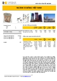
RReessuullttss GGeenneettiicc ssccrreeeenn ttoo iiddeennttiiffyy nneeggaattiivvee rreegguullaattoorrss ooff IIIISS We reasoned that the overexpression of a Dilp-binding protein that impinges on the ligand-receptor interaction should counteract the effects of receptor overexpression. dInR overexpression during eye development (by means of a GMR-Gal4 strain, in which the Gal4 protein is overexpressed in photoreceptor neurons, and a UAS-dInR, which expresses dInR when activated by Gal4) results in hyperplasia of the eyes, a phenotype that is sensitive to the levels of the Dilps [17]. A collection of enhancer-promoter (EP) elements, which allow the overexpression of nearby genes (F.W., W.B., H.S., D. Nellen, K. Basler and E.H., unpublished work), was screened for suppressors of the dInR-induced hyperplasia (Figure 1a). A strong suppressor (EP5.66, Figure 1b) carried an EP element 8.5 kb upstream of the Imp-L2 coding sequence (Figure 1f). Two different UAS transgenes, both containing the Imp-L2 coding sequence but varying in strength, confirmed that the suppression was caused by Imp-L2. Whereas the weaker UAS-Imp-L2 (containing 5’ sequences with three upstream open reading frames) only partially suppressed the dInR-induced overgrowth (Figure 1c), UAS-strong.Imp-L2 (UAS-s.Imp-L2, lacking the 5’ sequences) completely reversed the phenotype (Figure 1d). In addition, a point mutation in the Imp-L2 coding sequence (see below) abolished the suppressive effect of EP5.66 (Figure 1e). Imp- L2 is therefore a potent antagonist of dInR-induced growth.
the m-NSCs
causes
of
IIS is astonishingly well conserved in invertebrates. In Drosophila, IIS acts primarily to promote cellular growth, but it also affects metabolism, fertility and longevity [15,16]. Seven insulin-like peptides (Dilp1-7) homologous to vertebrate insulin and IGF-I have been identified as putative ligands of the Drosophila insulin receptor (dInR) [17]. These Dilps are expressed in a spatially and temporally controlled pattern, including expression in median neurosecretory cells (m-NSCs) of both brain hemispheres. The m-NSCs have axon terminals in the larval endocrine gland and on the aorta, where the Dilps are secreted into the hemolymph [17-19]. Ablation a developmental delay, growth retardation and elevated carbohydrate levels in the larval hemolymph [18,19], reminiscent of the phenotypes of starved or IIS- impaired flies.
Imp-L2 has previously been shown to be upregulated 8-10 hours after ecdysone treatment [20,21]. It encodes a secreted member of the immunoglobulin (Ig) superfamily contain- ing two Ig C2-like domains. Whereas several orthologs of Imp-L2 are present in invertebrates such as arthropods and nematodes, the homology in vertebrates is confined to the second Ig C2-like domain, which is homologous to the carboxyl terminus of human IGFBP-7 (Figure 1g). The carboxy-terminal part of IGFBP-7 differs considerably from the other IGFBPs, possibly accounting for the affinity of IGFBP-7 for insulin [6]. Interestingly, Imp-L2 has been shown to bind human insulin, IGF-I, IGF-II and proinsulin, and its homolog in the moth Spodoptera frugiperda, Sf-IBP, can inhibit insulin signaling through the insulin receptor [22].
The Drosophila genome does not encode an obvious homolog of the IGFBPs. Furthermore, genetic analyses of IIS in Drosophila and Caenorhabditis elegans have not revealed a functional insulin-binding protein so far. Here, we report the identification of the secreted protein Imp-L2 as a binding partner of Dilp2. Imp-L2 is not essential under standard conditions, but flies lacking Imp-L2 function are larger. Under adverse nutritional conditions, Imp-L2 is upregulated in the fat body and represses IIS activity in the entire organism, allowing the animal to endure periods of starvation.
OOvveerreexxpprreessssiioonn ooff IImmpp--LL22 iimmppaaiirrss ggrroowwtthh nnoonn--aauuttoonnoommoouussllyy To further assess the function of Imp-L2 as a secreted inhibitor of insulin signaling, we ectopically expressed Imp- L2 using various Gal4 drivers. Strong ubiquitous over- expression of Imp-L2 by Act-Gal4 led to lethality with both UAS transgenes. Whereas driving UAS-s.Imp-L2 by the weaker ubiquitous arm-Gal4 driver also resulted in lethality, driving UAS-Imp-L2 generated flies that were decreased in size and weight (-15% in males and -29% in females, data
10.2 Journal of Biology 2008, Volume 7, Article 10 Honegger et al. http://jbiol.com/content/7/3/10
Journal of Biology 2008, 77::10
http://jbiol.com/content/7/3/10 Journal of Biology 2008, Volume 7, Article 10 Honegger et al. 10.3
(c)
(a)
(b)
(d)
(e)
dInR
dInR, EP5.66MG2
dInR, EP5.66
dInR, IMPL2
dInR, s.IMPL2
EP5.66
GE24013 MG2
(f)
RA
RB
Imp-L2
Def-20 Def-42
Genomic rescue construct
0
1
2
3
4
5
6
7
8
9
10
11
12
13kb
(g)
** * **
*
*
*
not shown) but eclosed at the expected ratio and had wild- type appearance. By generating clones of cells that over- express Imp-L2, we confirmed that cell specification and patterning were normal in Imp-L2-overexpressing ommatidia (Figure 2a). However, a reduction of cell size was observed
in the clones. This reduction seemed to be non-autonomous because wild-type ommatidia close to the clone were also reduced in size. Given the convex nature of the eye we were unable to quantify the effects of Imp-L2 overexpression on more distantly located ommatidia. Eye-specific overexpression
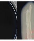
FFiigguurree 11 Imp-L2 overexpression suppresses dInR-induced growth. ((aa--ee)) Scanning electron micrographs of compound eyes. All flies (females) carry the GMR- Gal4 and UAS-dInRwt transgenes. The dInr-dependent big eye phenotype (a) is suppressed by EP5.66 (b). UAS-Imp-L2 (c) and the stronger UAS- s.Imp-L2 (d) also suppress, but EP5.66 driving the mutant Imp-L2MG2 allele can no longer suppress the dInR overexpression phenotype (e). ((ff)) Genomic organization of the Imp-L2 locus. The mutant alleles and P-element insertions used in this study are indicated. MG2 marks the point mutation in the EMS allele Imp-L2MG2 that generates a premature stop codon. ((gg)) Alignment of Imp-L2, its orthologs in invertebrates and the putative human ortholog IGFBP-7. Black and gray boxes indicate amino acid identity and similarity, respectively. The triangle marks the premature stop codon in Imp-L2MG2. Asterisks mark the cysteines forming the two disulfide bridges. The gray bars indicate the Ig domains. Dm, Drosophilamelanogaster Imp-L2; Ag, Anophelesgambiae CP2953; Sf, Spodopterafrugiperda IBP; Ce, Caenorhabditiselegans zig-4; Hs, Homosapiens IGFBP-7.
Journal of Biology 2008, 77::10
ppl-Gal4 x pp
GMR-Gal4 x
120
120
10.4 Journal of Biology 2008, Volume 7, Article 10 Honegger et al. http://jbiol.com/content/7/3/10
(a)
(c)
(b)
100
i
i
100
t h g e w
t h g e w
80
80
*
60
60
* *
40
40
20
20
l o r t n o c f o e g a t n e c r e P
0
0
l o r t n o c f o e g a t n e c r e P
P L 2
P L 2
P L 2
P L 2
G F P P
G F P
IM
IM
s.IM
s.IM
180180
55555 5
(d)
(e)
0180180808000111111180180180180180180180160160160160180801808018018001801808080180808018018080801801808080 1801801801601111111160160 180180160180 160 180
7 7
4 4
6 6
Control
140
1 1
12 12
2 3 2 3
8 910 8 910
11 11
12 12
120
IMPL2-/-
l
IMPL2-/-,GR
100
IMPL2+/-
80
IMPL2+/-,GR
60
40
o r t n o c f o e g a t n e c r e P
20
20 20
0
m:weight
Cell size
f:weight
Control
IMPL2-/-
Wing area
Cell number
Next, we assessed the effect of Imp-L2 overexpression on phosphatidylinositol(3,4,5)trisphosphate (PIP3) levels using a green fluorescent protein-pleckstrin homology domain fusion protein (tGPH) that specifically binds PIP3 and serves as a reporter for PIP3 levels in vivo [24]. The amount of membrane-bound tGPH reflects signaling activity in the phosphoinositide 3-kinase/protein kinase B (PI 3-kinase/ PKB) pathway. Overexpression of dInR resulted in a severe increase of membrane PIP3 levels (Additional data file 1, Figure S1A,B). Co-overexpression of Imp-L2 together with dInR reduced the PIP3 levels (Additional data file 1, Figure S1D), similar to the effect caused by PTEN (Additional data file 1, Figure S1C), a negative regulator of IIS. Therefore, Imp-L2 inhibits PI 3-kinase/PKB signaling upstream of PIP3, without affecting dInR levels (Additional data file 1, Figure S1B’,D’).
of both UAS-Imp-L2 and UAS-s.Imp-L2 by GMR-Gal4 led to a strong reduction in eye size (data not shown). Whereas the GMR-Gal4, UAS-Imp-L2 flies were of normal size, body weight was reduced by 38.3% and development was delayed by one day in GMR-Gal4, UAS-s.Imp-L2 male flies (Figure 2b). Next, we used the ppl-Gal4 driver to over- express Imp-L2 in the fat body, a tissue that can be expected to produce and secrete Imp-L2 more efficiently than the eye. Driving UAS-s.Imp-L2 by ppl-Gal4 was lethal, whereas ppl-Gal4, UAS-Imp-L2 flies showed a pronounced reduction in body size (Figure 2c) and were delayed by 2 days. Both the size decrease and the developmental delay are characteristic phenotypes of reduced IIS such as in chico mutants [23], supporting the hypothesis that Imp-L2 acts as a secreted negative regulator of this pathway.
FFiigguurree 22 Imp-L2 controls body and organ size. ((aa)) Tangential section through an adult eye containing an Imp-L2 overexpression clone marked by the lack of red pigment. Within the clone, the size of the ommatidia is reduced. Wild-type ommatidia close to the clone are also smaller (compare black circled areas). ((bb)) Eye-specific overexpression of UAS-s.Imp-L2 reduces male body weight (-38.3%, P = 7 x 10-42). ((cc)) Overexpression of UAS-Imp-L2 by ppl- Gal4 results in a 56.1% weight reduction in male flies, whereas ppl-Gal4 driven expression of UAS-s.Imp-L2 results in lethality (†). P = 3 x 10-47. ((dd)) Loss of Imp-L2 function increases body size in males (top) and females (bottom). ((ee)) Analyses of male and female weights. Wing area, cell number and cell size were assessed in female adult wings. GR indicates Imp-L2 genomic rescue construct. P-values are indicated by numbers as follows: 1, 2 x 10-33; 2, 8 x 10-18; 3, 9 x 10-16; 4, 6 x 10⁻⁷; 5, 3 x 10-46; 6, 1 x 10-24; 7, 8 x 10-31; 8, 2 x 10⁻⁷; 9, 4 x 10⁻⁴; 10, 4 x 10⁻⁷; 11, 3 x 10⁻⁷; 12, 1x 10⁻⁴. Genotypes: ‘control’ y, w/w; ‘Imp-L2-/-’ y, w; Imp-L2Def42/Imp-L2Def20; ‘Imp-L2+/-’ for the weight analysis (e) y, w; Imp-L2Def20/+; ‘Imp-L2+/-’ for the wing analysis (e) y, w; Imp-L2Def42/+; ‘Imp-L2-/-, GR’ y, w; Imp-L2Def42/Imp-L2Def20, GR-57; ‘Imp-L2+/-, GR’ y, w; Imp-L2Def20, GR-57/+. P-values were determined using unpaired Student’s t-test against the control except in (4) where the weight of IMP-L2+/- was compared to IMP-L2+/-, GR. n = 40 for the weight analysis in (b,c,e); n = 12 for the wing analysis in (e). Error bars represent s.d.
Journal of Biology 2008, 77::10
to generate
two strategies
suppressed
SSiizzee iinnccrreeaassee iinn IImmpp--LL22 mmuuttaannttss We used loss-of-function mutations in Imp-L2. First, we performed an ethylmethane- sulfonate (EMS) reversion screen in which we selected mutated chromosomes carrying EP5.66 that no longer suppressed the dInR overexpression phenotype (Figure 1a). One allele (Imp-L2MG2) containing a point mutation result- ing in a premature stop at amino acid 232 was identified in this way (Figure 1e,f). This truncation destroys the con- served cysteine bridge of the second Ig domain (Figure 1g). Overexpression of the truncated Imp-L2 version had no inhibitory effect on size (Figure 1e), suggesting that Imp-L2MG2 is a functional null allele.
resulting
lethality
the
weakly expressed in the seven m-NSCs that produce Dilp1, Dilp2, Dilp3 and Dilp5 (Figure 3b) and project their axons directly to the subesophageal ganglion, the CC, the aorta and the heart [19,26]. Thus, Imp-L2 potentially interacts with some of the Dilps directly at their source. We therefore tested for genetic interactions of Imp-L2 with the dilp genes. A deficiency (Df(3L)AC1) uncovering dilp1-5 not only dominantly the dInR-mediated big eye phenotype [17], but also dominantly enhanced the small eye phenotype caused by eye-specific overexpression of Imp-L2 (Additional data file 1, Figure S3). dilp2 is the most potent growth regulator of all dilp genes [18]. Weak ubiquitous overexpression of dilp2 by arm-Gal4 caused an increase in body and organ size [18], and this phenotype was dominantly enhanced by heterozygosity for Imp-L2 (Figure 3c). In homozygous Imp-L2 mutants, expression of dilp2 under the control of arm-Gal4 caused lethality, reminiscent of strong dilp2 expression [18]. Expressing Imp-L2 and dilp2 individually at high levels in the fat body also caused lethality, but coexpression resulted in viable flies of wild-type size (Figure 3d). Thus, Imp-L2 decreases the sensitivity to high insulin levels and is sufficient to rescue from dilp2-induced hyperinsulinemia.
It has previously been shown that Imp-L2 can bind human insulin and insulin-related peptides [22]. To address whether Imp-L2 binds Dilp2, we constructed a Flag-tagged version of Dilp2, which is functional (data not shown). Using in vitro translated, 35S-labeled Imp-L2 together with Flag-Dilp2 extracted from stably transfected S2 cells, we could show that Imp-L2 binds Dilp2 in vitro (Figure 3e). A truncated form of Imp-L2 lacking a functional second Ig domain (like that produced by the MG2 allele) failed to bind Dilp2 (Figure 3e).
Second, we generated additional Imp-L2 alleles by imprecise excision of GE24013 (GenExel), a P-element located 349 bp upstream of the ATG start codon of the Imp-L2-RB transcript (Figure 1f). We obtained Imp-L2 deletions (Def20, Def42) lacking the entire coding sequence. Heteroallelic combinations of the mutant alleles increased body size: whereas mutant males showed a 27% increase in body weight, mutant females were 64% heavier (Figure 2d,e). Introducing one copy of a genomic rescue construct (Figure 1f) [25] into homozygous mutant flies reverted the weight to the level of Imp-L2+/- flies, which were already heavier (+14% in males, +44% in females, Figure 2e) than the controls. By measuring the cell density in the wing, the size increase could be attributed primarily to an increase in the number of cells, because cell size was only slightly affected (Figure 2e). Apart from the size increase, the flies lacking Imp-L2 appeared completely normal, eclosed with the expected frequency and were not delayed. Thus, under standard conditions, Imp-L2 loss-of- function dominantly increases growth by augmenting cell number without perturbing patterning, developmental timing or viability.
them
in adipose
tissues, precluding
The weight difference was more pronounced in mutant females than in males, although the increases in wing area and cell number were similar (Figure 2e and data not shown). This differential effect was caused by enlarged ovaries in Imp-L2 mutant females (data not shown).
IImmpp--LL22 iiss eesssseennttiiaall uunnddeerr aaddvveerrssee nnuuttrriittiioonnaall ccoonnddiittiioonnss Despite being a potent inhibitor of Dilp2 action, Imp-L2 is not essential under standard conditions. Hyperactivation of the dInR pathway leads to increased accumulation of nutrients from circulating and thus resulting in starvation sensitivity at the organismal level [24]. We therefore tested whether Imp-L2 functions as an inhibitor of IIS under stress conditions. We exposed wild-type and Imp-L2 mutant early third instar larvae to various starvation conditions and scored for survival. Larvae lacking Imp-L2 showed a massive increase in mortality rate when exposed to 1% glucose or PBS for 24 hours (Figure 4c). To test whether the inability of the mutant larvae to cope with starvation was due to a failure in levels under these adjusting IIS, we monitored PIP3 conditions. Whereas control flies showed a decrease of PIP3 levels when exposed to complete starvation for 4 hours
IImmpp--LL22 bbiinnddss ttoo aanndd aannttaaggoonniizzeess DDiillpp22 The facts that Imp-L2 is a secreted protein and that removal of Imp-L2 function did not rescue either chico or PI3K mutant phenotypes (data not shown) are consistent with the hypothesis that Imp-L2 acts upstream of the intra- cellular IIS cascade at the level of the ligands. Immuno- histochemistry in larval tissues revealed that, besides strong expression in corpora cardiaca (CC) cells (Figure 3a and Additional data file 1, Figure S2D), Imp-L2 protein was also
http://jbiol.com/content/7/3/10 Journal of Biology 2008, Volume 7, Article 10 Honegger et al. 10.5
Journal of Biology 2008, 77::10
10.6 Journal of Biology 2008, Volume 7, Article 10 Honegger et al. http://jbiol.com/content/7/3/10
(a)
(c)
arm-Gal4, UAS-dilp2
160
P = 1x10⁻⁴
140
120
i
t h g e w
100
IMPL2
80
(b)
60
40
l o r t n o c f o e g a t n e c r e P
20
0
+/+
-/-
+/-
+/+
Imp-L2
+/-
-/-
160
IMPL2 dilp2-lacZ
-/-, GR
ppl-Gal4
120
(d)
(e)
100
t
+
Flag-dilp2
i
80
ImpL2-IVT
h g e w
t
l
60
ImpL2MG2-IVT
40
e g a n e c r e P
o r t n o c f
o
Flag pull-down
20
0
lacZ
dilp2 lacZ
s.IMPL2 lacZ
s.IMPL2 dilp2
downregulation of dilp3 and dilp5 at the transcriptional level [18].
(Figure 4a), Imp-L2 mutant larvae still contained PIP3 levels that were comparable to those of control larvae reared on normal food (Figure 4b), suggesting that Imp-L2 is necessary to adjust IIS under starvation conditions. The fact that PIP3 levels were also slightly reduced in Imp-L2 mutants upon starvation could be attributed to the
The dampening of IIS upon starvation could be achieved either by enhanced secretion of stored Imp- L2 or by an upregulation of Imp-L2 production.
FFiigguurree 33 Imp-L2 binds Dilp2 and counteracts its activity. ((aa,,bb)) Antibody staining of larval brains with an Imp-L2 antibody (green). (a) Specific neurons of both brain hemispheres, the subesophageal ganglion region (gray arrow) and the corpora cardiaca (white arrow) express Imp-L2 protein. The corpora allata are innervated by Imp-L2 expressing axons. White arrowheads mark the Dilp-producing m-NSCs. (b) In larvae carrying a dilp2-lacZ.nls transgene, co-staining with β-galactosidase and Imp-L2 antibodies reveals that the seven dilp-expressing m-NSCs also produce low levels of Imp-L2. ((cc)) The size increase of arm-Gal4, UAS-dilp2 flies is dominantly enhanced by reducing Imp-L2 levels. In an Imp-L2-/- background, dilp2 overexpression results in lethality, which can be rescued by a copy of the Imp-L2 genomic rescue construct (GR). ((dd)) Overexpression of dilp2 as well as of Imp-L2 at high levels by ppl-Gal4 causes lethality, whereas concomitant overexpression of dilp2 and Imp-L2 yields flies of wild-type size. The lacZ transgene was introduced to rule out a dosage effect of the UAS/Gal4-system. ((ee)) Imp-L2 binds Dilp2. In-vitro-translated, 35S-labeled wild-type (ImpL2-IVT, about 32 kDa) or mutant (ImpL2MG2-IVT, about 30 kDa) Imp-L2 (lane 1) was incubated with cell lysates of either non-transfected (lane 3) or stably transfected S2 cells expressing Flag-Dilp2 (lane 2). Imp-L2 could only be pulled down in the presence of Dilp2. The Imp-L2MG2 mutation abolished Dilp2 binding. Genotypes in (c): ‘Imp-L2+/-’ Imp-L2Def42/+; ‘Imp-L2-/-’ Imp-L2Def42/Imp-L2Def20; ‘Imp-L2-/-, GR’ Imp-L2Def42/Imp-L2Def20, GR-57; ‘control’ (black bar) arm-Gal4, UAS-GFP. P-values were determined using unpaired Student’s t-test (n = 40, except for bars 1-3 in (c): bar 1, n = 31; bars 2 and 3, n = 17). Error bars represent s.d.
Journal of Biology 2008, 77::10
100
http://jbiol.com/content/7/3/10 Journal of Biology 2008, Volume 7, Article 10 Honegger et al. 10.7
(a)
c) (c)
90
80
Yeast
70
60
50
tGPH
tGPH Hoechst
40
30
20
10
4h PBS
0
h 4 2 r e t f a e a v r a l d a e d f o e g a t n e c r e P
Yeast
PBS
20% glucose
1% glucose
IMPL2-/-
IMPL2-/-,GR
IMPL2+/-
Control
tGPH
tGPH Hoechst
(b)
(d)
Yeast
IMPL2-/- Yeast
tGPH
IMPL2
IMPL2 Hoechst
tGPH Hoechst
24h PBS
IMPL2-/- 4h PBS
IMPL2
IMPL2 Hoechst
tGPH
tGPH Hoechst
revealed a
larvae weaken
IIS by
conditions, Drosophila upregulating Imp-L2 expression in the fat body.
DDiissccuussssiioonn IIS signaling has evolved in animals to regulate growth and metabolism in accordance with environmental conditions. Appropriate IIS activity is ensured at several levels, including
Indeed, expression profiling slight upregulation of Imp-L2 after 12 hours complete starvation [27]. We could not detect a change in Imp-L2 protein expression in the brain, the ring gland or the gut after complete starvation for 24 hours (data not shown). However, Imp-L2 was induced in fat body cells, where it appeared in vesicle-like structures adverse nutritional (Figure 4d). Thus, under
FFiigguurree 44 Imp-L2 is necessary for blocking dInR signaling under starvation. ((aa,,bb)) tGPH fluorescence (green, showing PIP3 levels and thus indicating IIS activity) in the fat body of feeding third instar larvae under different nutritional conditions. Nuclear staining (Hoechst) is shown in blue in the right panels. (a) Under normal conditions (‘yeast’), IIS activity is high in wild-type feeding third instar larvae. Upon starvation, only little PIP3 localizes to the membranes of fat body cells. (b) In Imp-L2 mutants, IIS activity is higher than in control larvae and only slightly reduced after 4 h PBS starvation. ((cc)) Survival of Imp-L2Def42/Imp-L2Def20 early third instar larvae is severely compromised under starvation conditions. One copy of the genomic rescue construct (GR) suffices to restore viability. Heterozygous larvae were Imp-L2Def42/+, control larvae y,w/w. Larvae (40) were subjected for 24 h to 20% glucose, 1% glucose or PBS. The experiment was repeated twice. ((dd)) In starved larvae (y, w), Imp-L2 protein expression (green) is induced in fat body cells after 24 h PBS starvation. Imp-L2 is localized to vesicle-like structures but not detectable under normal nutritional conditions. Genotypes: (a,d) y, w; (b) y, w; Imp-L2Def42/Imp-L2Def20.
Journal of Biology 2008, 77::10
the controlled expression of binding partners of the extracellular ligands. Surprisingly, the well-characterized vertebrate IGFBPs have no obvious homologs in lower organisms. Here, we used a genetic strategy to search for negative regulators of IIS in Drosophila. Our approach led to the identification of Imp-L2 as a functional insulin-binding protein and antagonist of IIS.
[24], an organ that resembles the mammalian liver as the principal site of stored glycogen [30]. Even under adverse nutritional conditions, fat body cells with increased IIS activity continue stockpiling nutrients, thereby limiting the amount of circulating nutrients, which induces hyper- sensitivity to starvation of the larva [24]. Upon starvation, the expression of dilp3 and dilp5 is suppressed at the transcriptional level in the m-NSCs [18]. Our study reveals an additional layer of IIS regulation. Whereas Imp-L2 is not expressed in the fat body of fed larvae, starved animals induce Imp-L2 expression in the fat body to systemically dampen IIS activity. A lack of this control mechanism is lethal under unfavorable nutritional conditions, as Imp-L2 mutant larvae fail to cope with starvation.
Imp-L2 encodes a secreted peptide containing two Ig C2-like domains. Consistent with its secretion, the effects of Imp-L2 overexpression are non-autonomous. Tissue-specific over- expression of Imp-L2, for example in the larval fat body, results in a systemic response, and the entire animal is impaired in its capacity to grow. Conversely, the loss of Imp- L2 function produces larger animals. Our analysis of IIS activity (by means of the tGPH reporter in vivo) shows that Imp-L2 functions to downregulate IIS. We further show that wild-type Imp-L2 - but not a truncated version lacking the second Ig C2-like domain - binds Dilp2, consistent with previous findings that Imp-L2 binds human insulin, IGF-I, IGF-II and proinsulin [22].
CCoonncclluussiioonnss Our study provides the first functional characterization of an insulin-binding protein in invertebrates. We have identified Imp-L2 as a secreted antagonist of IIS in Drosophila. Given the sequence homology of their Ig domains, we propose that Imp-L2 is a functional homolog of vertebrate IGFBP-7. Because both Imp-L2 and IGFBP-7 are potent inhibitors of growth and Imp-L2 is essential for the endurance of periods of starvation, it is likely that the original function of the insulin-binding molecules was to keep IIS in check when nutrients were scarce. Thus, in accordance with several reports suggesting that IGFBP-7 acts as a tumor suppressor, loss of IGFBP-7 may provide tumor cells with a growth advantage under conditions of local nutrient deprivation, such as in prevascularized stages of tumorigenesis.
Thus, despite lacking any clear ortholog of the classical IGFBPs with their characteristic amino-terminal IGFBP motifs, invertebrates such as flies can regulate IIS activity at the level of the ligands as a result of Imp-L2 expression. Orthologs of Imp-L2 are present in C. elegans, Apis mellifera, Anopheles gambiae, Spodoptera frugiperda and Drosophila pseudoobscura. Importantly, the second Ig C2-like domain of Imp-L2 also has sequence homology to the carboxyl terminus of IGFBP-7, which is the only IGFBP that, besides binding to IGFs, also binds insulin (although this binding could not be detected in a different assay [7]). We speculate that Imp-L2 resembles an ancestral insulin-binding protein and that IGFBP-7 evolved from such an ancestor molecule by replacing the amino-terminal Ig C2-like domain with the IGFBP motif.
MMaatteerriiaallss aanndd mmeetthhooddss FFllyy ssttoocckkss The following fly stocks and transgenes have been used: y w; w1118; arm-Gal4; Act5C-Gal4; UAS-GFP; UAS-lacZ (all from the Bloomington Drosophila stock center); GMR-Gal4 (a gift of M. Freeman); ppl-Gal4 (a gift of M. Pankratz); UAS-dInR [17]; Df(3L)AC1 [17]; tGPH [24]; GMR>w+>Gal4 [17]; UAS- dPTEN [31]; UAS-dilp2 [17]; GE24013 (GenExel). All crosses were performed at 25°C unless stated otherwise.
Interestingly, Dilp2 and Imp-L2 are found in a complex with dALS (acid-labile subunit [28]). In vertebrates, most of the circulating IGFs are part of ternary complexes consisting of an IGF, IGFBP-3 and ALS [29]. These ternary complexes prolong the half-lives of the IGFs and restrict them to the vascular system, because the 150 kDa complexes cross the capillary barrier very poorly. IGFs can also be found in binary complexes of about 50 kDa with several IGFBP species but there is only little (< 5%) free circulating IGF [29]. Thus, it will be interesting to analyze the composition and bioactivities of Dilp2/Imp-L2/ALS complexes in Drosophila.
EEPP ssccrreeeenn aanndd iissoollaattiioonn ooff IImmpp--LL22 aalllleelleess The EP screen that led to the identification of Imp-L2 will be described elsewhere (F.W., W.B., H.S., D. Nellen, K. Basler and E.H., unpublished work). A double-headed EP element (containing ten Gal4-binding sites at each end) suppressing the GMR-Gal4, UAS-InR big eye phenotype was identified in the Imp-L2 locus. Plasmid rescue of EP5.66 revealed that it was inserted 6,969 bp upstream of the first exon of the Imp- L2-RB (CG15009-RB) transcript.
IIS coordinates nutritional status with growth and metabolism in developing Drosophila. It has been shown that IIS regulates the storage of nutrients in the fat body
10.8 Journal of Biology 2008, Volume 7, Article 10 Honegger et al. http://jbiol.com/content/7/3/10
Journal of Biology 2008, 77::10
CCeellll ccuullttuurree Drosophila embryonic S2 cells were grown at 25°C in Schneider’s Drosophila medium (Gibco/Invitrogen) supple- mented with 10% heat-inactivated fetal-calf serum (FCS), penicillin and streptomycin.
To obtain loss-of-function alleles of Imp-L2, we performed an EMS mutagenesis screen in which we selected mutated chromosomes carrying EP5.66 that could no longer suppress the dInR overexpression phenotype in the eye. EP5.66 males were fed with 25 mM EMS and subsequently crossed to GMR-Gal4, UAS-dInR virgins. 39,000 F1 flies were screened for a reversion of the suppressive effect of EP5.66 on the growth phenotype caused by GMR-Gal4, UAS-dInR. Only one of the identified reversion lines, Imp- L2MG2, could be confirmed. Sequencing the genomic DNA of Imp-L2MG2 revealed a point mutation that resulted in a truncation (Trp232Stop).
For the construction of the stably expressing Flag-dilp2 cell line, S2 cells were co-transfected with UAS-Flag-dilp2, Act- Gal4 and a third vector containing a blasticidin-resistance gene, using effectene transfection reagent (Qiagen). Two days after the transfection, the selection medium (Schneider’s containing 10% FCS and 25 µg/ml blasticidin) was added to the cells. After 10 days the selection medium was replaced by Schneider’s containing 10% FCS and 10 µg/ml blasticidin.
In order to generate additional Imp-L2 mutants, the P- element GE24013 (marked with white+) inserted 102 bp upstream of the first exon of the Imp-L2-RC transcript was mobilized by supplying ∆2-3 transposase. Jump starter males were mated with balancer females, and single F1 w- males were recrossed to balancer virgins. Stocks (350) were established and molecularly tested for deletions by single- fly PCR using several primer pairs, leading to the identifi- cation of the alleles Imp-L2Def42, Imp-L2Def20, Imp-L2Def35, Imp-L2Def223 and Imp-L2Def29.
CCoonnssttrruuccttiioonn ooff ppllaassmmiiddss In order to generate the UAS-Imp-L2 construct, a BglII/XhoI fragment of Imp-L2 was excised from the Imp-L2-RB containing cDNA clone LP06542 and inserted into pUAST [32]. To obtain UAS-s.Imp-L2, the second and third exons of Imp-L2 were amplified by PCR from genomic DNA. The fragment was subcloned into pCRII-Topo (Invitrogen). The insert was then excised with EcoRI and cloned into pUAST [32]. Because of the lack of the first exon of the Imp-L2-RB transcript (containing three upstream open reading frames), UAS-s.Imp-L2 has a stronger phenotype than UAS-Imp-L2. The EP element contains ten UAS sites, whereas the UAS transgenes contain only five.
IInn vviittrroo ppuullllddoowwnn aassssaayy S2 cells expressing Flag-dilp2 were grown to confluence in 175 cm2 culture flasks, washed with ice-cold PBS and extracted in immunopreciptiation (IP) buffer (120 mM NaCl, 50 mM Tris pH 7.5, 20 mM NaF, 1 mM benzamidine, 1 mM EDTA, 6 mM EGTA, 15 mM Na4P2O7, 0.5% Nonidet P-40, 30 mM β-glycerolphosphate, 1x Complete Mini protease inhibitor (Roche)). After incubation for 15 min on an orbital shaker at 4°C, solubilized material was recovered by centrifugation at 13,000 rpm for 15 min and super- natants were collected. Anti-Flag antibody (5 µg, Sigma M2, F3165) was added and incubated over night at 4°C while rotating. Protein G sepharose beads (Amersham Biosciences) were added for 2 h and the beads were washed four times with IP buffer. Cell lysate from native S2 cells was subjected to the same procedure and the resulting beads were used as control. To verify the immunoprecipitation, a fraction of the beads was incubated with SDS loading buffer (62.5 mM Tris-HCl pH 6.8, 20 mM DTT, 2% SDS, 25% glycerol, 0.02% bromophenol blue) for 5 min at 90°C and the proteins were separated by SDS-PAGE. The presence of Flag-Dilp2 was confirmed by immunoblotting.
For the generation of the genomic rescue construct, the genomic fragment L2G314 (kindly provided by J. Natzle) was used. The fragment (5 kb of genomic sequence upstream of the first exon of the Imp-L2-RB transcript and 1 kb downstream of the third exon) was excised with BamHI and Asp718 and inserted into the pCaSpeR-4 trans- formation vector [33].
Flag-dilp2
construct was
For the in vitro translation the Imp-L2-RC cDNA (SD23735) was cloned into pCRII.1 (Invitrogen) downstream of the SP6 polymerase promoter. As a control, the point mutation encoding a non-functional, truncated version of Imp-L2 (identified in the EMS reversion mutagenesis) was inserted into Imp-L2-RC (in pCRII.1 see above) using the Quick- Change site-directed mutagenesis protocol (Stratagene). Both the Imp-L2 and the Imp-L2MG2 constructs were trans- lated in vitro using the TNT Quick coupled transcription/ translation system (Promega) according to the manu- facturer’s protocol. Briefly, 2 µg of DNA was incubated with 20 µCi [35S]methionine and 20 µl TNT Quick Master Mix in a total volume of 25 µl for 90 min at 30°C. The product (2.5 µl) was used in the in vitro pulldown assay together with Flag-Dilp2 bound to beads or with control beads in IP
created by PCR The amplification of the dilp2 coding sequence without the signal peptide sequence from the full-length cDNA clone, EST GH11579 (obtained from Research Genetics). The resulting PCR product was then equipped with the hemagglutinin signal peptide sequence and a Flag tag and inserted into pUAST [32].
http://jbiol.com/content/7/3/10 Journal of Biology 2008, Volume 7, Article 10 Honegger et al. 10.9
Journal of Biology 2008, 77::10
buffer containing 0.05% NP-40. The reaction was rotated overnight at 4°C, the beads were washed six times with IP buffer (0.05% NP-40) and incubated with SDS loading buffer containing 100 mM DTT for 10 min at 80°C. The dissociated proteins were separated using SDS-PAGE and detected by autoradiography.
AAcckknnoowwlleeddggeemmeennttss We thank P. Léopold for openly communicating results before publica- tion, J. Natzle for the Imp-L2 antibody and the plasmid used for the genomic rescue construct, Ch. Hugentobler, A. Baer, A. Straessle, P. Gast and B. Bruehlmann for technical support, J. Reiling for critical reading of the manuscript, E. Brunner and the members of the Hafen lab for helpful discussions and valuable suggestions, and GenExel and the Bloomington stock center for fly stocks. This work was supported by grants from the Swiss National Science Foundation and the Kanton of Zürich.
PPhheennoottyyppiicc aannaallyysseess Freshly eclosed flies were collected, separated according to sex, placed on normal fly food for 3 days and anesthetized for 1 min with ether before weighing. Weight was deter- mined using a Mettler Toledo MX5 microbalance. Wing size was analyzed as described [34]. ImageJ 1.32j software was used to determine the pixels of the wing area. Scanning electron microscope pictures were taken from adult flies that were critical-point dried and coated with gold.
AAddddiittiioonnaall ddaattaa ffiilleess The following file is available: Additional data file 1 contains three figures. Figure S1 shows that the over- expression of Imp-L2 results in reduced PIP3 levels in vivo. In Figure S2, the dynamic expression pattern of Imp-L2 during development is shown. Figure S3 demonstrates that a reduction in Dilp levels enhances the growth-inhibitory effect of Imp-L2.
RReeffeerreenncceess 1.
Heat-shock induced overexpression clones (y, w, hs-Flp; GMR>w+>Gal4) were induced 24-48 h after egg-laying by a 1 h heat shock at 37°C. Tangential sections of adult eyes were generated as described [35].
10.10 Journal of Biology 2008, Volume 7, Article 10 Honegger et al. http://jbiol.com/content/7/3/10
Saltiel AR, Kahn CR: IInnssuulliinn ssiiggnnaalllliinngg aanndd tthhee rreegguullaattiioonn ooff gglluuccoossee aanndd lliippiidd mmeettaabboolliissmm.. Nature 2001, 441144::799-806. 2. Nakae J, Kido Y, Accili D: DDiissttiinncctt aanndd oovveerrllaappppiinngg ffuunnccttiioonnss ooff
3. iinnssuulliinn aanndd IIGGFF--II rreecceeppttoorrss.. Endocr Rev 2001, 2222::818-835. Efstratiadis A: GGeenneettiiccss ooff mmoouussee ggrroowwtthh.. Int J Dev Biol 1998, 4422:: 955-976.
5.
SSttaarrvvaattiioonn eexxppeerriimmeennttss For all starvation experiments, eggs were collected for 2 h on apple agar plates supplemented with yeast. After 72 h, larvae were quickly washed in PBS and transferred either to a new apple agar plate with yeast (normal food, called ‘yeast’ henceforth), a solution containing 20% glucose in PBS, or a filter paper soaked with 1% glucose in PBS or PBS only. After 24 h, dead larvae were counted.
6.
7.
For the tGPH reporter analysis under starvation, the ‘PBS’ or ‘yeast’ conditions were used (see above). After 4 h starva- tion, larvae were dissected in PBS, fixed and stained with Hoechst. Pictures were taken using a Leica SP2 confocal laser scanning microscope.
8.
9.
4. Hwa V, Oh Y, Rosenfeld RG: TThhee iinnssuulliinn--lliikkee ggrroowwtthh ffaaccttoorr-- bbiinnddiinngg pprrootteeiinn ((IIGGFFBBPP)) ssuuppeerrffaammiillyy.. Endocr Rev 1999, 2200::761-787. Jones JI, Clemmons DR: IInnssuulliinn--lliikkee ggrroowwtthh ffaaccttoorrss aanndd tthheeiirr bbiinnddiinngg pprrootteeiinnss:: bbiioollooggiiccaall aaccttiioonnss.. Endocr Rev 1995, 1166::3-34. Yamanaka Y, Wilson EM, Rosenfeld RG, Oh Y: IInnhhiibbiittiioonn ooff iinnssuulliinn rreecceeppttoorr aaccttiivvaattiioonn bbyy iinnssuulliinn--lliikkee ggrroowwtthh ffaaccttoorr bbiinnddiinngg pprrootteeiinnss.. J Biol Chem 1997, 227722::30729-30734. Vorwerk P, Hohmann B, Oh Y, Rosenfeld RG, Shymko RM: BBiinnddiinngg pprrooppeerrttiieess ooff iinnssuulliinn--lliikkee ggrroowwtthh ffaaccttoorr bbiinnddiinngg pprrootteeiinn--33 ((IIGGFFBBPP--33)),, IIGGFFBBPP--33 NN-- aanndd CC--tteerrmmiinnaall ffrraaggmmeennttss,, aanndd ssttrruucc-- ttuurraallllyy rreellaatteedd pprrootteeiinnss mmaacc2255 aanndd ccoonnnneeccttiivvee ttiissssuuee ggrroowwtthh ffaaccttoorr mmeeaassuurreedd uussiinngg aa bbiioosseennssoorr.. Endocrinology 2002, 114433::1677-1685. Lopez-Bermejo A, Khosravi J, Fernandez-Real JM, Hwa V, Pratt KL, Casamitjana R, Garcia-Gil MM, Rosenfeld RG, Ricart W: IInnssuulliinn rreessiissttaannccee iiss aassssoocciiaatteedd wwiitthh iinnccrreeaasseedd sseerruumm ccoonncceennttrraattiioonn ooff IIGGFF--bbiinnddiinngg pprrootteeiinn--rreellaatteedd pprrootteeiinn 11 ((IIGGFFBBPP--rrPP11//MMAACC2255)). Diabetes 2006, 5555::2333-2339. Burger AM, Leyland-Jones B, Banerjee K, Spyropoulos DD, Seth AK: EEsssseennttiiaall rroolleess ooff IIGGFFBBPP--33 aanndd IIGGFFBBPP--rrPP11 iinn bbrreeaasstt ccaanncceerr.. Eur J Cancer 2005, 4411::1515-1527.
10. Chen Y, Pacyna-Gengelbach M, Ye F, Knosel T, Lund P, Deutschmann N, Schluns K, Kotb WF, Sers C, Yasumoto H, Usui T, Petersen I: IInnssuulliinn--lliikkee ggrroowwtthh ffaaccttoorr bbiinnddiinngg pprrootteeiinn--rreellaatteedd pprrootteeiinn 11 ((IIGGFFBBPP--rrPP11)) hhaass ppootteennttiiaall ttuummoouurr--ssuupppprreessssiivvee aaccttiivviittyy iinn hhuummaann lluunngg ccaanncceerr.. J Pathol 2007, 221111::431-438.
IImmmmuunnoohhiissttoocchheemmiissttrryy aanndd iinn ssiittuu hhyybbrriiddiizzaattiioonn The antibody against Imp-L2 was described earlier [25] and kindly provided by J. Natzle (Department of Molecular and Cellular Biology, University of California, Davis, USA). Antibody staining against Imp-L2 was performed using the following dilutions: rat anti-Imp-L2 (1:500), donkey anti- rat-FITC (1:200, Jackson). Other antibodies used were: anti- β-galactosidase (1:2,000, polyclonal, rabbit), an antibody against the carboxyl terminus of dInR (INRcT, 1:10,000) [36]. Nuclei were either stained with 4’,6-diamidino-2- phenylindole (DAPI) or Hoechst. Pictures were taken using a Leica SP2 confocal laser scanning microscope.
11. Ye F, Chen Y, Knosel T, Schluns K, Pacyna-Gengelbach M, Deutschmann N, Lai M, Petersen I: DDeeccrreeaasseedd eexxpprreessssiioonn ooff iinnssuulliinn--lliikkee ggrroowwtthh ffaaccttoorr bbiinnddiinngg pprrootteeiinn 77 iinn hhuummaann ccoolloorreeccttaall ccaarrcciinnoommaa iiss rreellaatteedd ttoo DDNNAA mmeetthhyyllaattiioonn.. J Cancer Res Clin Oncol 2007, 113333::305-314.
12. Lin J, Lai M, Huang Q, Ma Y, Cui J, Ruan W: MMeetthhyyllaattiioonn ppaatttteerrnnss ooff IIGGFFBBPP77 iinn ccoolloonn ccaanncceerr cceellll lliinneess aarree aassssoocciiaatteedd wwiitthh lleevveellss ooff ggeennee eexxpprreessssiioonn.. J Pathol 2007, 221122::83-90.
RNA in situ hybridization using digoxigenin-labeled probes was performed as described [17]. The probes against Imp-L2 were derived from s.Imp-L2 in a pBluescript SK+ vector.
13. Ruan WJ, Lin J, Xu EP, Xu FY, Ma Y, Deng H, Huang Q, Lv BJ, Hu H, Cui J, Di MJ, Dong JK, Lai MD: IIGGFFBBPP77 ppllaayyss aa ppootteennttiiaall ttuummoorr ssuupp-- pprreessssoorr rroollee iinn ccoolloorreeccttaall ccaarrcciinnooggeenneessiiss.. Cancer Biol Ther 2007, 66::354-359.
14. Wajapeyee N, Serra RW, Zhu X, Mahalingam M, Green MR: OOnnccooggeenniicc BBRRAAFF iinndduucceess sseenneesscceennccee aanndd aappooppttoossiiss tthhrroouugghh
Journal of Biology 2008, 77::10
http://jbiol.com/content/7/3/10 Journal of Biology 2008, Volume 7, Article 10 Honegger et al. 10.11
ppaatthhwwaayyss mmeeddiiaatteedd bbyy tthhee sseeccrreetteedd pprrootteeiinn IIGGFFBBPP77. Cell 2008, 113322::363-374. 15. Garofalo RS: GGeenneettiicc aannaallyyssiiss ooff iinnssuulliinn ssiiggnnaalliinngg iinn DDrroossoopphhiillaa.. Trends Endocrinol Metab 2002, 1133::156-162. 16. Hafen E: CCaanncceerr,, ttyyppee 22 ddiiaabbeetteess,, aanndd aaggeeiinngg:: nneewwss ffrroomm fflliieess aanndd wwoorrmmss.. Swiss Med Wkly 2004, 113344::711-719.
18.
17. Brogiolo W, Stocker H, Ikeya T, Rintelen F, Fernandez R, Hafen E: AAnn eevvoolluuttiioonnaarriillyy ccoonnsseerrvveedd ffuunnccttiioonn ooff tthhee DDrroossoopphhiillaa iinnssuulliinn rreecceeppttoorr aanndd iinnssuulliinn--lliikkee ppeeppttiiddeess iinn ggrroowwtthh ccoonnttrrooll.. Curr Biol 2001, 1111::213-221. Ikeya T, Galic M, Belawat P, Nairz K, Hafen E: NNuuttrriieenntt--ddeeppeennddeenntt eexxpprreessssiioonn ooff iinnssuulliinn--lliikkee ppeeppttiiddeess ffrroomm nneeuurrooeennddooccrriinnee cceellllss iinn tthhee CCNNSS ccoonnttrriibbuutteess ttoo ggrroowwtthh rreegguullaattiioonn iinn DDrroossoopphhiillaa.. Curr Biol 2002, 1122::1293-1300.
19. Rulifson EJ, Kim SK, Nusse R: AAbbllaattiioonn ooff iinnssuulliinn--pprroodduucciinngg nneeuurroonnss iinn fflliieess:: ggrroowwtthh aanndd ddiiaabbeettiicc pphheennoottyyppeess.. Science 2002, 229966::1118-1120.
20. Osterbur DL, Fristrom DK, Natzle JE, Tojo SJ, Fristrom JW: GGeenneess eexxpprreesssseedd dduurriinngg iimmaaggiinnaall ddiissccss mmoorrpphhooggeenneessiiss:: IIMMPP--LL22,, aa ggeennee eexxpprreesssseedd dduurriinngg iimmaaggiinnaall ddiisscc aanndd iimmaaggiinnaall hhiissttoobbllaasstt mmoorr-- pphhooggeenneessiiss.. Dev Biol 1988, 112299::439-448.
21. Natzle JE, Hammonds AS, Fristrom JW: IIssoollaattiioonn ooff ggeenneess aaccttiivvee dduurriinngg hhoorrmmoonnee--iinndduucceedd mmoorrpphhooggeenneessiiss iinn DDrroossoopphhiillaa iimmaaggiinnaall ddiissccss.. J Biol Chem 1986, 226611::5575-5583.
22. Sloth Andersen A, Hertz Hansen P, Schaffer L, Kristensen C: AA nneeww sseeccrreetteedd iinnsseecctt pprrootteeiinn bbeelloonnggiinngg ttoo tthhee iimmmmuunnoogglloobbuulliinn ssuuppeerrffaammiillyy bbiinnddss iinnssuulliinn aanndd rreellaatteedd ppeeppttiiddeess aanndd iinnhhiibbiittss tthheeiirr aaccttiivviittiieess.. J Biol Chem 2000, 227755::16948-16953.
23. Bohni R, Riesgo-Escovar J, Oldham S, Brogiolo W, Stocker H, Andruss BF, Beckingham K, Hafen E: AAuuttoonnoommoouuss ccoonnttrrooll ooff cceellll aanndd oorrggaann ssiizzee bbyy CCHHIICCOO,, aa DDrroossoopphhiillaa hhoommoolloogg ooff vveerrtteebbrraattee IIRRSS11--44. Cell 1999, 9977::865-875.
24. Britton JS, Lockwood WK, Li L, Cohen SM, Edgar BA: DDrroossoopphhiillaa’’ss iinnssuulliinn//PPII33--kkiinnaassee ppaatthhwwaayy ccoooorrddiinnaatteess cceelllluullaarr mmeettaabboolliissmm wwiitthh nnuuttrriittiioonnaall ccoonnddiittiioonnss.. Dev Cell 2002, 22::239-249.
25. Garbe JC, Yang E, Fristrom JW: IIMMPP--LL22:: aann eesssseennttiiaall sseeccrreetteedd iimmmmuunnoogglloobbuulliinn ffaammiillyy mmeemmbbeerr iimmpplliiccaatteedd iinn nneeuurraall aanndd eeccttooddeerr-- mmaall ddeevveellooppmmeenntt iinn DDrroossoopphhiillaa.. Development 1993, 111199::1237- 1250. 26. Cao C, Brown MR: LLooccaalliizzaattiioonn ooff aann iinnssuulliinn--lliikkee ppeeppttiiddee iinn bbrraaiinnss ooff ttwwoo fflliieess.. Cell Tissue Res 2001, 330044::317-321.
27. Zinke I, Schutz CS, Katzenberger JD, Bauer M, Pankratz MJ: NNuuttrrii-- eenntt ccoonnttrrooll ooff ggeennee eexxpprreessssiioonn iinn DDrroossoopphhiillaa:: mmiiccrrooaarrrraayy aannaallyyssiiss ooff ssttaarrvvaattiioonn aanndd ssuuggaarr--ddeeppeennddeenntt rreessppoonnssee.. EMBO J 2002, 2211::6162-6173.
28. Arquier N, Géminard C, Bourouis M, Jarretou G, Honegger B, Paix A, Léopold P: DDrroossoopphhiillaa AALLSS rreegguullaatteess ggrroowwtthh aanndd mmeettaabboo-- lliissmm tthhrroouugghh ffuunnccttiioonnaall iinntteerraaccttiioonn wwiitthh iinnssuulliinn--lliikkee ppeeppttiiddeess.. Cell Metabolism 2008, 77::333-338.
29. Boisclair YR, Rhoads RP, Ueki I, Wang J, Ooi GT: TThhee aacciidd--llaabbiillee ssuubbuunniitt ((AALLSS)) ooff tthhee 115500 kkDDaa IIGGFF--bbiinnddiinngg pprrootteeiinn ccoommpplleexx:: aann iimmppoorrttaanntt bbuutt ffoorrggootttteenn ccoommppoonneenntt ooff tthhee cciirrccuullaattiinngg IIGGFF ssyysstteemm.. J Endocrinol 2001, 117700::63-70. 30. Wigglesworth VB: TThhee uuttiilliizzaattiioonn ooff rreesseerrvvee ssuubbssttaanncceess iinn DDrroossoopphhiillaa dduurriinngg fflliigghhtt.. J Exp Biol 1949, 2266::150-163.
31. Huang H, Potter CJ, Tao W, Li DM, Brogiolo W, Hafen E, Sun H, Xu T: PPTTEENN aaffffeeccttss cceellll ssiizzee,, cceellll pprroolliiffeerraattiioonn aanndd aappooppttoossiiss dduurriinngg DDrroossoopphhiillaa eeyyee ddeevveellooppmmeenntt.. Development 1999, 112266::5365-5372. 32. Brand AH, Perrimon N: TTaarrggeetteedd ggeennee eexxpprreessssiioonn aass aa mmeeaannss ooff aalltteerriinngg cceellll ffaatteess aanndd ggeenneerraattiinngg ddoommiinnaanntt pphheennoottyyppeess.. Develop- ment 1993, 111188::401-415. 33. Thummel CS, Pirrotta V: NNeeww ppCCaaSSppeeRR PP eelleemmeenntt vveeccttoorrss.. Dros Inf Serv 1992, 7711::150.
34. Reiling JH, Hafen E: TThhee hhyyppooxxiiaa--iinndduucceedd ppaarraallooggss SSccyyllllaa aanndd CChhaarryybbddiiss iinnhhiibbiitt ggrroowwtthh bbyy ddoowwnn--rreegguullaattiinngg SS66KK aaccttiivviittyy uuppssttrreeaamm ooff TTSSCC iinn DDrroossoopphhiillaa.. Genes Dev 2004, 1188::2879-2892.
35. Basler K, Christen B, Hafen E: LLiiggaanndd--iinnddeeppeennddeenntt aaccttiivvaattiioonn ooff tthhee sseevveennlleessss rreecceeppttoorr ttyyrroossiinnee kkiinnaassee cchhaannggeess tthhee ffaattee ooff cceellllss iinn tthhee ddeevveellooppiinngg DDrroossoopphhiillaa eeyyee.. Cell 1991, 6644::1069-1081.
36. Fernandez R, Tabarini D, Azpiazu N, Frasch M, Schlessinger J: TThhee DDrroossoopphhiillaa iinnssuulliinn rreecceeppttoorr hhoommoolloogg:: aa ggeennee eesssseennttiiaall ffoorr eemmbbrryy-- oonniicc ddeevveellooppmmeenntt eennccooddeess ttwwoo rreecceeppttoorr iissooffoorrmmss wwiitthh ddiiffffeerreenntt ssiiggnnaalliinngg ppootteennttiiaall.. EMBO J 1995, 1144::3373-3384.
Journal of Biology 2008, 77::10