T
ẠP CHÍ KHOA HỌC
TRƯ
ỜNG ĐẠI HỌC SƯ PHẠM TP HỒ CHÍ MINH
Tập 21, Số 7 (2024): 1167-1176
HO CHI MINH CITY UNIVERSITY OF EDUCATION
JOURNAL OF SCIENCE
Vol. 21, No. 7 (2024): 1167-1176
ISSN:
2734-9918
Websit
e: https://journal.hcmue.edu.vn https://doi.org/10.54607/hcmue.js.21.7.4344(2024)
1167
Research Article*
SYNTHESIS AND CHARACTERIZATION OF LANTHANUM-
DOPED COBALT FERRITE NANOPARTICLES PREPARED VIA
SIMPLE CO-PRECIPITATION
Le Kim Chung, Nguyen Hoang Huy,
Vo Cong Minh, Le Thi Viet Hoa, Truong Chi Hien*
Ho Chi Minh City University of Education, Vietnam
*Corresponding author: Truong Chi Hien – Email: hientc@hcmue.edu.vn
Received: June 12, 2024; Revised: June 27, 2024; Accepted: July 10, 2024
ABSTRACT
In this study, spinel ferrite nanoparticles CoFe2-xLaxO4 (x = 0, 0.025, and 0.05) were
successfully synthesized via a simple co-precipitation method using a 5% NaOH solution as a
precipitating agent. The physicochemical properties of the materials, annealed at 850 °C for one
hour, were characterized using powder X-ray diffraction (PXRD), energy-dispersive X-ray
spectroscopy (EDX), transmission electron microscopy (TEM), and vibrating-sample magnetometry
(VSM) at room temperature. The average crystallite size, calculated from PXRD, and the particle
size, determined by TEM, of the CoFe2-xLaxO4 samples ranged from 20 to 30 nm and decreased with
increasing La3+ ion doping percentage. The La-doped CoFe2O4 nanomaterials exhibited high
coercivity (Hc = 902.00–1045.26 Oe) and high saturation magnetization (Ms = 83.33–65.17 emu·g-
1), making them ideal for use in magnetic recording materials such as hard drives, magnetic tapes,
and the production of permanent magnets.
Keywords: Cobalt ferrite; co-precipitation method; La-doping; magnetic characteristics;
nanoparticles
1. Introduction
One category of inorganic nanomaterials currently attracting considerable research
interest consists of those with MFe2O4 ferrite spinel structures (M = Fe, Co, Ni, Cu, Zn)
(Dang et al., 2021; Elayakumar et al., 2019; Hoang et al., 2022; Lamouri et al., 2020; Nguyen
et al., 2017; Nguyen et al., 2021). In MFe2O4, the M2+ ion is located in the tetrahedral site
(A-site) while Fe3+ occupies the octahedral site (B-site). Due to their unique properties,
MFe2O4 spinels are attracting attention in various fields, including adsorption, catalysis,
electrode materials, electromagnetic materials, and magneto-optical materials (Chung et al.,
Cite this article as: Le Kim Chung, Nguyen Hoang Huy, Vo Cong Minh, Le Thi Viet Hoa, & Truong Chi Hien
(2024). Synthesis and characterization of lanthanum-doped Cobalt ferrite nanoparticles prepared via simple co-
precipitation . Ho Chi Minh City University of Education Journal of Science, 21(7), 1167-1176.

HCMUE Journal of Science
Le Kim Chung et al.
1168
2023; Dang et al., 2021; Fabricio et al., 2020; Kumar et al., 2016; Ngo et al., 2018; Wang et
al., 2012). Additionally, compared to metals and alloys, spinel ferrites offer advantages such
as thermal stability, long-term durability, and low cost.
Among spinel ferrites, cobalt ferrite (CoFe2O4) stands out as a promising hard magnetic
material for permanent magnets due to its high coercivity and moderate saturation magnetization
(Ngo et al., 2018; Rachidi et al., 2019). Additionally, cobalt ferrite exhibits high chemical
stability and mechanical hardness. The structural characteristics and properties of spinel cobalt
ferrite are influenced by its chemical composition, the distribution of cations within the crystal
lattice, particle size and morphology, synthesis method, and dopant concentration (Lamouri et
al., 2020; Maaz et al., 2007; Patankar et al., 2017; Zhao et al., 2014).
Doped CoFe2O4 and CoFe2O4 nanoparticles have been synthesized using various
methods such as hydrothermal, sol-gel, gel combustion, and sol-gel complexing. These
methods offer advantages such as uniformly distributed precursors, low sintering
temperatures, reduced particle sizes, and uniform particle size distributions (Maaz et al.,
2007; Ngo et al., 2018). However, the sol-gel technique presents limitations, particularly in
selecting the appropriate gelling organic compound and determining the correct molar ratio
between the gelling agent and the total metal ions. Additionally, it is essential to strictly
control factors such as temperature, time, and pH value during the gelling process.
In the studies by Nguyen et al. (2021, 2023) and Truong et al. (2022), spinel ferrites
MFe2O4 (M = Fe, Co) and orthoferrite AFeO3 (A = Ho, Eu) nanoparticles have been
successfully synthesized with sizes ranging from 20 to 50 nm using a simple co-precipitation
method. This method involves the hydrolysis of M(II), A(III), and Fe(III) cations in boiling
water without the introduction of surfactants. The hydrolysis of M(II), A(III), and Fe(III)
cations at high temperatures, followed by cooling, results in stable sol particles and limits
the increase in particle size compared to precipitation at room temperature (Nguyen et al.,
2017). To date, there have been no reports in the literature on the study of lanthanum-doped
cobalt spinel ferrite using this simple co-precipitation method. The significance of rare-earth
elements in various applications has motivated further investigation into the role of La3+ ion
substitution in the structural and magnetic characteristics of CoFe2O4.
2. Experiments and research methods
Chemicals used to synthesize CoFe2-xLaxO4 (x = 0, 0.025, and 0.05) nanomaterials
include lanthanum nitrate hexahydrate (La(NO3)3·6H2O, 99.9% purity, Sigma-Aldrich),
cobalt nitrate hexahydrate (Co(NO3)2·6H2O, 99.9% purity, Sigma-Aldrich), iron (III) nitrate
nonahydrate (Fe(NO3)3·9H2O, 99.9% purity, Sigma-Aldrich), sodium hydroxide (NaOH,
98% purity, Sigma-Aldrich), double-distilled water, filter paper, and pH indicator paper. All
chemicals were of analytical grade and used directly without further purification.
La-doped CoFe2O4 nanomaterials were synthesized via co-precipitation following the
procedure described in previous publications (Chung et al., 2023; Nguyen et al., 2023;

HCMUE Journal of Science
Vol. 21, No. 7 (2024): 1167-1176
1169
Truong et al., 2022;). A mixture of Co(NO3)2·6H2O, Fe(NO3)3·9H2O, and La(NO3)3·6H2O
was weighed in the appropriate molar ratios, dissolved in 50 mL of distilled water, and stirred
for 15 minutes to ensure complete dissolution. The resulting saline solution was then slowly
added dropwise into 500 mL of boiling distilled water on a heating magnetic stirrer,
maintaining the temperature at approximately 95 °C. After the complete addition of the salts,
the mixture was heated and stirred for an additional five minutes to ensure complete
hydrolysis and then allowed to cool to room temperature (~30 °C).
A 5% NaOH solution was added dropwise to the system until reaching a pH of 9
(Chung et al., 2023; Nguyen et al., 2023) to precipitate all Co2+, Fe3+, and La3+ cations. The
resulting precipitate was stirred for about 45 minutes, allowed to settle for 15 minutes, and
then filtered using a vacuum filtration setup. The precipitate was washed three times with
distilled water and allowed to dry naturally at room temperature until a constant volume was
reached, which took approximately five days. The dried precipitate was finely ground and
then calcined at 850 °C for one hour. The selection of the calcination temperature and
duration was based on the study by Chung et al. (2023) for the Y-doped CoFe2O4 system.
Powder X-ray diffraction (PXRD) analysis of the obtained CoFe2-xLaxO4 samples was
conducted using an EMPYREAN X-ray diffractometer (CuKα radiation, λ = 0.15406 nm,
angle range of 2θ = 10-80°). The average crystallite size (Dhkl, nm) of the CoFe2-xLaxO4
samples was calculated using the Debye-Scherrer equation (Nguyen et al., 2017).
kλ
D = β cosθ
hkl
hkl
⋅
⋅
(1)
where βhkl is the full-width at half maximum (FWHM, radian), and θ is the corresponding
diffraction angle of the maximum reflection (in degrees); k is the shape factor (for the
orthorhombic structure, k = 0.89).
The X-ray patterns were used to calculate the lattice parameter (a) from the d-spacing
using equation (2), for a cubic structure (Chung et al., 2023; Nguyen et al., 2017):
222
(h )
hkl
ad k l= ++
. (2)
where (h, k, and l) represent the Miller’s indices.
The quantitative and qualitative composition of the samples was determined by energy-
dispersive X-ray spectroscopy (EDX-analysis) using an FE-SEM S-4800 scanning electron
microscope.
The morphology and particle size of the obtained CoFe2-xLaxO4 samples were
determined by transmission electron microscopy (TEM) using a Joel JEM-1400 microscope.
The magnetic properties, including the coercivity (Hc, Oe), remanent magnetization
(Mr, emu∙g-1), and saturation magnetization (Ms, emu∙g-1) of the CoFe2-xLaxO4 samples (x =
0, 0.025, and 0.05) were investigated using a vibrating sample magnetometer MICROSENE
EV11. Their hysteretic loop was obtained by varying the magnetic field from – 16.000 to +
16.000 Oe at room temperature.
HCMUE Journal of Science
Le Kim Chung et al.
1170
3. Results and discussion
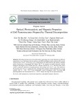
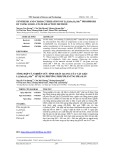
Figure 1 depicts the powder X-ray diffraction (PXRD) pattern of the dried precipitate
samples following calcination at 850 °C for one hour. The PXRD spectra in Figure 1 reveal
peaks corresponding to the positions of the CoFe2O4 standard peaks in the reference
database, indicating a cubic structure belonging to the Fd3m space group (JCPDS 22-1086)
(Chung et al., 2023). The Miller indices (hkl), including (111), (220), (311), (400), (422),
(511), (440), and (533), are recorded (Figure 1). All three samples exhibited flat baselines,
indicating high crystallinity with no observable peaks corresponding to impurity phases.
Additionally, a shift in the 2θ angular position was observed with changes in the x value
(Table 1), indicating partial replacement of Fe3+ ions by La3+ ions. This observation is
consistent with findings in other spinel ferrite systems synthesized using various methods,
such as CoFe2-xYxO4 (Chung et al., 2023), CuFe2-xHoxO4 (Hoang et al., 2022), CuFe2-xCexO4
(Elaykumar et al., 2019), CoFe2-xYxO4 (Patankar et al., 2017), and CoFe2-xLaxO4
(Fabricio et al., 2020).
Figure 1. PXRD patterns of La-doped CoFe2O4 nanoparticles annealed at 850 °C
Table 1. PXRD characteristics of CoFe2-xLaxO4 nanocrystals annealed at 850 °C for 1h
CoFe
2-x
La
x
O
4
2θ
(311)
, °
FWHM, °
D
hkl
, nm
a, Å
x = 0
35.757
0.154
54.2
8.32
x = 0.025
35.524
0.312
26.7
8.36
x = 0.05
35.558
0.343
24.3
8.39
A noticeable reduction in the 2θ angle corresponding to the (311) peak occurs as x
increases from 0 to 0.025 (Δ2θ = 0.233°), followed by a slight increase as x further increases
to 0.05 (Δ2θ = 0.034°) (Table 1). This observation confirms the substitution of Fe3+ ions for
La3+ ions (Chung et al., 2023; Fabricio et al., 2020). Moreover, an expansion of the peak
width is observed as x increases from 0 to 0.05 (Figure 1 and Table 1), indicating a gradual
decrease in the average crystal size (Dhkl, nm) calculated using equation (1) while the cubic
lattice parameter (a, Å) determined by equation (2) slightly increases (Table 1). Similar
trends of decreasing crystal size and increasing cubic lattice parameters with increasing
HCMUE Journal of Science
Vol. 21, No. 7 (2024): 1167-1176
1171
dopant content have been reported in doped spinel ferrite systems such as CuFe2-xCexO4 and
CuFe2-xHoxO4 (Elaykumar et al., 2019; Hoang et al., 2022).
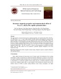
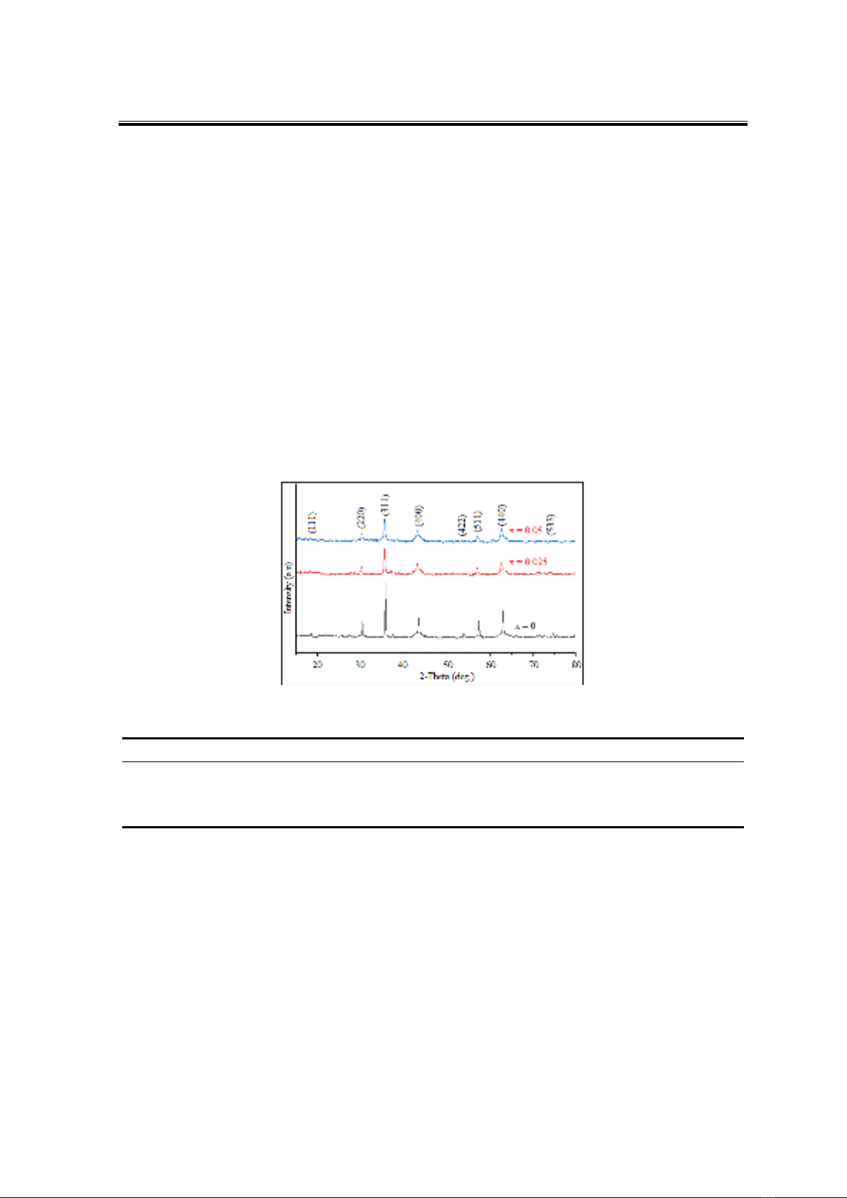
To determine the qualitative and quantitative composition of the elements present in
the sample, EDX analysis was performed on the CoFe1.95La0.05O4 sample. The results are
presented in Figure 2 and Table 2.
Figure 2. EDX and EDX-mapping images of CoFe1.95La0.05O4 nanoparticles annealed at 850 °C
The EDX diagram reveals peaks corresponding only to the elements present in the
sample, namely La, Fe, Co, and O, with no peaks observed for the impurity element Na. The
mass percentage and atomic percentage compositions of Co, Fe, La, and O closely match
their expected proportions in the CoFe1.95La0.05O4 formula, with deviations of less than 5%
(Table 2). This indicates a uniform distribution of Co, Fe, La, and O atoms within the
CoFe1.95La0.05O4 crystal (Figure 2).