
Lee et al. Journal of Inflammation 2010, 7:31
http://www.journal-inflammation.com/content/7/1/31
Open Access
RESEARCH
© 2010 Lee et al; licensee BioMed Central Ltd. This is an Open Access article distributed under the terms of the Creative Commons At-
tribution License (http://creativecommons.org/licenses/by/2.0), which permits unrestricted use, distribution, and reproduction in any
medium, provided the original work is properly cited.
Research
The role of Qa-2, the functional homolog of HLA-G,
in a Behcet's disease-like mouse model induced by
the herpes virus simplex
Meeyoung Lee
†1
, Bunsoon Choi
†1
, Hyuk Jae Kwon
1
, Ju A Shim
1
, Kyung Sook Park
2
, Eun-So Lee
3
and
Seonghyang Sohn*
1,4
Abstract
Background: It has been suggested that the HLA-G molecule is a genetic risk factor for Behcet's disease (BD). In this
study, we evaluated the level of Qa-2, a murine nonclassical class I MHC molecule and possible functional homolog of
HLA-G, to determine if it was associated with various symptoms of BD-like mice. In addition, we investigated siRNA
(small interfering RNA) treatment to determine if it inhibited Qa-2 expression, thereby changing the symptoms of mice.
Methods: RNA interference (RNAi) and vector transfection were employed to manipulate gene expression in vivo in
mice. siRNA (small interfering RNA) or Qa-2 expression vector was applied to inhibit or up-regulate Qa-2 expression,
respectively.
Results: The Qa-2 levels in granulocytes were lower in BD-like mice than in normal controls. The silencing of Qa-2 by
intravenous injection of siRNA (500 nmol/mouse, 4 times at 3-day intervals) specifically reduced the Qa-2 levels and
worsened the BD-like symptoms.
Conclusions: Silencing Qa-2 by injecting siRNA into mice resulted in deterioration of symptoms in BD-like mice.
Background
Since HLA-G (human leukocyte antigen-G) was first
detected by Geraghty et al. [1], it has been reported that
HLA-G protein is expressed at the feto-maternal inter-
face during pregnancy [2] and on a subset of thymic epi-
thelial cells [3], and that it is also involved in maintenance
of tolerance of the maternal immune system toward the
semi-allogeneic fetus. HLA-G is also expressed in other
tissues such as intestinal mucosa [4] and PBMC [5].
Numerous studies have evaluated the relevance of HLA-
G under pathologic conditions such as transplantation,
autoimmunity, cancer, and hematologic malignancies [6].
HLA-G interacts with different natural killer (NK) cell
receptors and is able to inhibit NK and T-cell cytotoxicity,
as well as T-cell proliferation [7]. Interestingly, HLA-G
has been described as a unique ligand of the killer cell
inhibitory receptor, KIR2DL4, which is expressed on the
surface of all NK cells [8]. Furthermore, HLA-G inhibits
the transendothelial migration of NK cells [9], shifts the
cytokine balance toward Th2 dominance [10], and sup-
presses the proliferation of allogeneic CD4+ T lympho-
cytes [11,12]. Taken together, HLA-G exerts specific
inhibitory effects against immune cells. In addition,
recent studies indicate unexpected expression of HLA-G
proteins in chronic cutaneous inflammatory diseases,
such as psoriasis [13] and atopic dermatitis [14].
Behcet's disease (BD) is a chronic multi-systemic disor-
der that involves the gastrointestinal, mucocutaneous,
ocular, vascular, central nervous, and articular systems.
BD has a chronic course that includes periodic exacerba-
tions and progressive deterioration [15]. Although the
etiology of BD is unclear, viral infection has long been
postulated as one of its main factors. The viral hypothesis
has been verified by detection of the virus in saliva [16],
intestinal ulcers [17], and genital ulcers [18] of patients
with BD since it was first proposed by Hulûsi Behçet [19].
Furthermore, inoculation of the earlobe of ICR mice with
herpes simplex virus (HSV) enables development of a
* Correspondence: sohnsh@ajou.ac.kr
1 Laboratory of Cell Biology, Ajou University Institute for Medical Sciences,
Suwon, Korea
† Contributed equally
Full list of author information is available at the end of the article

Lee et al. Journal of Inflammation 2010, 7:31
http://www.journal-inflammation.com/content/7/1/31
Page 2 of 12
BD-like animal model [20]. Manifestations in mice fol-
lowing HSV inoculation involve multiple symptoms such
as oral ulcers, genital ulcers, skin ulcers, eye symptoms,
gastrointestinal ulcers, arthritis, and neural involvement,
as well as skin crusting. The frequency of these symptoms
is similar to that of patients with BD [21]. In addition to
viral causes of BD, several studies have identified lympho-
cyte dysfunction as a possible cause [22,23]. Thus, atten-
tion has been focused on the T helper (Th) 1 and Th2
cytokines, with Th1 cells perhaps playing a more impor-
tant role in the immunopathogenesis of BD [24]. When
the Th2 adjuvant, aluminium hydroxide (alum), was
mixed with ovalbumin (OVA) and injected into mice suf-
fering from BD, their cutaneous symptoms were
improved [25].
Park et al. [26] reported that the frequency of haplo-
types containing a HLA-G 3741_3754 14 base pair inser-
tion and 1597*delC was increased in BD patients.
Moreover, individuals who were homozygous with the
3741_3754*ins14/*ins14 genotype were found to have a
risk of BD that was 2.7-times greater than that of the con-
trols. The HLA-G 3741*+14bp induces a significantly
lower expression level than the complete HLA-G mRNA
isoforms. In addition, the HLA-G 3741_3754 14-base
pair insertion allele was found to occur significantly more
frequently in BD patients with ocular, arthritis, and CNS
symptoms than in controls, and this insertion was found
to be related to the lower serum level of HLA-G [26]. The
authors who presented these findings suggested that
these HLA-G allelic variants are genetic risk factors for
BD. In addition, the HLA-G*010101 alleles have been
shown to have a significantly lower frequency in BD
patients than in control subjects [27].
As a result, it is important to determine if HLA-G con-
tributes to the pathogenesis of BD. To accomplish this,
Qa-2 expression, the functional homolog of HLA-G in
mice, was identified and modulated by small interfering
RNA (siRNA) and the Qa-2 expression vector. The results
of this study confirmed that decreased Qa-2 levels are
related to changes in the disease pattern and deteriora-
tion of BD-like symptoms.
Methods
Animals, induction of BD-like symptoms, and scoring of BD
activity
Five-week-old ICR male mice were used in this study. To
induce a BD-like disease in mice, their earlobes were
scratched with a needle and then inoculated with 1.0 ×
106 plaque forming units/ml of HSV type 1 (F strain).
Virus inoculation was performed twice with a 10-day
interval, after which the mice were observed for 30
weeks. Mice were housed in conventional temperature-
and light-controlled rooms (20-22°C, 12 h light cycle
starting at 8:00 a.m.) and had free access to food and
water. During the experiment, the animals were observed
closely. Mice were handled in accordance with the proto-
cols approved by our institutional animal care committee.
Manifestations in mice after HSV inoculation involved
multiple symptoms including oral ulcers, genital ulcers,
skin ulcers, eye symptoms, intestinal ulcers, arthritis, and
neural involvement, as well as skin crusting. Oral, genital,
and other skin ulcers (including bulla and crust), and eye
symptoms were all classified as major symptoms, while
other symptoms were classified as minor symptoms [20].
Overall, 15% of the HSV-injected mice developed BD-like
symptoms. The disappearance of symptoms and decrease
in lesion size constituted an improvement, similar to in
human patients.
The animals were observed once a week after HSV
inoculation, at which time the severity of BD was deter-
mined according to the BD activity index, as outlined in
the Behcet's Disease Current Activity Form 2006 http://
www.behcet.ws/pdf/BehcetsDiseaseActivityForm.pdf.
The occurrence of the following symptoms in the mouse
model were selected for analysis: mouth ulceration, geni-
tal ulceration, erythema, skin pustules, skin ulceration,
joints-arthritis, diarrhea, red eye (right, left), reduced
vision (right, left), loss of balance, discoloration, and
swelling of the face. The score of each symptom was one,
and the total score before and after treatment was used to
determine the severity of BD. Mice exhibiting signifi-
cantly reduced symptoms were photographed to docu-
ment improvement after treatment.
Synthesis and in vitro test of siRNA
Qa-2 siRNA oligonucleotides with the following sense
and anti-sense sequences were designed and synthesized
by Dharmacon (Chicago, IL, USA). The Qa-2 protein was
encoded by four genes in the Q region, Q6, Q7, Q8 and
Q9. These genes have a typical class I MHC gene struc-
ture involving exon 1 (leader peptide), exon 2 (α1
domain), exon 3 (α2 domain), exon 4 (α3 domain), exon 5
(transmembrane domain), and exons 6, 7 and 8 (cytoplas-
mic domains). As shown in Table 1, we selected four
sequences located in each domain to synthesize siRNA.
To confirm the function of interference, the synthesized
siRNA was tested in vitro in peripheral blood mononu-
clear cells (PBMC). To accomplish this, PBMCs were iso-
lated from 5-6 week-old ICR mice and cultured at 1 × 10⁵
cells/ml in DMEM medium with 1% antibiotics and 10%
FBS. siRNA (200 nM) was incubated with 3 μL of oligo-
fectamin (Gibco-Invitrogen, Rockville, MD) in 200 μL of
DMEM medium. After 24 h of treatment with siRNA, the
PBMCs were harvested and subjected to RT-PCR.
In vivo siRNA injection
For application to mice, 500 nM of siRNA in 200 μL of 5%
glucose, including transfection reagent jetPEI (Polyplus,

Lee et al. Journal of Inflammation 2010, 7:31
http://www.journal-inflammation.com/content/7/1/31
Page 3 of 12
France, Illkirchcedex), was intravenously injected into
mice one to four times with a three day interval between
injections. Two-days after the last injection, mice were
photographed and the PBMCs were analyzed using a flu-
orescence-activated cell sorter (FACS). The control group
was injected with 200 μL of 5% glucose. Qa-2 leader pep-
tide domain siRNA did not down-regulate the Qa-2
mRNA level in in vitro PBMC cultures when compared to
other domains; therefore, the leader peptide domain
siRNA was injected as a control. For in vivo administra-
tion to mice, 1.5 μL of transfection reagent was mixed
with 5% glucose and siRNA. The Qa-2 siRNA was mixed
with α3 domain, transmembrane domain and cytoplas-
mic domain in equal amounts, after which it was admin-
istered to mice.
Flow cytometry
To analyze the Qa-2 expression, cells were harvested and
fixed with 4% formaldehyde in 1% fetal bovine serum
containing PBS for 20 min at room temperature, after
which they were incubated with FITC-conjugated anti-
Qa-2 antibody (eBioscience, San Diego, CA, USA).
Stained cells were analyzed in FACS Vantage using the
Cell Quest software (Becton Dickinson, Franklin Lakes,
NJ, USA) by collecting at least 10,000 gated lymphocytes
[7].
Reverse transcription PCR (RT-PCR)
Total RNA was isolated using TRIzol (Life Technologies,
Helgerman, CT) according to the manufacturer's recom-
mendations. Two μg of total RNA were used as a template
for cDNA synthesis, which was conducted using a Super-
Script III First-Strand Synthesis System for RT-PCR kit
(Invitrogen, Carlsbad, CA). The cDNA was amplified by
PCR using the following primers: Qa-2, Sense: 5' -
AGGTCTTAT GGTGCTGTCAC-3', Anti sense: 5'- TGT
GTAATTCTGCTCCTTCC -3'; β-actin, Sense: 5'-TG
GAATCCTGTGGCATCCATGAAAC -3', Antisense: 5'-
TAAAACGCAGCTCAGTAACAGTCCG-3'; IFNγ,
Sense: 5'-AGCGGCTGACTGAACTCAGATTGTAG
CTTGTACCTTTACTTCACTG-3', Antisense: 5'-GTC
ACAGTTTTCA GCTGTATAGGG-3'. Amplified PCR
products were visualized on 1.2% agarose gels.
Real Time PCR
For real-time SYBR Green RT-PCR, a 20-μl reaction con-
taining 10 μl of 2× Quantitect SYBR Green Master Mix
(Qiagen, Valencia, CA, USA) was employed. The master
mix was composed of hot start Taq polymerase, a 0.4 μL
mix of 2 reverse transcriptases, 0.5 μL (10 ng/μL) of tem-
plate and 0.8 μL of primers. An ABI 7900 HT thermal
cycler (Lab Centraal B.V., Haarlem, The Netherlands) was
used for all real-time RT-PCR assays. Reverse transcrip-
tion was conducted at 50°C for 30 min, followed by dena-
turation at 95°C for 15 min. DNA was amplified by
subjecting the samples to 40 cycles of 95°C (30 s), 55°C
(30 s), and 72°C (30 s). Real-time RT-PCR data were col-
lected for 15 sec at 75°C to avoid non-specific fluores-
cence due to the formation of primer dimers at low
template concentrations. For generation of standard
quantitation curves, the cycle threshold values were plot-
ted proportionally against the logarithm of the input copy
numbers. Negative controls were included in each run.
Qa-2 vector construction
Qa-2 cDNA was amplified from total RNA extracted
from ICR mice lymph nodes by reverse transcriptase -
polymerase chain reaction (RT-PCR) using the following
primers: sense 5'-CGGGATCCCGATGGCTCTAACAA
TGCTGC-3', antisense 5'-CGGAATTCCGCTTCGTGT-
GAAAGTATGGAG-3'. The sense primer included the
BamH1 restriction site and the antisense primer included
the EcoR1 restriction site. The cDNA was subsequently
digested with BamHI and EcoRI and then inserted into
eukaryotic expression vector pcDNA3.1 (Invitrogen,
Carlsbad, CA, USA). Verification of the recombinant
construct was performed by DNA sequencing. The
empty vector pcDNA3.1 was used as a control. All plas-
mids were purified by two rounds of passage through
Endo-Free columns (Qiagen, Chatsworth, CA, USA), as
described elsewhere [28].
Qa-2 vector transfection to HeLa cells
HeLa cells were maintained in Dulbecco's modified Eagle
medium (DMEM) supplemented with 2 mM glutamine,
100 units/ml penicillin, 100 μg/ml streptomycin, and 5%
(v/v) dextran-charcoal-treated fetal bovine serum at 37°C
in 5% CO2. Cells were plated at 106 cells/10 cm dish the
day before transfection, after which they were transfected
using a lipofectimine kit (Invitrogen, Paisley, UK) accord-
Table 1: Qa-2 siRNA oligonucleotide sequences
Qa-2 domain siRNA oligonucleotides sequences
Leader peptide
domain
5'-CAACACUCGCAAUAUU-3'(sense)
3'-GUUGUGAGCGACGUUAUAA-5'(antisense)
α3 domain 5'-AGGUCUUAUGGUGCUGUCAUU-3'(sense)
3'-UUUCCAGAAUACCACGACAGU-
5'(antisense)
Transmembrane
domain
5'-UGUGAUGAAUAGGAGGUGAUU-3'(sense)
3'-UUACACUACUUAUCCUCCACU-
5'(antisense)
Cytoplasmic
membrane domain
5'-UAGAGCUCUGAUAGAUCUCUU-3'(sense)
3'-UUAUCUCGAGACUAUCUAGAG-
5'(antisense)
Lee et al. Journal of Inflammation 2010, 7:31
http://www.journal-inflammation.com/content/7/1/31
Page 4 of 12
ing to the manufacturer's instructions. The vector
pcDNA3.1 was transfected into HeLa cells as a control.
Administration of Qa-2 vector to mice
Normal and BD mice were intraperitoneally injected once
with 50 ng of pcDNA 3.1 or pcDNA 3.1 Qa-2 vector per
mouse, and their splenocytes or macrophages were iso-
lated three days later and analyzed by flow cytometry.
Vector mixed with transfection reagent jetPEI was
injected into mice and the frequency of Qa-2 protein
expression was analyzed by FACS.
Statistical analysis
All data are presented as the mean ± SE. Statistical differ-
ences between groups were determined using a Student's
t test and the Bonferroni correction. Statistical analysis
was conducted using MedCalc® version 9.3.0.0.
Results
Qa-2 mRNA and Qa-2 positive PBMCs were lower in BD
symptomatic mice than in normal healthy mice
RT-PCR revealed that Qa-2 mRNA expression in periph-
eral blood mononuclear cells (PBMC) of mucocutaneous
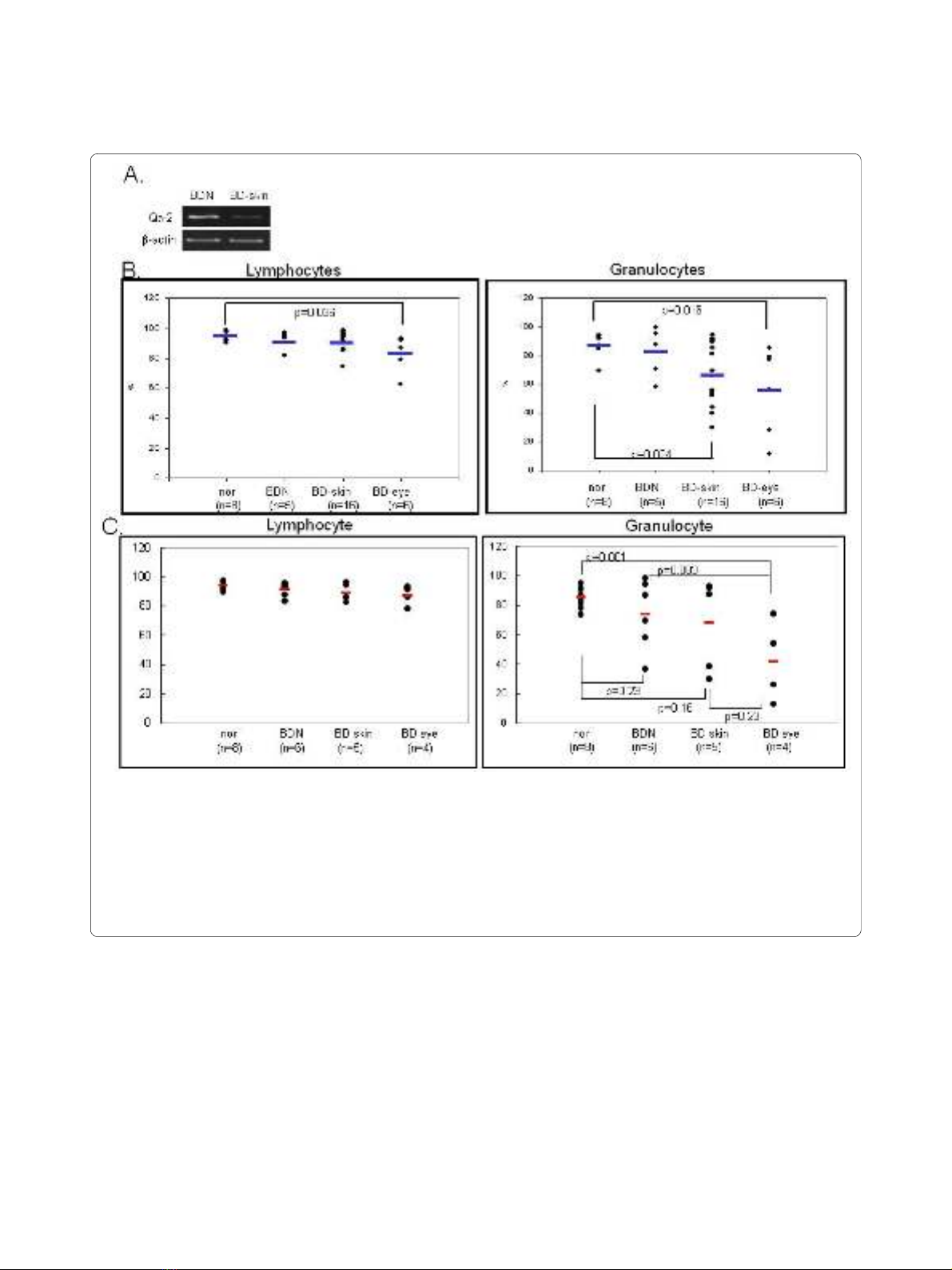
Figure 1 Qa-2 expression in PBMC of BD. A. RT-PCR demonstrated that mRNA expression was lower in PBMC of BD skin than in BD normal mice. B.
The frequency of Qa-2 in PBMC of normal healthy controls, BD asymptomatic (BD normal) mice, BD mucocutaneous symptomatic mice (BD skin), and
BD mucocutaneous and ocular symptomatic mice (BD eye) as determined by FACS analysis. In lymphocytes, the Qa-2 levels in BD eye mice were sig-
nificantly lower than in normal healthy mice (p = 0.036). These levels were also lower than in BD skin mice, although this difference was not significant.
In granulocytes, the Qa-2 levels in BD eye mice were significantly lower than in normal healthy mice (p = 0.016). The Qa-2 levels in BD eye mice were
lower than in normal and BD skin mice, although this difference was not statistically significant. Qa-2 levels in BD skin were significantly lower than in
normal controls (p = 0.024). C. The portion of Qa-2 positive cells in lymphocytes or granulocytes. The frequency of Qa-2 positive cells in the granulo-
cytes of BD skin and BD eye mice was lower than in normal controls and BD normal mice (BDN). The frequencies of Qa-2 positive cells in BD eye mice
were significantly lower than those in normal controls (p = 0.001).
Lee et al. Journal of Inflammation 2010, 7:31
http://www.journal-inflammation.com/content/7/1/31
Page 5 of 12
symptomatic BD mice was down-regulated when com-
pared to asymptomatic BD mice, despite HSV inocula-
tion (BD normal, BDN) (Figure 1A). Next, Qa-2 levels in
PBMCs obtained from normal healthy mice, BD asymp-
tomatic mice (BDN), BD skin symptomatic mice (BD
skin), and BD eye symptomatic mice (BD eye) were ana-
lyzed by flow cytometry. The symptoms of BD skin con-
sisted of typical mucocutaneous symptoms in mice
without ocular symptoms, while those of BD eye mice
consisted of ocular symptoms with mucocutaneous
symptoms. After FACS staining, lymphocytes and granu-
locytes were separated by gating. In lymphocytes, Qa-2
positive cells accounted for 94.78 ± 3.56% in normal
healthy mice, 92.56 ± 6.13% in BD normal mice, 91.73 ±
5.96% in BD skin, and 84.49 ± 11.95% in BD eye mice. BD
eye mice were found to have a statistically lower number
of Qa-2 positive cells than normal healthy mice (p =
0.036). In granulocytes, Qa-2 positive cells were 87.01 ±
7.97% in normal healthy mice, 82.29 ± 17.47% in BD nor-
mal mice, 67.9 ± 21.42% in BD skin mice, and 56.00 ±
30.49% in BD eye mice. BD skin and BD eye mice showed
significantly lower levels of Qa-2 positive cells than nor-
mal healthy mice (p = 0.024, p = 0.016 each) (Figure 1B).
The portion of Qa-2 positive cells in the granulocytes of
BD skin and BD eye mice was lower than that of normal
control and BD normal (BDN) mice. The portion of Qa-2
positive cells in the granulocytes of BD eye mice was sig-
nificantly lower than that of normal controls (p = 0.001)
(Figure 1C). As shown in Figure 1, the decreased level of
Qa-2 was related to the BD symptoms.
RNA interference of Qa-2 transcription in vitro; Qa-2 siRNA
reduced Qa-2 mRNA levels in PBMCs of normal mice
PBMCs isolated from normal mice were transfected for
24 h with Qa-2 siRNA with different domains, after
which the expression of Qa-2 was determined by reverse
transcriptase-PCR. siRNA for the α3 domain, transmem-
brane domain, and cytoplasmic domain inhibited the Qa-
2 level; however, the leader peptide domain did not.
Mixed siRNA consisting of equal amounts each of these
four domains did not downregulate the Qa-2 mRNA
level. Flow cytometric analysis also showed a decreased
frequency of Qa-2 expression in the Qa-2 siRNA domain-
treated groups, except for the leader peptide domain (Fig-
ure 2).
Downregulation of Qa-2 by intravenous injection of siRNA
into BD mice
Next, an siRNA mixture composed of the siRNA of the
α3 domain, transmembrane domain and the cytoplasmic
domain was injected into BD mice. Five to six individual
BD mice in each group were intravenously injected once
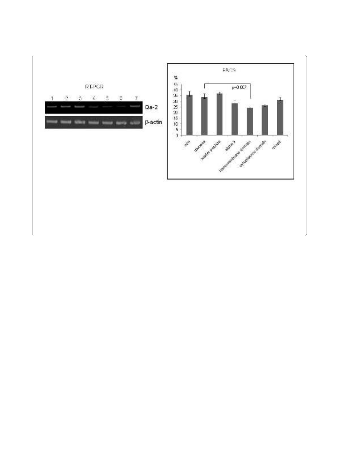
Figure 2 Qa-2 siRNA reduced Qa-2 mRNA and the frequency of Qa-2 positive cells in PBMC of normal mice. PBMC isolated from mice were
transfected with Qa-2 siRNAs with different Qa-2 domains for 24 hrs, and the expression of Qa-2 was then determined by reverse transcriptase-PCR
and FACS analysis. Lanes 4, 5 and 6 (α3 domain, transmembrane domain, and cytoplasmic domain, respectively) showed that siRNA effectively re-
duced the Qa-2 mRNA levels. Lane 3 (leader peptide) did not decrease the Qa-2 level. Lane 7 (a mixture of leader peptide, α3 domain, transmembrane
domain, and cytoplasmic domain) also did not decrease the Qa-2 level. Lane 1, control (not treated); Lane 2, 5% glucose treated; Lane 3, leader peptide
200 nmole; Lane 4, α3 domain 200 nmole; Lane 5, transmembrane domain 200 nmole; Lane 6, cytoplasmic domain 200 nmole; Lane 7, mixed 200
nmole (leader peptide + α3 domain+ transmembrane domain + cytoplasmic domain).


%20--%3e%3cdefs%3e%3cstyle%3e%20.st0%20{%20fill:%20%23fff;%20}%20.st1%20{%20fill:%20%237800fa;%20}%20%3c/style%3e%3c/defs%3e%3cpath%20class='st1'%20d='M117.78,12.18H43.11c2.9,3.47,4.65,7.94,4.65,12.82,0,5.6-2.3,10.66-6.01,14.29h76.02l7.22-13.56-7.22-13.56Z'/%3e%3cg%3e%3cpath%20class='st0'%20d='M53.58,26.17h-.59v-1.46h.59v-4.96h2.83c1.78,0,2.67.94,2.67,2.82v5.76c0,1.87-.89,2.81-2.67,2.81h-2.83v-4.96ZM55.36,21.37v3.34h1.1v1.46h-1.1v3.34h1.01c.61,0,.91-.37.91-1.1v-5.93c0-.74-.3-1.1-.91-1.1h-1.01Z'/%3e%3cpath%20class='st0'%20d='M65.99,31.14h-1.8l-.31-2.07h-2.19l-.31,2.07h-1.64l1.82-11.39h2.62l1.82,11.39ZM65.28,18.04c-.25.46-.51.77-.75.94-.21.15-.47.22-.79.22-.26,0-.57-.07-.92-.22l-.38-.15c-.14-.05-.26-.07-.37-.07-.3,0-.53.18-.71.54l-.91-.68c.25-.46.51-.77.75-.94.21-.14.48-.21.79-.21.26,0,.57.07.92.21l.38.15c.14.05.26.07.37.07.3,0,.53-.18.71-.54l.91.68ZM61.91,27.52h1.73l-.87-5.76-.87,5.76Z'/%3e%3cpath%20class='st0'%20d='M74.53,26.89v1.52c0,1.91-.89,2.86-2.67,2.86s-2.67-.95-2.67-2.86v-5.93c0-1.91.89-2.86,2.67-2.86s2.67.95,2.67,2.86v1.11h-1.69v-1.22c0-.75-.31-1.12-.93-1.12s-.93.37-.93,1.12v6.15c0,.74.31,1.11.93,1.11s.93-.37.93-1.11v-1.63h1.69Z'/%3e%3cpath%20class='st0'%20d='M81.4,31.14h-1.8l-.31-2.07h-2.19l-.31,2.07h-1.64l1.82-11.39h2.62l1.82,11.39ZM75.9,19.2l1.52-1.91h1.71l1.51,1.91h-1.61l-.76-.95-.75.95h-1.61ZM77.32,27.52h1.73l-.87-5.76-.87,5.76ZM83.1,15.99l-1.76,1.91h-1.26l1.17-1.91h1.86Z'/%3e%3cpath%20class='st0'%20d='M84.86,19.75c1.78,0,2.67.94,2.67,2.82v1.48c0,1.87-.89,2.81-2.67,2.81h-.85v4.28h-1.79v-11.39h2.64ZM84.01,21.37v3.86h.85c.58,0,.87-.36.87-1.08v-1.71c0-.71-.29-1.07-.87-1.07h-.85Z'/%3e%3cpath%20class='st0'%20d='M93.51,19.75c1.78,0,2.67.94,2.67,2.82v1.48c0,1.87-.89,2.81-2.67,2.81h-.85v4.28h-1.79v-11.39h2.64ZM92.66,21.37v3.86h.85c.58,0,.87-.36.87-1.08v-1.71c0-.71-.29-1.07-.87-1.07h-.85Z'/%3e%3cpath%20class='st0'%20d='M98.8,31.14h-1.79v-11.39h1.79v4.88h2.03v-4.88h1.83v11.39h-1.83v-4.88h-2.03v4.88Z'/%3e%3cpath%20class='st0'%20d='M105.36,24.55h2.46v1.62h-2.46v3.34h3.09v1.63h-4.88v-11.39h4.88v1.63h-3.09v3.18ZM108.17,17.29l-1.76,1.91h-1.26l1.17-1.91h1.86Z'/%3e%3cpath%20class='st0'%20d='M112.2,19.75c1.78,0,2.67.94,2.67,2.82v1.48c0,1.87-.89,2.81-2.67,2.81h-.85v4.28h-1.79v-11.39h2.64ZM111.35,21.37v3.86h.85c.58,0,.87-.36.87-1.08v-1.71c0-.71-.29-1.07-.87-1.07h-.85Z'/%3e%3c/g%3e%3ccircle%20class='st1'%20cx='25'%20cy='25'%20r='20'/%3e%3cpath%20class='st0'%20d='M32.78,19.27c2.92,0,4.43,2.55,5.28,5.33l.71,2.17c.14.38-.33.75-.71.75h-5.61c.19-.33.24-.71.09-1.08l-.75-2.45c-.43-1.32-.99-2.64-1.79-3.77.75-.57,1.65-.94,2.78-.94h0ZM25,18.38c3.25,0,4.9,2.78,5.89,5.89l.76,2.45c.14.42-.33.8-.8.8h-11.69c-.42,0-.94-.38-.8-.8l.75-2.45c.99-3.11,2.64-5.89,5.89-5.89h0ZM25,11.35c1.74,0,3.11,1.37,3.11,3.11s-1.37,3.11-3.11,3.11-3.11-1.41-3.11-3.11,1.41-3.11,3.11-3.11h0ZM17.27,19.27c1.08,0,1.98.38,2.73.94-.8,1.13-1.37,2.45-1.74,3.77l-.8,2.45c-.14.38-.05.75.09,1.08h-5.56c-.42,0-.9-.38-.75-.75l.71-2.17c.9-2.78,2.41-5.33,5.33-5.33h0ZM17.27,12.91c1.51,0,2.78,1.27,2.78,2.83s-1.27,2.83-2.78,2.83-2.83-1.27-2.83-2.83,1.27-2.83,2.83-2.83h0ZM32.78,12.91c1.56,0,2.78,1.27,2.78,2.83s-1.23,2.83-2.78,2.83-2.83-1.27-2.83-2.83,1.27-2.83,2.83-2.83h0ZM27.07,28.56v.09c0,.57-.24,1.08-.61,1.46h0v.05c-.38.33-.9.57-1.46.57s-1.08-.24-1.46-.61h0c-.38-.38-.61-.9-.61-1.46v-.09h1.41v.09c0,.19.05.38.19.47v.05c.09.09.28.19.47.19s.38-.09.47-.19v-.05c.14-.09.24-.28.24-.47t-.05-.09h1.41ZM30.99,28.56v.09c0,1.65-.66,3.16-1.74,4.24-1.08,1.08-2.59,1.79-4.24,1.79s-3.16-.71-4.24-1.79l-.05-.05c-1.04-1.08-1.7-2.55-1.7-4.2v-.09h1.41v.09c0,1.27.47,2.4,1.27,3.25h.05c.85.85,1.98,1.37,3.25,1.37s2.4-.52,3.25-1.37c.85-.8,1.37-1.98,1.37-3.25v-.09h1.37ZM34.99,28.56v.09c0,2.78-1.13,5.28-2.92,7.07-1.79,1.79-4.29,2.92-7.07,2.92s-5.23-1.13-7.07-2.92c-1.79-1.79-2.92-4.29-2.92-7.07v-.09h1.41v.09c0,2.4.94,4.53,2.5,6.08,1.56,1.56,3.72,2.5,6.08,2.5s4.52-.94,6.08-2.5c1.56-1.56,2.5-3.68,2.5-6.08v-.09h1.41Z'/%3e%3c/svg%3e)