JOURNAL OF SCIENCE AND TECHNOLOGY DONG NAI TECHNOLOGY UNIVERSITY
127
Special Issue
CHALLENGES AND OPPORTUNITIES IN AI-DRIVEN MEDICAL IMAGE ANALYSIS: A COMPREHENSIVE REVIEW
1Faculty of Information Technology, Dong Nai Technology University, Bien Hoa City, Vietnam 2Universiti Malaysia Pahang Al-Sultan Abdullah, Pahang City, Malaysia *Corresponding author: trinhdinhthang@dntu.edu.vn
Trinh Dinh Thang1*, Firdaus Hassan2, Hoang Quoc Ky1, Nguyen Duc Manh1
GENERAL INFORMATION
Received date: 27/03/2024
Revised date: 16/05/2024
Accepted date: 11/07/2024
KEYWORD
AI;
Analysis;
Challenges;
Medical Imaging;
Opportunities.
ABSTRACT The intricacies and limitations of AI-driven medical image analysis are the focus of this paper, particularly challenges in acquiring, preprocessing, and analyzing medical imaging data. We explore how Artificial Intelligence (AI) presents significant potential to revolutionize this field: improving diagnostic accuracy; streamlining workflows–even enhancing patient outcomes. Our investigation delves deep into two key aspects – integrating AI technologies into existing systems like Picture Archiving and Communication Systems (PACS), discussing their implications not only for clinical practice but also for elevating patient care. Our goal: to confront present difficulties and unleash the complete potential of Artificial Intelligence in revolutionizing medical image analysis; we intend this pursuit through a multi-disciplinary strategy--one that encourages cooperation among healthcare professionals, data scientists, and computer engineers.
1. INTRODUCTION in various
and computations,
productivity and
The fourth industrial revolution (4IR) of the 21st century is marked by the convergence of artificial intelligence (AI) and computer-based information communication technology (ICT), bringing about remarkable advancements in productivity across various sectors. At the heart of this evolution, AI surpasses human capabilities to process large datasets, thereby enabling swift and accurate decision-making. Consequently, AI emerges as a vital solution for addressing enhancing challenges across diverse fields (Neethi et al., 2024) .
AI is critical for data-driven decision- industries, especially making healthcare. Massive amounts of data, rapidly advancing high- performance models validated against natural images are delivering results that exceed the interpretive capabilities of diagnostic and doctors. Using AI resource increase to utilization, operational performance, and quality of service in healthcare operations can significantly improve care outcomes and increase patient satisfaction while reducing costs. A study by Bennett et al. reported that using AI algorithms to diagnose diseases improved diagnostic performance by 41.9%
128
Special Issue
JOURNAL OF SCIENCE AND TECHNOLOGY DONG NAI TECHNOLOGY UNIVERSITY
2.1 AI
and reduced healthcare costs by 58.5%. (Jafar et al., 2022).
shifting
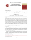
Figure 1: illustrates the distinction between machine learning (ML) and deep learning (DL)
(AI) Artificial Intelligence
In the medical imaging field, machine learning (ML) and deep learning (DL) are applied to analyze medical images of organs, and this field constitutes an active area of research. DL algorithms and model structures have been introduced to DL-based medical image analysis, and studies on lesion detection, quantification, and classification are ongoing. Indeed, healthcare towards is personalized approaches, such as proactive diagnosis and prevention of individual diseases. Therefore, the collection and analysis of large- scale big data such as genetic, medical, and living information by applying AI are essential. In Korea, research on AI medical software termed Doctor Answer has been ongoing since 2018 with the participation of governments, hospitals, and companies to identify a medical approach suitable for the Korean population. The development of Doctor Answer 2.0 is currently in progress (Habuza et al., 2021).
technology encompasses various subfields, prominently including machine learning (ML) and deep learning (DL). ML involves algorithms that learn from data and make predictions or decisions, while DL is a subset of ML that utilizes neural networks with multiple layers to process complex data. Figure 1 illustrates the distinction between ML and DL (Panayides et al., 2020).
classification, detection, In the context of medical imaging, ML and DL techniques have demonstrated remarkable interpreting in analyzing and capabilities medical images, facilitating tasks such as disease and segmentation.
2.2 Medical Field and AI
challenges
Figure 2 depicts the significant impact of AI on the medical field, particularly in disease analysis and diagnosis. In this paper, we investigate the challenges and limitations associated with medical image data in AI-driven medical image analysis. Our key findings indicate that the quality of medical imaging data, pixel size, and image resolution significantly impact the performance of AI models. Additionally, the integration of clinical data, compensating for biological variability, and addressing ethical concerns related to patient privacy are critical for the successful implementation of AI in medical imaging. By analyzing the configuration of medical information systems for transmitting medical image data using AI, we aim to provide insights and these into overcoming improving the efficacy of AI applications in healthcare.
2. RELATED WORK
JOURNAL OF SCIENCE AND TECHNOLOGY DONG NAI TECHNOLOGY UNIVERSITY
129
Special Issue
diagnosis
the potential for imaging, hold medical revolutionizing and disease treatment. They offer not only enhanced accuracy and efficiency but also improved patient outcomes (Panayides et al., 2020).
Figure 2: AI-driven healthcare
lung
Several AI-driven tools have emerged in the field of medical image analysis, each with its own set of advantages and disadvantages. Google AI for lung cancer detection is notable for its high accuracy in detecting lung cancer and reducing false positives. However, its focus is primarily on lung cancer, and it requires extensive computational resources, limiting its application in settings with fewer resources. In contrast, our proposed tool aims to be more versatile, applicable across multiple disease types, and optimized for resource utilization in diverse clinical environments.
tomography imaging (CT)
imaging
The integration of AI into medical practices has yielded notable advancements in disease detection and diagnosis. For instance, a 2019 STAT News report highlighted Google AI's performance cancer, in detecting surpassing the accuracy of six radiologists by identifying 5% more cancers and reducing false positive rates by 11%. Furthermore, research published in 2019 in Nature Medicine demonstrated the effectiveness of AI-based in computed detecting lung cancer cases with an impressive accuracy of 94%. Similarly, magnetic (MRI) exhibited an resonance accuracy exceeding 85% in diagnosing brain tumors (Mohammad Amini et al., 2023).
Google's AI algorithms, DeepMind's technology for breast cancer diagnosis boasts high accuracy and can potentially outperform human radiologists in identifying breast cancer. Despite this, it is limited to breast cancer diagnosis and raises ethical concerns regarding patient data privacy. The proposed tool addresses these privacy issues with robust measures and is designed for broader application across various cancers and diseases, ensuring a wider impact on patient care.
IBM Watson Health Imaging integrates well with existing medical imaging systems and can analyze large datasets, making it a powerful tool for healthcare facilities. However, its high cost and dependency on IBM's ecosystem can be prohibitive for many institutions. Our proposed tool, in contrast, focuses on providing cost-effective solutions and offers greater flexibility in integrating with different systems, making advanced AI-driven medical image analysis accessible to a broader range of healthcare facilities. furthermore, achieved remarkable accuracies: 99% for breast cancer diagnosis; and 95% for lung cancer detection—this underscores their potential to elevate disease-detection rates. Also--in terms of diagnosing metastatic cancer--AI-based methods have demonstrated promise with considerably superior accuracy rates when juxtaposed against conventional techniques. Google's DeepMind algorithm showcased its potential superiority over human doctors in identifying breast cancers—a transformative impact of AI on healthcare underscored by a groundbreaking Nature study published in 2020. AI technologies, when integrated into
130
Special Issue
JOURNAL OF SCIENCE AND TECHNOLOGY DONG NAI TECHNOLOGY UNIVERSITY
flipping, and scaling may be employed to enhance the robustness and diversity of the dataset.
3.2 Model Development and Training
Doctor Answer 1.0, developed in Korea, represents a collaborative effort involving the government and hospitals, tailored specifically to the Korean population. While it is a promising initiative, it is still in the early stages of development and has limited international applicability. Our proposed tool, being more advanced in development, aims for global applicability, making it relevant and useful in various international healthcare settings.
and planning
tuning
Microsoft InnerEye is well-supported for integrates radiotherapy seamlessly with cloud-based platforms for scalability. However, it is primarily focused on radiotherapy and requires internet connectivity and cloud services, which might not be feasible in all settings. Our proposed tool offers offline capabilities, ensuring functionality even in areas with limited internet connectivity, and supports a broader range of medical imaging applications beyond radiotherapy.
3. METHODOLOGY
3.1 Data Collection and Preparation The development of AI models for medical image analysis involves the implementation of deep learning architectures tailored to the specific imaging task. Convolutional neural networks (CNNs) are commonly utilized due to their effectiveness in extracting features from medical images. The training process begins by splitting the dataset into training, validation, and testing sets to evaluate model performance. Transfer learning techniques may be employed, leveraging pre-trained models on large-scale image datasets to expedite training and improve convergence. Hyperparameter is conducted to optimize model performance, including adjustments to learning rates, batch sizes, and network architectures. The training process involves iterative epochs, where the model learns to accurately classify or segment medical images based on annotated ground truth labels (Latif et al., 2020).
3.3 Evaluation and Validation
institutions and image analysis include
labeled by expert radiologists robustness to The methodology for this study involves the collection and preparation of medical image datasets for AI-driven analysis. Medical imaging datasets are sourced from various repositories, healthcare encompassing a diverse range of imaging modalities such as X-ray, CT, MRI, and ultrasound. The datasets include annotated images to facilitate supervised learning tasks (Panayides et al., 2020).
to The trained AI models are evaluated using various metrics to assess their performance in tasks. Common medical evaluation metrics accuracy, sensitivity, specificity, and area under the receiver operating characteristic curve (AUC- ROC). Cross-validation techniques may be employed and ensure generalization of the models across different subsets of the dataset. Additionally, external validation on independent datasets may be conducted validate model further performance and generalizability.
To ensure data quality and consistency, rigorous preprocessing steps are undertaken. This includes standardizing image formats, resolving any artifacts or inconsistencies in the data, and anonymizing patient information to comply with privacy regulations. Additionally, data augmentation techniques such as rotation, The validated AI models undergo rigorous testing against clinical standards and guidelines to assess their clinical utility and efficacy.
JOURNAL OF SCIENCE AND TECHNOLOGY DONG NAI TECHNOLOGY UNIVERSITY
131
Special Issue
particularly valuable for imaging the brain, spinal cord, joints, and soft tissues.
Clinical validation involves comparison with gold and standard diagnostic methods evaluation by domain experts to ascertain the accuracy and reliability of the AI-driven analyses.
Ultrasound: Relies on high-frequency sound waves to generate real-time images of internal organs, blood flow, and fetal development. Ultrasound is widely used for obstetric, abdominal, cardiac, and musculoskeletal imaging.
4. MEDICAL IMAGING SYSTEM
facilitating healthcare,
hardware and
abnormalities,
Positron Emission Tomography (PET): Involves the injection of radioactive tracers that emit positrons, which are detected by a PET scanner to produce three-dimensional images of metabolic activity within the body. PET is valuable for cancer staging, detecting brain and assessing cardiac function.
anatomical structures of
Each
imaging modality
offers
and
functionalities imaging
unique advantages and is selected based on the clinical question, anatomical region of interest, patient characteristics, and imaging requirements.
Medical imaging systems play a pivotal role in modern the acquisition, storage, transmission, and analysis of medical image data for diagnostic and therapeutic purposes. These systems comprise software sophisticated components designed to capture high-quality images and physiological processes within the human body. In this section, we delve into the of components a comprehensive medical system (Panayides et al., 2020).
4.1 Medical Imaging Equipment 4.2 Picture Archiving and Communication System (PACS)
Medical imaging equipment encompasses a diverse array of modalities tailored to specific imaging needs and clinical applications (Najjar, 2023; Tiwari et al., 2019). Some of the commonly utilized imaging modalities include:
ionizing
X-ray: Utilizes
radiation
for detecting bone
to produce images of internal structures, particularly useful lung fractures, abnormalities, and soft tissue calcifications.
information and
facilitating
Computed Tomography (CT): Generates cross-sectional images of the body by combining multiple X-ray projections, providing detailed anatomical the diagnosis of various conditions such as tumors, fractures, and vascular abnormalities.
Magnetic Resonance
enabling the
Imaging
(MRI): Utilizes strong magnetic fields and radio waves to produce detailed images of soft tissues, organs, and blood vessels without ionizing radiation. MRI is
The Picture Archiving and Communication System (PACS) is a cornerstone technology in modern radiology, revolutionizing the way medical images are stored, retrieved, and shared across healthcare facilities. PACS is designed to manage digital images generated by various medical imaging modalities such as X-ray, CT, MRI, ultrasound, and nuclear medicine. The primary functions of PACS include image acquisition, storage, retrieval, and distribution, thereby enabling efficient and seamless access to medical images and related data throughout a healthcare organization. The Picture Archiving and Communication System (PACS) serves as the cornerstone of modern radiology departments, efficient management and distribution of medical image data across healthcare facilities. PACS comprises several integrated components, including:
132
Special Issue
JOURNAL OF SCIENCE AND TECHNOLOGY DONG NAI TECHNOLOGY UNIVERSITY
User
Interface
Image Acquisition Devices: These devices capture medical images using various imaging modalities such as X-ray machines, CT scanners, MRI machines, and ultrasound systems.
and Image Display interpretation, result image and
and Workflow Management: Intuitive user interfaces enable radiologists, technologists, and clinicians to access and interact with medical image data efficiently. Workflow management tools report streamline generation, dissemination, optimizing clinical productivity and patient care delivery.
tools allow
Data Storage and Archive: PACS employs robust storage infrastructure to archive medical image data securely and cost- effectively. Redundant storage systems, backup mechanisms, and disaster recovery strategies ensure data availability and integrity.
Processing Workstations: Radiologists and clinicians utilize dedicated workstations equipped with specialized software for viewing, analyzing, and interpreting medical images. Advanced visualization for multiplanar reconstruction, 3D rendering, and quantitative analysis.
to facilitate the to
Data Communication and Networking Infrastructure: PACS relies on robust network infrastructure seamless transmission of medical image data between acquisition devices, workstations, and storage servers. High-speed networks ensure rapid access real-time images and support collaboration among healthcare professionals.
to
diagnostic accuracy,
PACS facilitates rapid image retrieval, remote access images, and seamless integration with electronic health records information (EHRs) and other healthcare systems. By streamlining image management and enhancing workflow efficiency, PACS reduces improves turnaround times, and enhances patient care outcomes.
Advantages of PACS:
Efficiency: PACS significantly reduces the time required to access and share medical images, faster diagnosis and treatment.
Cost Savings: Eliminating the need for physical films and related storage reduces costs and space requirements.
leading to
Figure 3. The Picture Archiving and Communication System (PACS)
Database System and
Enhanced Patient Care: Faster access to tools
Improved Collaboration: PACS allows for easy sharing of images among healthcare enhancing multidisciplinary professionals, collaboration.
improved diagnostic and images contribute to better patient outcomes.
Information Management: PACS stores medical image data in a centralized database, organized according to patient identifiers, examination details, and imaging modalities. Advanced database management systems ensure data integrity, security, and compliance with regulatory requirements.
JOURNAL OF SCIENCE AND TECHNOLOGY DONG NAI TECHNOLOGY UNIVERSITY
133
Special Issue
5.3 Data Diversity and Representativeness 5. PROBLEMS OF MEDICAL IMAGING DATA Medical imaging datasets often
Medical imaging datasets are crucial for training deep learning (DL) models to classify specific diseases. However, these datasets come with several significant challenges that impact the performance and applicability of AI in medical imaging.
5.1 Challenges in Obtaining High-Quality Data lack diversity, which can limit the generalizability of AI models. Datasets may not represent the full spectrum of patient demographics, including age, gender, ethnicity, and comorbidities. This lack of diversity can lead to biases in AI models, resulting in reduced performance in underrepresented populations (Ghassemi et al., 2020).
5.4 Ethical and Legal Issues
images legal ethical
in annotations can lead
Insurance Portability One of the primary challenges in medical imaging is obtaining large volumes of labeled data. Annotating medical requires professional expertise, which is both time- consuming and resource-intensive. Furthermore, the variability to inconsistencies in training data, affecting the reliability of AI models (Litjens et al., 2017).
Image Sizes and High impose strict requirements on 5.2 Large Dimensionality
The use of medical imaging data raises significant concerns, and particularly regarding patient privacy and data security. Regulations such as the General Data Protection Regulation (GDPR) in Europe and the Health and Accountability Act (HIPAA) in the United States the handling of patient data. Ensuring compliance with these regulations while enabling the use of data for AI research is a complex challenge (Kaushal et al., 2020) . processing, and 6. SUGGESTIONS AND DISCUSSION
Medical images, especially 3D images from modalities like CT and MRI, have large file sizes and high dimensionality. This makes storage, analysis computationally intensive. The large size of these images can also lead to longer processing times and higher costs for data storage infrastructure (Razzak et al., 2018).
Table 2: Limitations of Medical Imaging Data
Clause
Quality
Pixel size
the accuracy and
Addressing Data Quality and Annotation: To improve data quality and consistency, it is essential to develop standardized protocols for image annotation. Leveraging crowd-sourced platforms and semi-automated annotation tools can help alleviate the burden on medical professionals and accelerate the annotation process (Irvin et al., 2019) . Additionally, implementing quality control mechanisms can reliability of ensure annotations.
Small object size
Large Image Managing
Compensating
Additional information
Content Difficulty in obtaining large amounts of labeled data Many 3D images with large sizes Object size is relatively small Requires correction for age in the same tissue image of the same disease Use other information such as sex, smoking, and drinking for classification analysis
Sizes: Advancements in data compression techniques and the use of efficient storage solutions such as cloud-based platforms can help manage the large sizes of medical images. Employing parallel processing and distributed computing
134
Special Issue
JOURNAL OF SCIENCE AND TECHNOLOGY DONG NAI TECHNOLOGY UNIVERSITY
the expertise can also reduce the time required for image processing and analysis (Coudray et al., 2018) .
inclusive and
advancements in
necessitate of medical professionals, which is both time-consuming and costly. Furthermore, the large size and high dimensionality of medical images, particularly 3D images from modalities like CT and MRI, impose substantial demands on storage and computational resources. Addressing these challenges involves developing standardized protocols for data annotation and leveraging semi-automated tools to streamline the process. Additionally, data compression and cloud-based storage solutions can mitigate the burdens associated with large image sizes. Enhancing Data Diversity: To address the issue of data diversity, it is crucial to compile datasets that are representative of diverse patient populations. Collaborative efforts between institutions globally can facilitate the sharing of data and resources, leading to more datasets. comprehensive Synthetic data generation techniques, such as GANs (Generative Adversarial Networks), can also be explored to augment existing datasets with diverse examples (Frid-Adar et al., 2018) Ensuring Ethical Compliance the diversity Secondly,
lack existing datasets
to potential biases
Developing robust data anonymization techniques can help protect patient privacy while enabling the use of medical images for AI research. Establishing clear guidelines and frameworks for data sharing that comply with is essential. legal and ethical standards Engaging with and bodies regulatory stakeholders to create policies that balance innovation with privacy can foster a more conducive environment for AI in healthcare (McKinney et al., 2020).
7. CONCLUSION
and representativeness of medical imaging datasets are crucial for the generalizability of AI models. Many sufficient representation of diverse patient populations, leading in AI-driven diagnoses. Collaborative efforts to compile from multiple datasets comprehensive institutions globally, along with synthetic data generation techniques, can help enhance the diversity and inclusivity of training data. This, in turn, will improve the robustness and fairness of AI models across different demographic groups.
Ethical and
learning (DL)
In this paper, we examined the challenges and complexities associated with medical image analysis using artificial intelligence (AI) and deep techniques. The integration of AI in medical imaging holds immense potential to revolutionize healthcare by enhancing diagnostic accuracy, improving patient outcomes, and reducing healthcare costs. However, realizing this potential requires addressing several critical issues related to the quality, diversity, and ethical management of medical imaging data. legal considerations are paramount when dealing with medical imaging data. Ensuring patient privacy and data security while enabling AI research requires robust anonymization techniques and adherence to regulatory frameworks such as GDPR and HIPAA. Developing clear guidelines and fostering collaboration between regulatory bodies, healthcare providers, and researchers can create an environment that balances innovation with ethical responsibility.
Firstly, the acquisition and annotation of high-quality medical imaging data remain significant obstacles. Accurate annotations The implementation of Picture Archiving and Communication Systems (PACS) plays a pivotal role in managing and utilizing medical
JOURNAL OF SCIENCE AND TECHNOLOGY DONG NAI TECHNOLOGY UNIVERSITY
135
Special Issue
Literature Review and Meta-Analysis on Scalable Blockchain-Based Electronic Voting Systems. Sensors, 22(19), 7585. https://doi.org/10.3390/s22197585 the
technical and interoperability issues other
essential are imaging data. PACS facilitates the efficient acquisition, storage, retrieval, and distribution of digital images, thus enhancing workflow and collaboration among medical professionals. However, initial setup and ongoing maintenance of PACS require significant expertise. investment and Overcoming hospital integrating PACS with information for systems maximizing its benefits.
and protocols
Latif, S., Usman, M., Manzoor, S., Iqbal, W., Qadir, J., Tyson, G., Castro, I., Razi, A., Boulos, M. N. K., Weller, A., & Crowcroft, J. (2020). Leveraging Data to Combat COVID-19: A Science Comprehensive IEEE Review. Transactions on Artificial Intelligence, 1(1), 85–103. https://doi.org/10.1109/TAI.2020.30205 21
D., Alves,
efficient management the To address the identified challenges, we propose several key strategies. Firstly, adopting standardized leveraging advanced technologies for data annotation and the quality and improve processing can imaging datasets. consistency of medical Secondly, promoting data and sharing collaboration across institutions can enhance dataset diversity representativeness. and Thirdly, robust anonymization techniques and clear ethical guidelines are necessary to ensure patient privacy and data security. Finally, continuous investment in infrastructure, such as PACS and cloud-based storage solutions, will and support utilization of medical imaging data. Mohammad Amini, M., Jesus, M., Fanaei P., Sheikholeslami, Hassanzadeh Benam, A., & Hariri, F. (2023). Artificial Intelligence Ethics and Challenges in Healthcare Applications: A Comprehensive Review in the Context of the European GDPR Mandate. Machine Learning and Knowledge Extraction, 5(3), 1023–1035. https://doi.org/10.3390/make5030053 REFERENCE
in Medical 13(17),
Najjar, R. (2023). Redefining Radiology: A Review Intelligence of Artificial Integration Imaging. 2760. Diagnostics, https://doi.org/10.3390/diagnostics13172 760
Informatics 24,
Habuza, T., Navaz, A. N., Hashim, F., Alnajjar, F., Zaki, N., Serhani, M. A., & Statsenko, Y. (2021). AI applications in robotics, diagnostic image analysis and precision medicine: Current limitations, future trends, guidelines on CAD systems for in Medicine medicine. Unlocked, 100596. https://doi.org/10.1016/j.imu.2021.10059 6
Jafar, U., Ab Aziz, M. J., Shukur, Z., & Hussain, H. A. (2022). A Systematic Neethi, A. S., Kannath, S. K., Kumar, A. A., Mathew, J., & Rajan, J. (2024). A comprehensive review and experimental comparison of deep learning methods for automated detection. hemorrhage Engineering Applications of Artificial 108192. Intelligence, 133,
136
Special Issue
JOURNAL OF SCIENCE AND TECHNOLOGY DONG NAI TECHNOLOGY UNIVERSITY
https://doi.org/10.1016/j.engappai.2024. 108192
Ghassemi, M., Oakden-Rayner, L., & Beam, A. L. (2020). The false hope of current approaches to explainable artificial intelligence in health care. The Lancet Digital Health, 2(9), e469-e470.
to fix
Kaushal, A., Altman, R., & Langlotz, C. (2020). Health care AI systems are biased. We that. Harvard Business need Review.
Panayides, A. S., Amini, A., Filipovic, N. D., Sharma, A., Tsaftaris, S. A., Young, A., Foran, D., Do, N., Golemati, S., Kurc, T., Huang, K., Nikita, K. S., Veasey, B. P., Zervakis, M., Saltz, J. H., & Pattichis, C. in Medical Imaging S. (2020). AI Informatics: Current Challenges and Future Directions. IEEE Journal of Informatics, Biomedical and Health 24(7), 1837–1857. https://doi.org/10.1109/JBHI.2020.2991 043 Irvin, J., et al. (2019). CheXpert: A large chest radiograph dataset with uncertainty labels and expert comparison. Proceedings of the AAAI Conference on Artificial Intelligence, 33, 590-597.
Coudray, N., et al. (2018). Classification and mutation prediction from non–small cell lung cancer histopathology images using deep learning. Nature Medicine, 24(10), 1559-1567.
Tiwari, A., Chaudhari, M., & Rai, A. (2019). Multidisciplinary Approach of Artificial Intelligence over Medical Imaging: A Review, Recent Challenges, Opportunities for Research. 2019 Third International Conference on I-SMAC (IoT in Social, Mobile, Analytics and 237–242. (I-SMAC), Cloud) https://doi.org/10.1109/I- SMAC47947.2019.9032566 Frid-Adar, M., et al. (2018). GAN-based synthetic medical image augmentation for increased CNN performance in liver lesion classification. Neurocomputing, 321, 321-331.
Litjens, G., et al. (2017). A survey on deep image analysis. in medical
learning Medical Image Analysis, 42, 60-88.
McKinney, S. M., et al. (2020). International evaluation of an AI system for breast cancer screening. Nature, 577(7788), 89- 94.
Razzak, M. I., Naz, S., & Zaib, A. (2018). Deep learning for medical image processing: Overview, challenges and the future. Classification in BioApps, 323-350.