Nguyễn Hoàng Hải, Đại học Quốc gia Hà Nội, 2022
Khoa học đương đại và
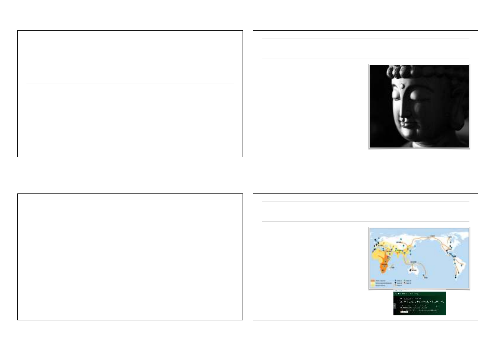
Phật giáo
Giác quan và tín hiệu thần kinh hoạt động như
thế nào?
1
Nội dung chính
1. Các nguyên lí quan trọng của Phật giáo.
2. Tứ diệu đế.
3. Nguyên lí trung đạo.
4. Nguyên lí duyên khởi.
2
Các nguyên lí quan trọng của Phật giáo
3
Chế độ đẳng cấp
❖Loài người thông minh (sapiens) đặt chân lên
Ấn Độ khoảng 70—60 ky BP.
❖Người Aryan chiếm lĩnh bán đảo Ấn Độ từ 4
ky BP—1,2 ky BP.
❖Chế độ đẳng cấp hình thành từ 3,5 ky BP—
nay: Brahmin, Kshatriya, Vaishya, Shudra.
Ngoài ra còn có Paria (Dalit).
❖1893, Risley đưa ra lí thuyết không lai hoá.
❖2013, các nghiên cứu cho thấy sự lai hoá xảy ra
từ 4 ky BP—1,9 ky BP.
4
Ấn độ giáo khi Phật giáo ra đời
1. Tồn lại linh hồn, một tự ngã (atman) thường
hằng, bất biến luân hồi (samsara) trong các thể
xác, tách biệt khỏi thể xác.
2. Hệ quả của luân hồi là thuyết nhân quả và
thuyết tái sinh thông qua nghiệp (karma).
3. Con người luôn muốn được giải thoát (moksha)
khỏi luân hồi thông qua thực hành chính đạo
(dharma).
4. Chính đạo có ba cách: hiểu biết tri thức, hành
động đúng đắn, tôn kính chư thần.
5. Có hàng triệu vị thần nhưng có ba thần quan
trọng nhất: Brahma, Vishnu, Shiva. Ấn độ giáo
không thừa nhận có thượng đế toàn năng.
5
Phật giáo và giá trị mới
1. Tứ diệu đế (catvāri āryasatyāni).
2. Nguyên lí trung đạo: chính đạo là trung đạo, không thiên về thái cực hưởng lạc
hoặc khổ hạnh. Trung đạo được thể hiện ở bát chính đạo: tuệ, giới, định.
3. Nguyên lí duyên khởi (pratitya samutpada): sự phụ thuộc lẫn nhau của mọi sự vật
và hiện tượng. Ba đặc trưng tam pháp ấn: vô thường, khổ đau, vô ngã.
4. Xoá bỏ đẳng cấp, bất kì sinh vật nào cũng có Phật tính, thực hành trung đạo sẽ đạt
giải thoát. Trạng thái giải thoát là Niết bàn (Nirvana).
6
Trung quán tông
❖Tam bảo: Phật (Buddha), Pháp (Dharma), Tăng (Sanga).
❖Pháp dựa trên nguyên lí trung đạo và nguyên lí duyên khởi.
❖Phật giáo nguyên thuỷ: duyên khởi là tự ngã của từng pháp riêng biệt.
❖Phật giáo phát triển (Trung quán tông): duyên khởi là sự tương hỗ giữa các pháp
nên các pháp không thể tồn tại độc lập, chúng không có tự tính. Đó chính là tính
không, thể hiện tính tương đối của sự vật và hiện tượng.
7
Các giác quan
8
Ý thức
❖Thế giới bên ngoài tác động lên các giác quan
để tạo thành các tín hiệu thần kinh.
❖Các tín hiệu thần kinh là các tín hiệu điện tạo
thành từ sự chênh lệch điện tích (ion) giữa các
vùng khác nhau của tế bào thần kinh.
❖Tín hiệu thần kinh được lan truyền từ tế bào
này sang tế bào khác thông qua các khớp nối
thần kinh để đến bộ não.
❖Tín hiệu được xử lí ở nhiều vùng não trước
khi đến vỏ não. Ở đó, ý thức về thế giới được
hình thành.
9
Lục căn
❖Theo Phật giáo, con người có lục căn: tai, mắt, mũi, lưỡi, thân, ý.
❖Aristotle: tai, mắt, mũi, lưỡi, xúc giác.
❖Khoa học cận đại
❖Năm 1830, Charles Bell đề xuất giác quan thứ sáu là tự nhận thức được cơ thể
trong không gian.
❖Thế kỉ 20, Charles Sherrington đề xuất giác quan thứ sáu là hệ cảm giác thân thể
(somatosensory).
❖Giác quan và ý thức liên quan đến hoạt động của hệ thần kinh khoa học thần kinh.
⇒
Tứ diệu đế—khổ
1. Sự tồn tại là khổ.
2. Nguyên nhân của sự khổ.
3. Sự diệt khổ—Niết bàn.
4. Con đường diệt khổ.
11
KHỔ
Ý thức
Sướng
Đau
Vô minh
Giác
quan
Ý thức là gì?
❖Từ lâu, nghiên cứu về ý thức là chủ đề của các nhà
triết học. Tuy nhiên giờ đây nó là chủ đề của các nhà
khoa học thực nghiệm.
❖TK17, René Descartes: cơ thể và ý thức tạo thành bởi
những thứ hoàn toàn khác nhau. Cơ thể tồn tại trong
không gian và thời gian. Ý thức tồn tại trong thời gian.
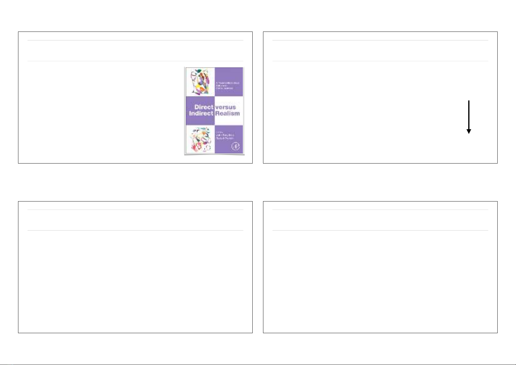
❖1995, David Chalmers đưa “Bài toán khó về ý thức”
(the hard problem of consciousness): chúng ta nhận
thức thế giới như chính bản thân nó (trực tiếp) hay thế
giới là một bản copy của bộ não (gián tiếp)?
❖Tham khảo: G. Miller, What Is the Biological Basis of
Consciousness? Science 309 (5731), 79 (2005). J. Morgan,
The hard problem of consciousness: understanding our reality,
The Lancet Neurology 17 (5), 403 (2018).
Bài toán khó chưa có lời giải
❖John Smythies and Robert French, Direct versus Indirect Realism: A
Neurophilosophical Debate on Consciousness, Academic Press (2018).
1. Các nhà triết học có xu hướng theo thực tại trực tiếp, các nhà
thần kinh thì theo thực tại gián tiếp.
2. Thực tại gián tiếp: phủ nhận sự tồn tại của kiến thức tiên nghiệm
vì toàn bộ kiến thức của con người đến từ các giác quan.
3. Thực tại trực tiếp: kinh nghiệm nhận thức không phải là thế giới
thực nhưng là một hình ảnh thu nhỏ được sao chép ở bên ngoài.
❖Do đó, việc nghiên cứu triết học, tôn giáo cần có sự hiểu biết của khoa
học thần kinh.
Bốn lực cơ bản
Giác quan của con người
chỉ thu nhận được lực điện
từ!
14
Lực
Hạt tác động
Tầm tác dụng
Cường độ
Hấp dẫn
Tất cả các hạt có
khối lượng
Vô hạn
Yếu
Lực hạt nhân yếu
Quark, lepton
Ngắn
Điện từ
Các hạt mang điện
Vô hạn
Lực hạt nhân mạnh
Quark, gluon
Ngắn
Mạnh
Học thuyết thần kinh
❖Golgi: các dây thần kinh kết nối với nhau tạo thành các mạch thần kinh. Ông phát minh ra phương
pháp nhuộm (stain) tế bào thần kinh nhưng phủ nhận sự tồn tại của tế bào thần kinh. Các cơ quan,
các mô đều được tạo thành từ các tế bào.
❖Ramón y Cajal: Học thuyết thần kinh (neuron doctrine) cho rằng hệ thần kinh được tạo bởi các tế
bào thần kinh độc lập. Giữa chúng có kết nối với nhau để tạo ra các mạch thần kinh.
❖Charles Sherrington: cho rằng giữa các tế bào thần kinh có kết nối với nhau thông qua các khớp nối
thần kinh (synapse).
Các giác quan
❖Dù có năm giác quan nhưng các tế bào thần
kinh thu nhận tín hiệu thông qua 4 cách
1. Chuyển đổi tín hiệu hoá học thành tín hiệu
điện: vị giác, khứu giác.
2. Chuyển đổi tín hiệu nhiệt thành tín hiệu
điện: xúc giác.
3. Chuyển đổi tín hiệu ứng suất cơ học thành
tín hiệu điện: xúc giác.
4. Chuyển đổi tín hiệu quang học thành tín
hiệu điện: thị giác.
Giác quan
Kích thích
Cường độ
Vị giác
Hoá chất (lỏng)
Nồng độ
Khứu giác
Hoá chất (khí)
Nồng độ
Thính giác
Âm thanh
Độ lớn âm
Xúc giác
Nhiệt, ứng suất
Nhiệt độ, áp suất
Thị giác
Ánh sáng
Độ sáng
16
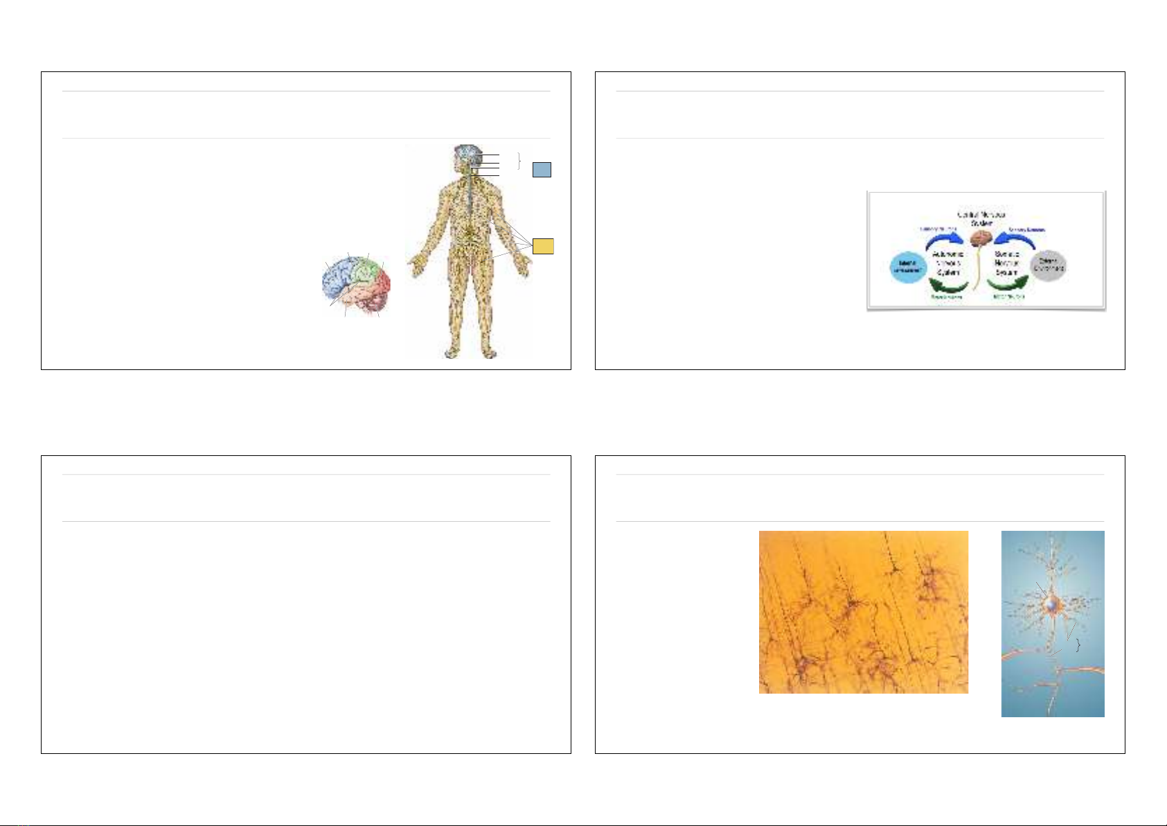
Phân chia theo giải phẫu
❖Phân chia theo giải phẫu:
1. Hệ thần kinh trung ương: não, tuỷ sống.
Central nervous system: brain and spinal
cord.
2. Hệ thần kinh ngoại biên: các tế bào thần
kinh cảm giác và tế bào thần kinh vận
động.
Peripheral nervousSsystem: sensory and
motor neurons that connect to the central
nervous system.
Cerebrum
Cerebellum Brain
Brain stem Central
nervous
system
Peripheral
nervous
system
Spinal cord
▲
FIGURE 1.7
The basic anatomical subdivisions of the nervous system.
The nervous system consists of two divisions, the central nervous
system (CNS) and the peripheral nervous system (PNS). The CNS
consists of the brain and spinal cord. The three major parts of
the brain are the cerebrum, the cerebellum, and the brain stem.
The PNS consists of the nerves and nerve cells that lie outside
the brain and spinal cord.
Frontal
lobe
Parietal
lobe
Central
sulcus
Sylvian
fissure
Occipital
lobe
Temporal lobe Cerebellum
▲FIGURE 1.8
The lobes of the cerebrum. Notice the
deep Sylvian fissure, dividing the frontal
lobe from the temporal lobe, and the
central sulcus, dividing the frontal lobe
from the parietal lobe. The occipital lobe
lies at the back of the brain. These land-
marks can be found on all human brains.
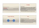
Hệ thần kinh ngoại biên
❖Hệ thần kinh ngoại biên được chia thành
1. Hệ thần kinh thân thể (somatomic nervous
system) quản lí hoạt động của các cơ vân,
hoạt động có ý muốn.
2. Hệ thần kinh tự chủ, còn gọi là hệ thần
kinh thực vật (autonomic nervous system)
quản lí hoạt động của các cơ quan nội
tạng không theo ý muốn.
Tế bào thần kinh
❖Tế bào thần kinh gồm hai loại: nơ ron (neuron) và tế bào đệm (glia).
❖Neuron là quan trọng nhất: thu nhận tín hiệu từ môi trường, trao đổi với nhau, ra lệnh cho cơ thể.
❖Glia có chức năng như một chất keo dính để làm bộ đệm, đỡ cho các neuron.
❖Có khoảng 100 tỉ neuron và 100 ngàn tỉ kết nối trong một cơ thể của người trưởng thành.
❖Diện tích bề mặt của các tế bào thần kinh là 25000 m2, bằng 4 lần diện tích sân bóng đá.
Cấu tạo của neuron
1. Thân tế bào (cell body,
soma).
2. Sợi trục (axon): dẫn
truyền tín hiệu từ thân tế
bào đến các tế bào khác.
3. Sợi nhánh (dendrite): là
phần mở rộng của thân tế
bào để tiếp nhận tín hiệu
từ tế bào khác.
Dendrites
Soma
Neurites
Axon
▲FIGURE 2.4
The basic parts of a neuron.
▲FIGURE 2.3
Golgi-stained neurons. (Source: Hubel, 1988, p. 126.)


![Bài giảng Tín ngưỡng và tôn giáo Việt Nam Trường Đại học Tân Trào [Chuẩn Nhất]](https://cdn.tailieu.vn/images/document/thumbnail/2025/20250814/kimphuong1001/135x160/58751755145071.jpg)


![Đề cương ôn tập Lịch sử văn minh thế giới có lời giải [chuẩn nhất]](https://cdn.tailieu.vn/images/document/thumbnail/2026/20260512/hoatulip0906/135x160/70401778724716.jpg)


![Đề cương ôn tập Văn hóa học đại cương [chuẩn nhất]](https://cdn.tailieu.vn/images/document/thumbnail/2026/20260507/hoahongxanh0906/135x160/90171778558833.jpg)

![Đề cương ôn tập Văn hóa các dân tộc thiểu số ở Việt Nam [mới nhất]](https://cdn.tailieu.vn/images/document/thumbnail/2026/20260507/hoahongxanh0906/135x160/79611778493384.jpg)


![Câu hỏi ôn tập Cơ sở văn hóa Việt Nam [mới nhất]](https://cdn.tailieu.vn/images/document/thumbnail/2026/20260506/camtucau2026/135x160/14481778123622.jpg)


%20--%3e%3cdefs%3e%3cstyle%3e%20.st0%20{%20fill:%20%23fff;%20}%20.st1%20{%20fill:%20%237800fa;%20}%20%3c/style%3e%3c/defs%3e%3cpath%20class='st1'%20d='M117.78,12.18H43.11c2.9,3.47,4.65,7.94,4.65,12.82,0,5.6-2.3,10.66-6.01,14.29h76.02l7.22-13.56-7.22-13.56Z'/%3e%3cg%3e%3cpath%20class='st0'%20d='M53.58,26.17h-.59v-1.46h.59v-4.96h2.83c1.78,0,2.67.94,2.67,2.82v5.76c0,1.87-.89,2.81-2.67,2.81h-2.83v-4.96ZM55.36,21.37v3.34h1.1v1.46h-1.1v3.34h1.01c.61,0,.91-.37.91-1.1v-5.93c0-.74-.3-1.1-.91-1.1h-1.01Z'/%3e%3cpath%20class='st0'%20d='M65.99,31.14h-1.8l-.31-2.07h-2.19l-.31,2.07h-1.64l1.82-11.39h2.62l1.82,11.39ZM65.28,18.04c-.25.46-.51.77-.75.94-.21.15-.47.22-.79.22-.26,0-.57-.07-.92-.22l-.38-.15c-.14-.05-.26-.07-.37-.07-.3,0-.53.18-.71.54l-.91-.68c.25-.46.51-.77.75-.94.21-.14.48-.21.79-.21.26,0,.57.07.92.21l.38.15c.14.05.26.07.37.07.3,0,.53-.18.71-.54l.91.68ZM61.91,27.52h1.73l-.87-5.76-.87,5.76Z'/%3e%3cpath%20class='st0'%20d='M74.53,26.89v1.52c0,1.91-.89,2.86-2.67,2.86s-2.67-.95-2.67-2.86v-5.93c0-1.91.89-2.86,2.67-2.86s2.67.95,2.67,2.86v1.11h-1.69v-1.22c0-.75-.31-1.12-.93-1.12s-.93.37-.93,1.12v6.15c0,.74.31,1.11.93,1.11s.93-.37.93-1.11v-1.63h1.69Z'/%3e%3cpath%20class='st0'%20d='M81.4,31.14h-1.8l-.31-2.07h-2.19l-.31,2.07h-1.64l1.82-11.39h2.62l1.82,11.39ZM75.9,19.2l1.52-1.91h1.71l1.51,1.91h-1.61l-.76-.95-.75.95h-1.61ZM77.32,27.52h1.73l-.87-5.76-.87,5.76ZM83.1,15.99l-1.76,1.91h-1.26l1.17-1.91h1.86Z'/%3e%3cpath%20class='st0'%20d='M84.86,19.75c1.78,0,2.67.94,2.67,2.82v1.48c0,1.87-.89,2.81-2.67,2.81h-.85v4.28h-1.79v-11.39h2.64ZM84.01,21.37v3.86h.85c.58,0,.87-.36.87-1.08v-1.71c0-.71-.29-1.07-.87-1.07h-.85Z'/%3e%3cpath%20class='st0'%20d='M93.51,19.75c1.78,0,2.67.94,2.67,2.82v1.48c0,1.87-.89,2.81-2.67,2.81h-.85v4.28h-1.79v-11.39h2.64ZM92.66,21.37v3.86h.85c.58,0,.87-.36.87-1.08v-1.71c0-.71-.29-1.07-.87-1.07h-.85Z'/%3e%3cpath%20class='st0'%20d='M98.8,31.14h-1.79v-11.39h1.79v4.88h2.03v-4.88h1.83v11.39h-1.83v-4.88h-2.03v4.88Z'/%3e%3cpath%20class='st0'%20d='M105.36,24.55h2.46v1.62h-2.46v3.34h3.09v1.63h-4.88v-11.39h4.88v1.63h-3.09v3.18ZM108.17,17.29l-1.76,1.91h-1.26l1.17-1.91h1.86Z'/%3e%3cpath%20class='st0'%20d='M112.2,19.75c1.78,0,2.67.94,2.67,2.82v1.48c0,1.87-.89,2.81-2.67,2.81h-.85v4.28h-1.79v-11.39h2.64ZM111.35,21.37v3.86h.85c.58,0,.87-.36.87-1.08v-1.71c0-.71-.29-1.07-.87-1.07h-.85Z'/%3e%3c/g%3e%3ccircle%20class='st1'%20cx='25'%20cy='25'%20r='20'/%3e%3cpath%20class='st0'%20d='M32.78,19.27c2.92,0,4.43,2.55,5.28,5.33l.71,2.17c.14.38-.33.75-.71.75h-5.61c.19-.33.24-.71.09-1.08l-.75-2.45c-.43-1.32-.99-2.64-1.79-3.77.75-.57,1.65-.94,2.78-.94h0ZM25,18.38c3.25,0,4.9,2.78,5.89,5.89l.76,2.45c.14.42-.33.8-.8.8h-11.69c-.42,0-.94-.38-.8-.8l.75-2.45c.99-3.11,2.64-5.89,5.89-5.89h0ZM25,11.35c1.74,0,3.11,1.37,3.11,3.11s-1.37,3.11-3.11,3.11-3.11-1.41-3.11-3.11,1.41-3.11,3.11-3.11h0ZM17.27,19.27c1.08,0,1.98.38,2.73.94-.8,1.13-1.37,2.45-1.74,3.77l-.8,2.45c-.14.38-.05.75.09,1.08h-5.56c-.42,0-.9-.38-.75-.75l.71-2.17c.9-2.78,2.41-5.33,5.33-5.33h0ZM17.27,12.91c1.51,0,2.78,1.27,2.78,2.83s-1.27,2.83-2.78,2.83-2.83-1.27-2.83-2.83,1.27-2.83,2.83-2.83h0ZM32.78,12.91c1.56,0,2.78,1.27,2.78,2.83s-1.23,2.83-2.78,2.83-2.83-1.27-2.83-2.83,1.27-2.83,2.83-2.83h0ZM27.07,28.56v.09c0,.57-.24,1.08-.61,1.46h0v.05c-.38.33-.9.57-1.46.57s-1.08-.24-1.46-.61h0c-.38-.38-.61-.9-.61-1.46v-.09h1.41v.09c0,.19.05.38.19.47v.05c.09.09.28.19.47.19s.38-.09.47-.19v-.05c.14-.09.24-.28.24-.47t-.05-.09h1.41ZM30.99,28.56v.09c0,1.65-.66,3.16-1.74,4.24-1.08,1.08-2.59,1.79-4.24,1.79s-3.16-.71-4.24-1.79l-.05-.05c-1.04-1.08-1.7-2.55-1.7-4.2v-.09h1.41v.09c0,1.27.47,2.4,1.27,3.25h.05c.85.85,1.98,1.37,3.25,1.37s2.4-.52,3.25-1.37c.85-.8,1.37-1.98,1.37-3.25v-.09h1.37ZM34.99,28.56v.09c0,2.78-1.13,5.28-2.92,7.07-1.79,1.79-4.29,2.92-7.07,2.92s-5.23-1.13-7.07-2.92c-1.79-1.79-2.92-4.29-2.92-7.07v-.09h1.41v.09c0,2.4.94,4.53,2.5,6.08,1.56,1.56,3.72,2.5,6.08,2.5s4.52-.94,6.08-2.5c1.56-1.56,2.5-3.68,2.5-6.08v-.09h1.41Z'/%3e%3c/svg%3e)