
BioMed Central
Page 1 of 11
(page number not for citation purposes)
Retrovirology
Open Access
Research
Complementation of diverse HIV-1 Env defects through
cooperative subunit interactions: a general property of the
functional trimer
Karl Salzwedel1,2 and Edward A Berger*1
Address: 1Laboratory of Viral Diseases, National Institute of Allergy and Infectious Diseases, National Institutes of Health, Bethesda, MD 20892,
USA and 2Current address: Division of AIDS, NIAID, 6700-B Rockledge Drive, Room 4149, Bethesda, MD 20892, USA
Email: Karl Salzwedel - salzwedelkd@niaid.nih.gov; Edward A Berger* - edward_berger@nih.gov
* Corresponding author
Abstract
Background: The HIV-1 Env glycoprotein mediates virus entry by catalyzing direct fusion
between the virion membrane and the target cell plasma membrane. Env is composed of two
subunits: gp120, which binds to CD4 and the coreceptor, and gp41, which is triggered upon
coreceptor binding to promote the membrane fusion reaction. Env on the surface of infected cells
is a trimer consisting of three gp120/gp41 homo-dimeric protomers. An emerging question
concerns cooperative interactions between the protomers in the trimer, and possible implications
for Env function.
Results: We extended studies on cooperative subunit interactions within the HIV-1 Env trimer,
using analysis of functional complementation between coexpressed inactive variants harboring
different functional deficiencies. In assays of Env-mediated cell fusion, complementation was
observed between variants with a wide range of defects in both the gp120 and gp41 subunits. The
former included gp120 subunits mutated in the CD4 binding site or incapable of coreceptor
interaction due either to mismatched specificity or V3 loop mutation. Defective gp41 variants
included point mutations at different residues within the fusion peptide or heptad repeat regions,
as well as constructs with modifications or deletions of the membrane proximal tryptophan-rich
region or the transmembrane domain. Complementation required the defective variants to be
coexpressed in the same cell. The observed complementation activities were highly dependent on
the assay system. The most robust activities were obtained with a vaccinia virus-based expression
and reporter gene activation assay for cell fusion. In an alternative system involving Env expression
from integrated provirus, complementation was detected in cell fusion assays, but not in virus
particle entry assays.
Conclusion: Our results indicate that Env function does not require every subunit in the trimer
to be competent for all essential activities. Through cross-talk between subunits, the functional
determinants on one defective protomer can cooperatively interact to trigger the functional
determinants on an adjacent protomer(s) harboring a different defect, leading to fusion.
Cooperative subunit interaction is a general feature of the Env trimer, based on complementation
activities observed for a highly diverse range of functional defects.
Published: 11 August 2009
Retrovirology 2009, 6:75 doi:10.1186/1742-4690-6-75
Received: 4 July 2009
Accepted: 11 August 2009
This article is available from: http://www.retrovirology.com/content/6/1/75
© 2009 Salzwedel and Berger; licensee BioMed Central Ltd.
This is an Open Access article distributed under the terms of the Creative Commons Attribution License (http://creativecommons.org/licenses/by/2.0),
which permits unrestricted use, distribution, and reproduction in any medium, provided the original work is properly cited.

Retrovirology 2009, 6:75 http://www.retrovirology.com/content/6/1/75
Page 2 of 11
(page number not for citation purposes)
Background
The envelope glycoprotein (Env) of human immunodefi-
ciency virus type 1 (HIV-1) promotes virus entry by cata-
lyzing direct fusion between the virion membrane and the
target cell plasma membrane; similarly, Env-expressing
cells can fuse with target cells to form multinucleated
giant cells (syncytia). Env is synthesized as a gp160 pre-
cursor protein that assembles into homo-trimeric com-
plexes in the endoplasmic reticulum. During transport
through the secretory pathway, gp160 is cleaved in the
trans-Golgi network by a furin-like protease(s) to yield the
external gp120 subunit noncovalently associated with the
gp41 transmembrane subunit (derived from the N- and C-
regions of gp160, respectively) [1]. The functional Env
spike on mature virions of HIV-1 and the related simian
immunodeficiency virus consists of a homo-trimer of
gp120/gp41 hetero-dimers [2].
Env-mediated fusion involves a strict division of labor
between the two subunits: gp120 is responsible for
sequential binding to specific target cell receptors, first to
CD4 and then to the coreceptor (a specific chemokine
receptor, typically CCR5 or CXCR4); receptor binding
then triggers gp41 to promote membrane fusion. These
steps involve a tightly orchestrated series of conforma-
tional changes in both Env subunits that drive the fusion
process. The emerging understanding of the complexities
of HIV Env/receptor interactions and the subsequent
events leading to fusion/entry have been the central focus
of numerous review articles over the past decade [3-8]. X-
ray crystallographic analyses of gp120 from HIV-1 [9] and
the closely related simian immunodeficiency virus [10]
have revealed that CD4 binding induces a profound rear-
rangement of the relatively disordered gp120 subunit to
create a new surface consisting of four anti-parallel beta
strands derived from discontinuous regions of the linear
sequence; this highly conserved "bridging sheet", which is
not present in the unliganded pre-CD4-bound state, is
directly involved in binding to coreceptor [11] in conjunc-
tion with the third variable loop (V3) of gp120, which
determines coreceptor specificity [12,13]. Binding of
gp120 to coreceptor then triggers the fusogenic activity of
gp41 in a process believed to involve insertion of the gp41
N-terminal fusion peptide (FP) into the target cell plasma
membrane [14,15]. Detailed structural information is not
yet available for the native state of gp41, but the structure
of the final post-fusion state has been determined to be a
trimer of hairpins in the form of a six-helix coiled-coil
bundle [16-18]. A transient intermediate conformation is
thought to exist in which the gp41 subunits adopt an
extended triple-helix coiled-coil with the N-terminal FPs
inserted into the target cell membrane. The heptad repeat
(HR) segments near the external C-terminal region (HR2)
then fold to insert in anti-parallel fashion into the grooves
formed by the cluster of the three N-terminal heptad
repeat (HR1) segments; the resulting formation of a 6-
helix bundle brings the virion and target cell plasma
membranes together, and provides the driving force for
membrane fusion underlying HIV entry. The molecular
complexity of the HIV entry process presents a variety of
targets for novel antiviral agents [19-22]; the T-20 peptide
(enfuvirtide, Fuzeon) targeting the gp41 intermediate
conformation is the first-in-class HIV-1 fusion inhibitor
[23], and the recently approved maraviroc is the first-in-
class inhibitor that binds to the CCR5 coreceptor and
blocks the gp120 interaction [24].
While each gp120/gp41 hetero-dimeric complex contains
all the determinants required for fusion, it is possible that
molecular interactions between complexes within the
trimer influence Env function. In a previous study we used
a quantitative vaccinia expression-based cell fusion assay
to demonstrate that individual subunits within the Env
trimer can interact cooperatively during fusion [25]. By
coexpressing Env proteins with defects in different essen-
tial determinants, we found that functional complemen-
tation could occur between subunits within a mixed
trimer. In the present report, we show that subunit com-
plementation is a general capacity of the HIV-1 Env
trimer, though its efficiency and detectability are depend-
ent on the particular defective variants examined and the
assay systems employed. The results are discussed in terms
of potential biological implications for Env function and
HIV neutralization.
Methods
Construction and expression of Env variants
For vaccinia virus expression-based cell fusion assays,
HIV-1 Envs were transiently expressed from pSC59-based
plasmids under control of a strong synthetic vaccinia virus
early/late promoter [26]. Previously described plasmids
[27,28] were used to express wild-type Envs from the fol-
lowing HIV-1 strains: LAI [29] (LAV isolate, unless indi-
cated otherwise), plasmid pCB-41; SF162, plasmid pCB-
32; Ba-L, plasmid pCB-43, and CM235, plasmid pCB-52.
In addition, a Kpn I-Xho I fragment encoding wild-type
YU-2 Env was substituted into a variant of pCB-41 con-
taining a unique Xho I site at the 3' end of Env (pKS-9) to
create the plasmid pKS-10. As a negative control, an
uncleaveable (Unc) mutant form of LAI (IIIB isolate) was
used (plasmid pCB-16).
Plasmids pKS-3 and pKS-4 [25] encode mutants of LAI
Env (HXB2 isolate) with a D368R substitution in the CD4
binding site (BS) of gp120 that abolishes CD4 binding
(LAI-BS) and a Leu to Arg substitution at residue 26 of the
gp41 N-terminal fusion peptide (LAI-FP26) [30], respec-
tively. Additional site-directed mutations were introduced
(QuikChange kit, Stratagene, La Jolla, CA) into pCB-41
encoding wild-type LAI Env resulting in the following
plasmid constructs. See Fig. 1 Legend for descriptions: FP
mutants LAI-FP2 (plasmid pKS-13) and LAI-FP9 (plasmid
Retrovirology 2009, 6:75 http://www.retrovirology.com/content/6/1/75
Page 3 of 11
(page number not for citation purposes)
pKS-14); heptad repeat mutants LAI-HR1a (plasmid pKS-
15) and LAI-HR1e (plasmid pKS-16); V3 loop mutant
LAI-V3 (plasmid pKS-17). A Kpn I-Bam HI fragment
encoding the LAI-Δ665-856 mutant, previously referred to
as Δ192 [31], was substituted into a variant of pCB-41
(pKS-8) in which a Bam HI site upstream of the promoter
had been destroyed by cutting and then filling in with T4
DNA polymerase; the resulting plasmid was pKS-18. Kpn
I-Xho I fragments encoding the mutants LAI-HT-1 [32],
LAI-HT-2 [32], and LAI-Δ665-682 [33,34] were substi-
tuted into the plasmid pKS-8 to create the plasmids pKS-
19, pKS-20, pKS-21, and pKS-22, respectively. Previous
studies indicate that each of these variants is capable of
Env processing (except for Unc), surface expression
(except for LAI-Δ665-856, which is secreted), and CD4
binding (except for LAI-BS) [25,30,32-34].
For MAGI cell HIV infectivity assays, virus was expressed
from the pNL4-3 proviral clone [35] encoding wild-type
LAI Env (LAV isolate). pNL4-3 containing a frame-shift
mutation at the Nhe I site within Env (pNL4-3Δenv) [33]
was used as a negative control. Nhe I-Bam HI fragments
encoding the LAI-FP26 and LAI-BS mutants [25] were sub-
cloned into pNL4-3 to create pKS-11 (encoding NL4.3-
FP26) and pKS-12 (encoding NL4.3-BS), respectively. The
phenotype for each construct, both when expressed alone
and in complementation experiments, was confirmed
using two independent plasmid clones constructed in par-
allel.
Vaccinia virus-based cell fusion assay
Env-mediated cell fusion activity was measured using a
quantitative vaccinia-based reporter gene assay as
described previously [36,37]. Each vaccinia virus was used
at a multiplicity of infection of 10. Target cells were pre-
pared by co-infecting NIH 3T3 cells with vaccinia virus
recombinant vCB21R-LacZ containing the E. coli lacZ
reporter gene linked to the T7 promotor [38], plus vac-
cinia recombinants encoding the following cDNAs linked
to vaccinia early/late promoters: CD4, vCB-3 [39] and the
designated coreceptor CCR5, vHC-1 [40] or CXCR4,
vCBYF1-fusin [41]. Effector cells were prepared by trans-
fecting HeLa cells with the above-described plasmids con-
taining the Env genes linked to a strong synthetic vaccinia
early/late promoter and infecting with vaccinia recom-
binant vP11T7gene1 encoding bacteriophage T7 RNA
polymerase [42]. Transfection was performed with
DOTAP (Boehringer Mannheim, Indianapolis, IN); the
total amount of DNA was held constant at 5 μg DNA per
25 cm2 flask, in both single-transfection and cotransfec-
tion experiments. Effector and target cells were incubated
overnight at 31°C to allow expression of the recombinant
proteins. After these cells were washed by centrifugation,
they were mixed in equal numbers in duplicate wells of a
96-well plate (2 × 10⁵ of each per well) and incubated for
2.5 hr at 37°C. Fusion reactions were terminated by addi-
tion of nonidet-P40 (0.5% final) and quantified by spec-
trophotometric measurement of β-galactosidase activity
as described previously [36]. For each data point, error
bars indicate the standard errors of the mean of duplicate
samples; in cases where error bars appear to be absent, the
data points were so close that error bars are not visible. All
experiments were repeated at least twice; representative
data are shown for each experiment.
MAGI cell assays for cell fusion and virus entry
HIV-1 entry and Env-mediated cell fusion in the context of
HIV-1 provirus expression were measured using the HeLa-
CD4/LTR-β-gal (MAGI) indicator target cell line [43],
which was obtained from the NIH AIDS Research and Ref-
erence Reagent Program (originally contributed by M.
Emerman). BS-C-1 cells plated at 3 × 10⁵ per well in 6-well
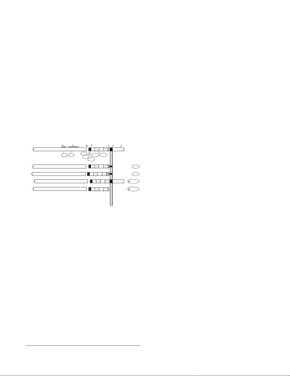
Mutations in HIV-1 EnvFigure 1
Mutations in HIV-1 Env. A schematic representation of
HIV-1 Env. Functional and structural domains within the
gp120 and gp41 subunits are labeled at the top: V3 loop (3rd
variable loop), CD4 BS (CD4 binding site), FP (fusion pep-
tide), TRR (tryptophan-rich region), TM (transmembrane
domain). For various inactivating mutants in the LAI Env (des-
ignations encircled), the approximate locations of specific
point mutants are indicated underneath, and the deletion
mutants are indicated on the right. The specific point muta-
tions are: V3 (R320G in the conserved GPGR motif at the tip
of the V3 loop); BS (D368R within the CD4 binding site); var-
ious positions in the fusion peptide including FP2 (Val→Glu),
FP9 (Leu→Arg) and FP26 (Leu→Arg); heptad repeat muta-
tions HR1e (V570R) and HR1a (I573P). HT-1 and HT-2 are
chimeric LAI Env/Thy-1.1 glycoproteins that are membrane-
associated via a glycosyl-phosphatidylinositol (GPI) anchor.
HT-1 contains the gp41 ectodomain minus the tryptophan-
rich region (K665 through I682), whereas HT-2 contains the
entire gp41 ectodomain; both constructs have 22 intervening
amino acid residues from the C-terminus of Thy1.1. The
Δ665-682 construct has a selective deletion of the TRR
(K665 through I682) and the Δ665-856 construct has an
introduced premature stop codon that results in deletion of
the C-terminal 192 aa of Env, including the TM and cytoplas-
mic domains. See Methods for construction and references.
HR1 HR2
NH
2
665-682
NH
2
HR1 HR2 HT-1
gp120 gp41
HR1 HR2
TM Cytoplasmic
Tail
FP
CleavageCD4 BS
NH
2
COOH
V3 loop TRR
ENV
V3
FP26
BS FP9
FP2 HR1e HR1a
HR1 HR2
NH
2
HT-2
HR1 HR2
NH
2
665-856

Retrovirology 2009, 6:75 http://www.retrovirology.com/content/6/1/75
Page 4 of 11
(page number not for citation purposes)
plates the previous day were transfected (or cotransfected)
with the designated pNL4-3-based proviral construct(s)
using FuGENE 6 (Boehringer Mannheim, Indianapolis,
IN) according to the manufacturer's protocol. The next
day, cells were washed and given fresh media (2 ml per
well) containing 10 mM HEPES. Three days post-transfec-
tion, the supernatants were removed, filtered through a
0.45 μm filter to remove cellular debris, and stored at 4°C.
For cell fusion assays, the cells were trypsinized, washed,
mixed 1:10 with MAGI cells, and replated in duplicate at
1 × 10⁵ total cells per well of a 24-well plate. Cells were
allowed to fuse overnight at 37°C and were then stained
with X-gal. Cell fusion was quantitated by counting the
total number of blue multi-nucleated syncytia per well
with the aid of a grid. For the virus entry assays, p24 levels
in the filtered supernatants were quantitated using the
HIV-1 p24 Antigen Assay (Coulter), and supernatant vol-
umes were normalized accordingly. MAGI cells were
infected in duplicate with 300 μl of filtered supernatant
per well of a 24-well plate and stained with X-gal 48 hrs
post-infection. Virus entry was quantitated by counting
the total number of blue foci per well. For complementa-
tion pairs, supernatants containing up to 2.35 ng of p24
per well were used (equivalent to 628 infectious units for
wild-type). This corresponds to approximately 15–20% of
the total supernatant from the cells. For each data point,
the standard errors of the mean of duplicate samples are
shown.
Results
To test the ability of fusion-inactive Env subunits to func-
tionally complement one another in the context of mixed
Env trimers, we first employed a vaccinia-based quantita-
tive cell fusion assay system wherein fusion between effec-
tor cells expressing Env and target cells expressing the
necessary receptors leads to reporter gene activation (β-
galactosidase production) [36,37]. We examined comple-
mentation between variants in gp120 that were inactive
due to inability to interact with CD4 (CD4 BS mutation)
or coreceptor (mismatched specificity, or mutation in the
V3 loop), as well as variants in gp41 with mutations at dif-
ferent points within the FP and HR1 regions, as well as
modifications of the membrane proximal tryptophan-rich
region (TRR) and the transmembrane (TM) domain (Fig.
1). Throughout these studies, target cells lacking corecep-
tor served as negative controls; where indicated, an
uncleaveable mutant Env (Unc) containing a mutation in
the gp120/gp41 cleavage site provided an additional neg-
ative control.
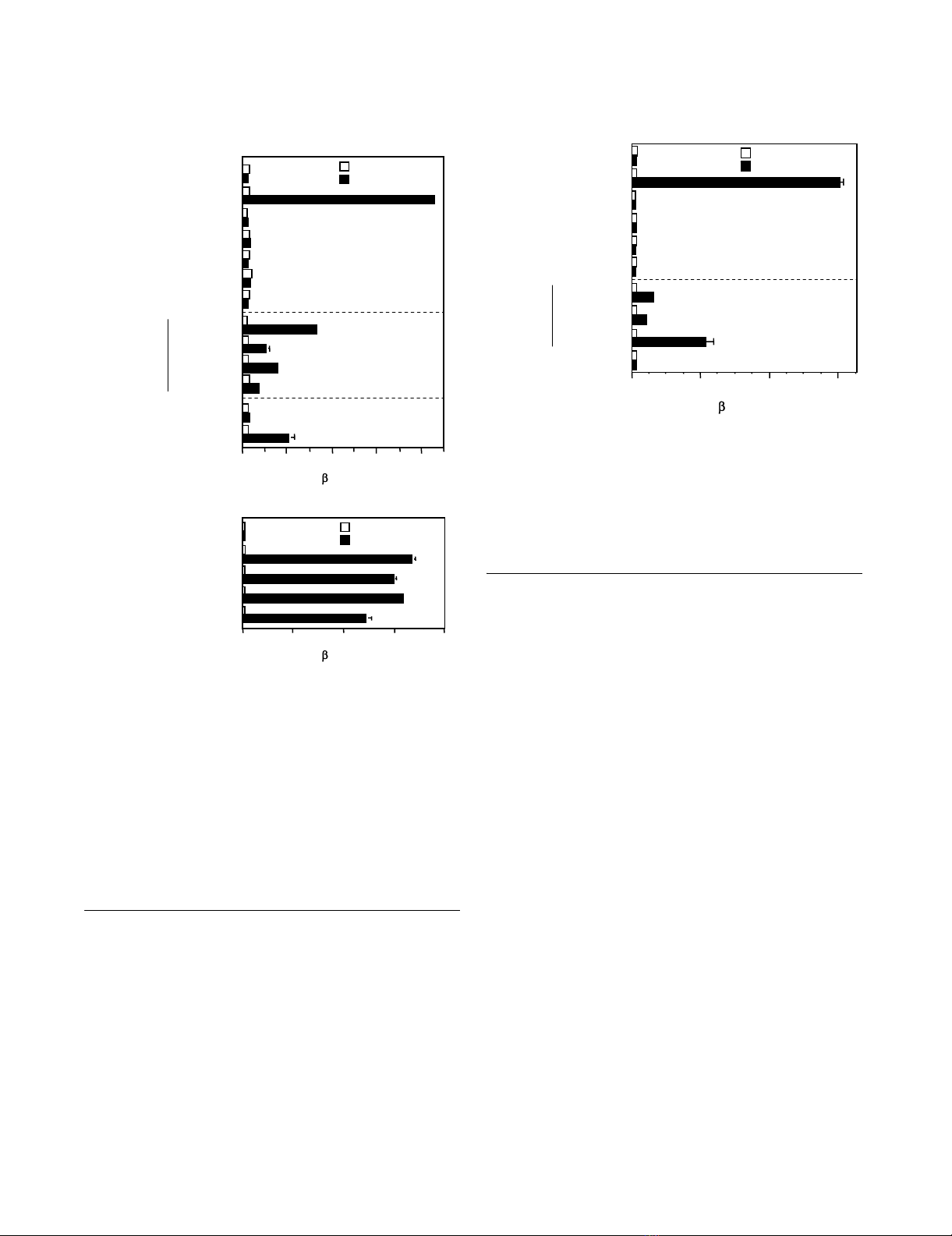
Complementation by Env subunits from HIV-1 primary
isolates of different genetic subtypes
Previously we demonstrated complementation between
Env constructs from two HIV-1 strains that were highly
laboratory-adapted and both clade B: LAI (X4, i.e. CXCR4-
specific) and SF162 (R5, i.e. CCR5-specific) [25]. To deter-
mine whether complementation potential is a more gen-
eral property of HIV-1 Envs, we analyzed the relative
complementation efficiencies of an LAI Env mutant con-
taining a defective FP (LAI-FP26) with Envs from diverse
R5 isolates in a CXCR4-dependent cell fusion assay. When
tested alone under these conditions, wild type LAI showed
potent activity whereas the LAI-FP26 mutant and all four
wild type R5 Envs were non-fusogenic (Fig. 2A, top sec-
tion). In coexpression experiments that enabled mixed
trimer formation, complementation of LAI-FP26 with the
R5 Envs occurred not only with SF162 as shown previ-
ously, but also with the laboratory-adapted Ba-L strain
(clade B) and the primary YU-2 (clade B) and CM235
(CRF01_AE recombinant) isolates (Fig. 2A, middle sec-
tion). The differences in the relative complementation
efficiencies of the various Envs correlated roughly with
their relative intrinsic fusogenicities in a CCR5-dependent
assay (Fig. 2B).
Complementation by Env subunits containing a
mutationally inactivated V3 loop
Our previous results [25] coupled with the data above
demonstrate that an Env with a mutational defect in gp41
can complement an Env containing a gp120 subunit inca-
pable of interacting with coreceptor due to mismatched
coreceptor specificity. We wished to extend this finding by
testing a gp120 subunit rendered inherently defective for
coreceptor interaction by site-directed mutation. The V3
loop, though highly variable, contains a conserved β-turn
motif at its crown (typically GPGR or GPGQ) that is essen-
tial for coreceptor binding activity [12,13]. We analyzed a
point mutant (LAI-V3) containing a G in place of the R
residue in the GPGR motif, which has been shown previ-
ously to abolish fusogenicity [44]. Our results demon-
strate that the fusion-defective LAI-V3 was able to
complement LAI-FP26 (Fig. 2A, bottom section). The
fusion activity was in the same range observed for the
coreceptor-mismatched Envs (Fig. 2A, middle section),
indicating that complementation efficiency was not lim-
ited by structural incompatibilities between Envs from
these different strains.
Previously we demonstrated complementation between
Envs containing different nonfunctional gp120 subunits
within a mixed trimer; functional mixed trimers were
formed when LAI-BS (defective for CD4 binding) was
coexpressed with wild-type SF162 (incapable of corecep-
tor interaction in a CXCR4-specific assay) [25]. To extend
this finding we tested the ability of LAI-BS to complement
LAI-V3, i.e. gp120 subunits incapable of interacting with
CD4 and coreceptor, respectively (Fig. 3). The efficiency
was similar to that observed for complementation
between LAI-BS and SF162, again indicating that there
were minimal structural incompatibilities in mixed trim-
ers between these two strains. As reported previously [25],
these examples of complementation between Envs with
Retrovirology 2009, 6:75 http://www.retrovirology.com/content/6/1/75
Page 5 of 11
(page number not for citation purposes)
distinct gp120 receptor binding deficiencies were some-
what less active than complementation between Envs
containing a defective gp120 and a defective gp41 (LAI-BS
+ LAI-FP26) (Fig. 3). As expected, no complementation
was observed upon coexpression of LAI-V3 with SF162,
since the gp120 from neither Env is capable of function-
ing with the CXCR4 coreceptor.
Varying complementation efficiencies of different point
mutations within the gp41 FP
Our previously described data and the experiments above
demonstrated functional complementation of a particular
gp41 FP mutation, i.e. substitution of Arg for Leu at the
26th position from the gp41 N-terminus (LAI-FP26). To
extend these analyses, we analyzed two additional FP
mutations previously shown by others to abolish
fusogenic activity without affecting Env processing or
CD4 binding [30]: LAI-FP2 substitutes Glu for Val at the
2nd position of the FP, and LAI-FP9 substitutes Arg for Leu
at the 9th position (Fig. 1). The LAI-FP2 mutant has been
shown to dominantly interfere with cell fusion when
coexpressed with wild-type Env, whereas the LAI-FP9
mutant reduced fusion two-fold and the LAI-FP26 mutant
had no negative effect when coexpressed with wild-type
Env [45]. The results of complementation experiments
with these gp41 FP mutations are shown in Fig. 4. Con-
sistent with previous reports, each mutation alone
strongly impaired cell fusion activity compared to wild
type (top sections in Figs. 4A–C). The relative efficiencies
of complementation, FP26 > FP9 > FP2 was observed
whether the complementation partner was LAI-BS (Fig.
4A), LAI-V3 (Fig. 4B), or SF162 (wt) (Fig. 4C).
Complementation with gp41 subunits lacking the normal
membrane anchoring and membrane proximal external
regions
Highly conserved regions close to the membrane are
known to be critical for Env function, including the 22
amino acid TM domain that anchors Env to the surface of
virions and infected cells, and the membrane-proximal
external region, generally defined as the last 24 C-terminal
residues of the gp41 ectodomain (L660 – K683) [15]. This
Complementation with laboratory-adapted and primary Envs from different cladesFigure 2
Complementation with laboratory-adapted and pri-
mary Envs from different clades. The vaccinia system
was employed to assay cell fusion between effector cells
expressing Envs and target cells expressing CD4 either with
(filled bars) or without (open bars) the indicated coreceptor.
A) CXCR4-dependent fusion. Effector cells expressed the
indicated Envs either individually (top section) or in combina-
tion with LAI-FP26 (middle section). Effector cells expressed
LAI-V3 individually or in combination with LAI-FP26 (bottom
section). B) CCR5-dependent fusion. The indicated wt Envs
were assayed for their intrinsic fusogenicity with target cells
expressing CD4 and CCR5.
Cell Fusion Activity ( -gal, OD/min x 1000)
B. CCR5-mediated fusion
SF162 (wt)
Ba-L (wt)
YU-2 (wt)
CM235 (wt)
Unc
0 50 100 150 200
CCR5
No coreceptor
A. CXCR4-mediated fusion
Cell Fusion Activity ( -gal, OD/min x 1000)
SF162 (wt)
Ba-L (wt)
YU-2 (wt)
CM235 (wt)
LAI (wt)
SF162 (wt)
Ba-L (wt)
YU-2 (wt)
CM235 (wt)
LAI-FP26
LAI-V3
LAI-V3
010203040
Unc CXCR4
No coreceptor
LAI-FP26 +
LAI-FP26 +
Complementation between gp120 variantsFigure 3
Complementation between gp120 variants. The vac-
cinia system was employed to assay cell fusion between effec-
tor cells expressing Envs and target cells expressing CD4
either with (filled bars) or without (open bars) CXCR4.
Effector cells expressed the indicated Envs either individually
(top section) or in combination with the indicated gp120-
defective Envs LAI-BS or LAI-V3 (bottom section).
Cell Fusion Activity (-gal, OD/min X 1000)
0204060
CXCR4
No coreceptor
Unc
LAI (wt)
LAI-BS
LAI-FP26
LAI-V3
SF162 (wt)
LAI-FP26
LAI-V3
SF162 (wt)
LAI-V3 + SF162 (wt)
LAI-BS +


%20--%3e%3cdefs%3e%3cstyle%3e%20.st0%20{%20fill:%20%23fff;%20}%20.st1%20{%20fill:%20%237800fa;%20}%20%3c/style%3e%3c/defs%3e%3cpath%20class='st1'%20d='M117.78,12.18H43.11c2.9,3.47,4.65,7.94,4.65,12.82,0,5.6-2.3,10.66-6.01,14.29h76.02l7.22-13.56-7.22-13.56Z'/%3e%3cg%3e%3cpath%20class='st0'%20d='M53.58,26.17h-.59v-1.46h.59v-4.96h2.83c1.78,0,2.67.94,2.67,2.82v5.76c0,1.87-.89,2.81-2.67,2.81h-2.83v-4.96ZM55.36,21.37v3.34h1.1v1.46h-1.1v3.34h1.01c.61,0,.91-.37.91-1.1v-5.93c0-.74-.3-1.1-.91-1.1h-1.01Z'/%3e%3cpath%20class='st0'%20d='M65.99,31.14h-1.8l-.31-2.07h-2.19l-.31,2.07h-1.64l1.82-11.39h2.62l1.82,11.39ZM65.28,18.04c-.25.46-.51.77-.75.94-.21.15-.47.22-.79.22-.26,0-.57-.07-.92-.22l-.38-.15c-.14-.05-.26-.07-.37-.07-.3,0-.53.18-.71.54l-.91-.68c.25-.46.51-.77.75-.94.21-.14.48-.21.79-.21.26,0,.57.07.92.21l.38.15c.14.05.26.07.37.07.3,0,.53-.18.71-.54l.91.68ZM61.91,27.52h1.73l-.87-5.76-.87,5.76Z'/%3e%3cpath%20class='st0'%20d='M74.53,26.89v1.52c0,1.91-.89,2.86-2.67,2.86s-2.67-.95-2.67-2.86v-5.93c0-1.91.89-2.86,2.67-2.86s2.67.95,2.67,2.86v1.11h-1.69v-1.22c0-.75-.31-1.12-.93-1.12s-.93.37-.93,1.12v6.15c0,.74.31,1.11.93,1.11s.93-.37.93-1.11v-1.63h1.69Z'/%3e%3cpath%20class='st0'%20d='M81.4,31.14h-1.8l-.31-2.07h-2.19l-.31,2.07h-1.64l1.82-11.39h2.62l1.82,11.39ZM75.9,19.2l1.52-1.91h1.71l1.51,1.91h-1.61l-.76-.95-.75.95h-1.61ZM77.32,27.52h1.73l-.87-5.76-.87,5.76ZM83.1,15.99l-1.76,1.91h-1.26l1.17-1.91h1.86Z'/%3e%3cpath%20class='st0'%20d='M84.86,19.75c1.78,0,2.67.94,2.67,2.82v1.48c0,1.87-.89,2.81-2.67,2.81h-.85v4.28h-1.79v-11.39h2.64ZM84.01,21.37v3.86h.85c.58,0,.87-.36.87-1.08v-1.71c0-.71-.29-1.07-.87-1.07h-.85Z'/%3e%3cpath%20class='st0'%20d='M93.51,19.75c1.78,0,2.67.94,2.67,2.82v1.48c0,1.87-.89,2.81-2.67,2.81h-.85v4.28h-1.79v-11.39h2.64ZM92.66,21.37v3.86h.85c.58,0,.87-.36.87-1.08v-1.71c0-.71-.29-1.07-.87-1.07h-.85Z'/%3e%3cpath%20class='st0'%20d='M98.8,31.14h-1.79v-11.39h1.79v4.88h2.03v-4.88h1.83v11.39h-1.83v-4.88h-2.03v4.88Z'/%3e%3cpath%20class='st0'%20d='M105.36,24.55h2.46v1.62h-2.46v3.34h3.09v1.63h-4.88v-11.39h4.88v1.63h-3.09v3.18ZM108.17,17.29l-1.76,1.91h-1.26l1.17-1.91h1.86Z'/%3e%3cpath%20class='st0'%20d='M112.2,19.75c1.78,0,2.67.94,2.67,2.82v1.48c0,1.87-.89,2.81-2.67,2.81h-.85v4.28h-1.79v-11.39h2.64ZM111.35,21.37v3.86h.85c.58,0,.87-.36.87-1.08v-1.71c0-.71-.29-1.07-.87-1.07h-.85Z'/%3e%3c/g%3e%3ccircle%20class='st1'%20cx='25'%20cy='25'%20r='20'/%3e%3cpath%20class='st0'%20d='M32.78,19.27c2.92,0,4.43,2.55,5.28,5.33l.71,2.17c.14.38-.33.75-.71.75h-5.61c.19-.33.24-.71.09-1.08l-.75-2.45c-.43-1.32-.99-2.64-1.79-3.77.75-.57,1.65-.94,2.78-.94h0ZM25,18.38c3.25,0,4.9,2.78,5.89,5.89l.76,2.45c.14.42-.33.8-.8.8h-11.69c-.42,0-.94-.38-.8-.8l.75-2.45c.99-3.11,2.64-5.89,5.89-5.89h0ZM25,11.35c1.74,0,3.11,1.37,3.11,3.11s-1.37,3.11-3.11,3.11-3.11-1.41-3.11-3.11,1.41-3.11,3.11-3.11h0ZM17.27,19.27c1.08,0,1.98.38,2.73.94-.8,1.13-1.37,2.45-1.74,3.77l-.8,2.45c-.14.38-.05.75.09,1.08h-5.56c-.42,0-.9-.38-.75-.75l.71-2.17c.9-2.78,2.41-5.33,5.33-5.33h0ZM17.27,12.91c1.51,0,2.78,1.27,2.78,2.83s-1.27,2.83-2.78,2.83-2.83-1.27-2.83-2.83,1.27-2.83,2.83-2.83h0ZM32.78,12.91c1.56,0,2.78,1.27,2.78,2.83s-1.23,2.83-2.78,2.83-2.83-1.27-2.83-2.83,1.27-2.83,2.83-2.83h0ZM27.07,28.56v.09c0,.57-.24,1.08-.61,1.46h0v.05c-.38.33-.9.57-1.46.57s-1.08-.24-1.46-.61h0c-.38-.38-.61-.9-.61-1.46v-.09h1.41v.09c0,.19.05.38.19.47v.05c.09.09.28.19.47.19s.38-.09.47-.19v-.05c.14-.09.24-.28.24-.47t-.05-.09h1.41ZM30.99,28.56v.09c0,1.65-.66,3.16-1.74,4.24-1.08,1.08-2.59,1.79-4.24,1.79s-3.16-.71-4.24-1.79l-.05-.05c-1.04-1.08-1.7-2.55-1.7-4.2v-.09h1.41v.09c0,1.27.47,2.4,1.27,3.25h.05c.85.85,1.98,1.37,3.25,1.37s2.4-.52,3.25-1.37c.85-.8,1.37-1.98,1.37-3.25v-.09h1.37ZM34.99,28.56v.09c0,2.78-1.13,5.28-2.92,7.07-1.79,1.79-4.29,2.92-7.07,2.92s-5.23-1.13-7.07-2.92c-1.79-1.79-2.92-4.29-2.92-7.07v-.09h1.41v.09c0,2.4.94,4.53,2.5,6.08,1.56,1.56,3.72,2.5,6.08,2.5s4.52-.94,6.08-2.5c1.56-1.56,2.5-3.68,2.5-6.08v-.09h1.41Z'/%3e%3c/svg%3e)