See discussions, stats, and author profiles for this publication at: https://www.researchgate.net/publication/354311618
advpub dmj Sari et al 2021-102
ArticleinDental Materials Journal · September 2021
DOI: 10.4012/dmj.2021-102
CITATIONS
11
READS
231
6 authors, including:
Mona Sari
Yogyakarta State University
33 PUBLICATIONS406 CITATIONS
SEE PROFILE
Rizki Amalina
Sultan Agung Islamic University
7 PUBLICATIONS32 CITATIONS
SEE PROFILE
Ika Dewi Ana
Universitas Gadjah Mada
114 PUBLICATIONS1,615 CITATIONS
SEE PROFILE
Yusril Yusuf
Universitas Gadjah Mada
157 PUBLICATIONS1,632 CITATIONS
SEE PROFILE
All content following this page was uploaded by Mona Sari on 28 July 2022.
The user has requested enhancement of the downloaded file.

INTRODUCTION
Dental caries represents a multifactorial chronic
infectious disease. Tooth decay is a very common
disease; although its prevalence has decreased in most
developed countries, caries remains a major public
health problem1-4). Susceptible teeth, bacterial plaques,
and the substrate are three main factors that determined
the development of a caries lesion3,5) . A goal of modern
dentistry is to manage non-cavitated carious lesions
non-invasively in an attempt to prevent further disease
progression and preserve the integrity of healthy tooth
substrate2).
The processes of demineralization and
remineralization is regulated by the degree of saturation
of the oral cavity (saliva and plaque) with apatite
minerals. The development of a caries lesion starts when
the saliva/plaque pH at the enamel surface reaches the
critical value of 5.5 and the organic acid from cariogenic
bacteria diffuses into the enamel. At low pH values, the
phosphate group exhibits protonation, releasing calcium
phosphate from the enamel’s surface and decreasing
the enamel microhardness. Visually, the demineralized
enamel appears as white spot lesions. These lesions can
either develop into cavities or be remineralized3). Given
an appropriate change in conditions, remineralization
can become the predominant process, thus leading
to lesion repair6). Remineralization of hard dental
tissues is defined as the process whereby calcium and
phosphate ions are supplied from a source external to
the tooth to promote ion deposition into crystal voids in
demineralized enamel, producing a net mineral gain2,7).
Hydroxyapatite (HA; (Ca10[PO4]6[OH]2), represents
the calcium phosphate family8,9), which is a major
component of human bones and teeth and is generally
added to medical procedures in orthopedics, dental,
and maxillofacial applications10,11). In the microscopic
structure of tooth enamel, HA fills the fine pores of the
tooth surface in almost the entire tooth enamel, with
the result that teeth are not brittle. The development of
restorative and preventive dentistry materials by adding
HA is a currently attracting a great deal of attention.
The addition of HA to teeth is expected to increase the
remineralization process in teeth6,12).
The effect of remineralization is expected to be
more pronounced if the HA particle size can be reduced
to smaller than micron size. With the introduction
of nanotechnology, several researchers have tested
the use of nanoparticles in restorative and preventive
dentistry5,6,12). One type of nanoparticles used in dentistry
is nano-HA. Nano-HA is considered promising because
of its similarity to bone and the mineral structure of
teeth, as well as because of its biocompatibility, and
bioactivity11). In addition, nano-HA acts as filler by
repairing small holes and depressions on the enamel
surface —a function enhanced by the small size of
the particles that compose it13) . The remineralization
characteristics of nano-HA particles have been reported
in studies in which nanoparticles were added to a glass
ionomer or other restorative materials5,14).
Various techniques have been developed to
synthesize nano-HA, such as the sol-gel procedure15,16),
Development of a hydroxyapatite nanoparticle-based gel for enamel
remineralization —A physicochemical properties and cell viability assay
analysis
Mona SARI1, Dewi Monica RAMADHANTI2, Rizki AMALINA2, Chotimah1, Ika Dewi ANA3 and Yusril YUSUF1
1 Department of Physics, Faculty of Mathematics and Natural Science, Universitas Gadjah Mada, Yogyakarta 55281, Indonesia
2 Department of Oral Biology, Faculty of Dentistry, Universitas Islam Sultan Agung, Semarang 50112, Indonesia
3 Department of Dental Biomedical Sciences, Faculty of Dentistry, Universitas Gadjah Mada, Yogyakarta 55281, Indonesia
Corresponding author, Yusril YUSUF; E-mail: yusril@ugm.ac.id
Nano-hydroxyapatite (nHA) was synthesized from abalone mussel shells (Haliotis asinina) using a precipitation method, and gel
HA-Abalone was developed using the carbomer materials with concentrations of 0, 10, 20, 30, and 40 wt%. The specimens used were
25 freshly extracted caries-free premolar teeth, and the treatment was done twice a day for 14 days. Gel HA-Abalone 20 wt%, with a
crystallite size of 14.70±1.21 nm, was the best concentration to achieve the best remineralization (~863 VHN) of the superficial layer.
Based on the results of cell viability assay on gel HA-Abalone 20 wt%, the growth of NIH/3T3 cells was inhibited beginning at a gel
concentration of 1,000 µg/mL, and the half maximal inhibitory concentration (IC50) value was 1,497 µg/mL. Based on to the one-way
analysis of variance (ANOVA), the result reflected statistically significant differences in the average of the cell viability and enamel
surface microhardness values (p<0.05).
Keywords: Nano-hydroxyapatite, Gel HA-Abalone, Enamel, Remineralization
Color figures can be viewed in the online issue, which is avail-
able at J-STAGE.
Received Mar 23, 2021: Accepted Jun 28, 2021
doi:10.4012/dmj.2021-102 JOI JST.JSTAGE/dmj/2021-102
Dental Materials Journal 2022; 41(1): 68–77
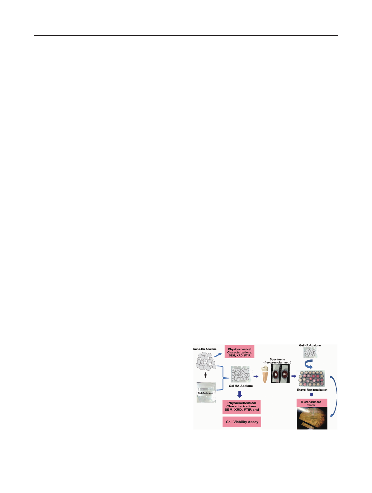
Fig. 1 Schematic of methods to fabricate and characterize
of nano-HA and gel HA-abalone, and the enamel
remineralization procedure.
precipitation from an aqueous solution10,11,17-20), and
hydrothermal21,22) and solid-state reactions23). In this
study, the precipitation method was selected to synthesize
nano-HA per several considerations. Specifically, nano-
HA is synthesized without the use of organic solvents (at
relatively low cost). This is a simple process with a large
output (87%), making the method suitable for large-scale
(i.e., industrial) production.
Nano-HA made by chemical synthesis is called
synthetic nano-HA. Synthetic nano-HA can be obtained
via the reaction of either synthetic or natural compounds
that are high in calcium. Some such natural materials
include cow bones, fish bones, cuttlefish, and mussel
shells10). In this study, abalone mussel shells (Haliotis
asinina) from Indonesia were used as the natural
compound for chemical synthesis; they are 90–95%
calcium carbonate24). Abalone mussel shells have been
developed as the basic material to fabricate nano-HA.
In previous research25), it was found that abalone meat
is a rare ingredient of traditional Chinese food and one
of the necessary dishes for Chinese banquets because of
its delicious taste and high nutritional values. However,
thousands of tons of shells were found abandoned around
a town. This resulted in a waste of natural resources
and polluted the environment. In a contrasting case in
Indonesia, the cultivation of abalone mussel shells has
been carried out at the Center for Marine Cultivation
Research and Fisheries Extension in Bali, Indonesia26).
The process of cultivating abalone mussel shells is
applied for research needs; moreover, the shells are sold
to craftspeople and the production of abalone shells is
usually 200 kg/month.
Mouthwash, toothpaste, gum, and gel are common
preparations as nano-HA carriers. A gel formulation
was chosen in this study because it also increased the
contact time between the active ingredients and the
tooth enamel3). The absorption process of a substance
in the body is influenced by the preparation and the
concentration of the materials. A high ion concentration
increases the remineralization potential many times
compared with saliva15). In this study, carbomer-based
gel preparations were used because they can easily flow
and enter the tooth enamel surface.
In this study, nano-HA was synthesized via the
co-precipitation method, using calcium carbonate
(CaCO3) from abalone mussel shells and a calcination
temperature of 1,000ºC to obtain calcium oxide; this
approach was adopted following a previous study27). The
characteristics of nano-HA made from abalone mussel
shells were observed. As mentioned at the beginning
of the introduction, the addition of HA to teeth is
expected to enhance the remineralization process. In
addition, nano-HA acts as a filler by repairing small
holes and depressions on the enamel surface. In this
study, nanocomposite HA with carbomer-based gel
is developed for the enamel remineralization process
because it can release active ingredients and diffuse
into tooth enamel tissue quickly. The synthesized nano-
HA was mixed with the carbomer for the gel fabrication
process, with concentrations of carbomer to nano-HA of
0, 10, 20, 30, and 40 wt%. The specimens used were 25
freshly extracted caries-free premolar teeth, following
the inclusion criteria. The physicochemical properties of
the gel HA-Abalone were characterized using scanning
electron microscopy (SEM), X-ray diffractometry
(XRD), and Fourier transform infrared spectroscopy
(FTIR). Evaluation based on enamel remineralization
parameters, including an enamel surface microhardness
test, was performed using a Vickers microhardness
(VHN) tester.
MATERIALS AND METHODS
The fabrication was divided into four main stages, which
were as follows: synthesizing nano-HA from abalone
mussel shells, fabrication of gel HA-Abalone, preparation
of freshly extracted caries-free premolar teeth, and
conducting the enamel remineralization procedure. The
schematic methods for this study are shown in Fig. 1.
Materials
The abalone mussel shells used as a source of CaCO3
were taken from Bali, Indonesia. The precursors of
diammonium hydrogen phosphate ([NH4]2HPO4)≥99.5%
and ammonium hydroxide (NH4OH) 25% solution
were purchased from Merck (NJ, USA). The gel HA-
Abalone was fabricated in the pharmacy laboratory of
Universitas Islam Agung Semarang, Indonesia. Bovine
calf serum 10% and phosphate-buffered saline (PBS)
were purchased from Sigma-Aldrich (St. Louis, MO,
USA). Penicillin-streptomycin, fungizone, and DMEM
high-glucose medium were purchased from Gibco (New
York, USA), whereas (3-(4,5-Dimethylthiazol-2-yl)-2,5-
diphenyltetrazolium bromide) (MTT) was purchased
from Biobasic (New York, USA), and dimethyl sulfoxide
(DMSO) was purchased from Merck KGaA (Darmstadt,
Germany). The specimens used were 25 freshly
extracted caries-free premolar teeth that were removed
for orthodontics reasons, following the inclusion criteria.
These samples obtained clearance from the Human
Ethics Review Committee of the Faculty of Dentistry,
Universitas Islam Sultan Agung, Semarang, Indonesia
69
Dent Mater J 2022; 41(1): 68–77

(No. 264/B.1-KEPK/SA-FKG/1/2021). Written informed
consent was obtained from all participants. Saline
solution, glycerin, propylene glycol, and demineralization
water were also obtained from the Faculty of Dentistry,
Universitas Islam Sultan Agung, Indonesia.
Preparation of calcium oxide (CaO) from abalone mussel
shells and synthesis of nano HA
The CaO and nano-HA were fabricated in previous
research27), so this study used those samples.
Fabrication of gel HA-Abalone
Fabrication of the gel was carried out using carbomer as
acrylic acid polymers materials, with concentrations of
carbomer to nano-HA of 0, 10, 20, 30, and 40 wt%. Nano-
HA powder was dissolved in 100 mL of distilled water
that had been heated to 50°C. Carbomer was added to
the nano-HA powder solution and distilled water and
stirred until homogeneous. Then, 10 mL of glycerin and
5 mL of propylene glycol were added to the nano-HA and
carbomer solution mixture and stirred until the solution
turned into a gel. The gel that formed was kept at room
temperature for 24 h.
Preparation of freshly extracted caries-free premolar
teeth
The specimens used were 25 freshly extracted caries-free
premolar teeth that had been removed for orthodontics
reasons, following the inclusion criteria. First premolars,
also called bicuspids, are the permanent teeth located at
upper jaw between first molars in the back of mouth and
canine teeth (cuspids) in the front. They are transitional
teeth, displaying some of the features of both canines and
molars. They have two cups on the buccal and palatal
parts, so they are called bicuspids. Caries-free premolar
teeth can be selected via visual selection; such teeth
have no white spots, no carious cavities, and attrition,
abrasion, erosion, or enamel structure anomalies. The
tooth cutting was done in the cementoenamel junction
area with a diamond bur, so the crown was left intact.
The cut teeth were planted in self-curing acrylic beams
measuring of 2×2 cm. The surface of the tooth enamel
was sanded using sandpaper (1,000 and 1,500 numbers).
The surface thickness of the sample was 0.5 mm, and
the samples was polished with a polishing tool bur with
alumina coating until smooth, flat, and shiny. The border
of the acrylic beam and the surface of the tooth enamel
were stained with the red nail polish. The surface of the
samples was rinsed in running water.
Enamel remineralization procedure
The specimens were randomly divided into the five
following groups: gel HA-Abalone 0 wt%, gel HA-Abalone
10 wt%, gel HA-Abalone 20 wt%, gel HA-Abalone 30
wt%, and gel HA-Abalone 40 wt%. Each gel was applied
to the tooth enamel surface for 10 min. The determined
time of 10 min was the average time for a person to eat.
The samples were rinsed with distilled water and soaked
with saline solution for 10 min. Then, the samples were
incubated for 10 min. At the same time, each gel was
also applied to the tooth enamel surface for 10 min. The
samples were rinsed with distilled water and soaked
with demineralization water for 10 min. Then, the
samples were incubated for 10 min. This treatment was
done twice a day for 14 days.
Characterization of gel HA-Abalone and enamel surface
1. Morphology, particle grain size, and composition
analysis
SEM (JSM-6510LA-1400, JOEL, Tokyo, Japan) was used
to observe the morphology of the gel HA-Abalone. The
particle grain size distribution of the gel HA-Abalone
was calculated according to the measurements of 100
randomly selected particles using ImageJ software.
2. Crystallographic analysis
The crystallographic properties of the gel HA-Abalone
were determined by XRD (PAN analytical Type X’Pert
Pro, Tokyo, Japan). The XRD data were recorded in the
range of 2θ: 10–80° using Cu-Kα radiation at λ=0.154
nm27).
3. FTIR analysis
The analysis of the functional groups of the gel HA-
Abalone were conducted using FTIR (Thermo Nicolet
iS10, Tokyo, Japan). Separately, the powder and gel
were ground and mixed with potassium bromide and
then passed into compact tablets. The FTIR instrument
was operated in the range of 400–1,000 cm-1 28).
4. Enamel surface microhardness test
Evaluation based on enamel remineralization
parameters, including an enamel surface microhardness
test (measuring baseline, after demineralization,
and after gel treatment), was conducted using a VHN
tester (ASTM E92, Buehler, IL, USA) by evaluating the
Vickers hardness number (VHN). The test on the 25
samples was set with a load of 100 gf. Data analysis was
performed using one-way analysis of variance (ANOVA).
A p-value less than 0.05 was considered statistically
significant2,3,5,6).
5. Cell viability assay of the gel HA-Abalone
1) Extraction solution of gel HA-Abalone
The gel HA-Abalone 20 wt% had the best results in terms
of physicochemical properties, so it was used in the cell
viability assay. An amount of 0.377 g of gel HA-Abalone
20 wt% was mixed with 94.2 mL of distilled water for
analysis to reach a concentration of 2,000 µg/mL. The
solution was then stirred at a temperature of 60ºC at a
velocity of 350 rpm until it turned into a homogeneous
solution. It was sonicated at a temperature of 60ºC for 1
h before the gel HA-Abalone solution was stored in the
refrigerator27,28).
2) Cell culture and seeding
Mouse fibroblast (NIH/3T3) cells were cultured in DMEM
high-glucose (Gibco)+10% Bovine Calf Serum (Sigma)
2% Penicillin-Streptomycin (Gibco)+0.5% Fungizone
(Gibco). The NIH/3T3 were seeded on the bottom of a
70
Dent Mater J 2022; 41(1): 68–77
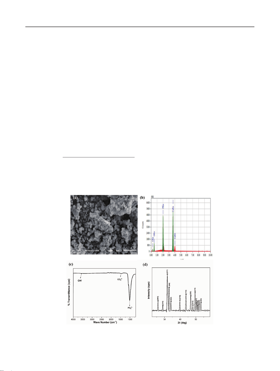
Fig. 2 Analysis of the psychochemical properties of nano-HA from abalone
mussel shell: (a) morphology, (b) composition, (c) FTIR spectrum, and
(d) XRD pattern27).
96-well plate at a density of 2×10⁴ cells/well. The cell
was incubated at 37°C in 5% CO2 for 24 h. A 100 µL
amount of scaffold solution was added to the cells. The
cell seeded on the scaffold was incubated at 37ºC in 5%
CO2 for 24 h. Prior to cell seeding, the gel HA-Abalone 20
wt% solution was stored in the refrigerator.
3) MTT (3-(4,5-Dimethylthiazol-2-yl)-2,5-
diphenyltetrazolium bromide) assay
MTT assay was performed to measure the cell viability
of NIH/3T3 cells and estimated through a color change
phenomenon from yellow tetrazolium salt to purple
formazan28,29). Cell viability was studied for an incubation
period of 48 h. The measurement was taken for the gel
HA-Abalone 20 wt% and a control (well without gel).
Then, the medium was discarded, 100 µL of MTT solution
with a concentration of 0.5 mg/mL was added to the well,
and the gel was incubated for 4 h. Then, DMSO was
added to the well at 100 µL/well. The absorbance was
recorded by Tecan Spark® (Tecan Trading, Zurich,
Switzerland) at 570 nm27). The cell viability was
calculated using the following equation:
absorbance of scaffold−absorbance of control media
Cell Viability (%)=
absorbance of control−absorbance of control media
×100.
(2.1)
Based on Eq. (2.1), cell viability was determined according
to the absorption value of the test cultures, expressed
as a percentage of absorption for unstimulated control
cultures27,30). Then, the IC50 value was analyzed via
nonlinear regression using GraphPad Prism software
version 7 (GraphPad Software, CA, USA).
6. Statistical analysis
All enamel surface microhardness and cell viability
assay data were presented as the mean±standard
deviation (SD) and one-way ANOVA was used to
analyze the obtained results, followed by Tukey’s test.
p-Values<0.05 were considered statistically significant.
These data were statistically analyzed using OriginPro
software version 2018 (OriginLab, Northampton, MA,
USA).
RESULTS
Nano-HA synthesis from abalone mussel shells
The psychochemical properties of nano-HA from abalone
mussel shells were determined using SEM-EDS, XRD,
and FTIR, as shown in Fig. 2. The results of these
analyses have been studied in previous research27),
where nano-HA based on abalone mussel shells had a
Ca/P molar ratio of 1.67. The crystallite size, microstrain,
and X-ray density of the synthesized nano-HA were
33.91±7.5 nm, 0.00373, and 10.46 g/cm3, respectively.
The distance between the crystal planes of the nano-
HA was determined using the Scherrer equation and
calculated to be 2.81Å27). This result is close to the crystal
71
Dent Mater J 2022; 41(1): 68–77