
JST: Engineering and Technology for Sustainable Development
Volume 35, Issue 1, March 2025,001-008
1
Polysaccharide from Sargassum Oligocystum Algae:
Isolation, Antioxidant and Antibacterial Activities
Hoang Thi Ngoc Nhon, Nguyen Ngoc Thu, Le Thi Hong Anh*
Food Science and Technology Faculty, Ho Chi Minh City University of Industry and Trade,
Ho Chi Minh City, Vietnam
*Corresponding author email: anhlth@huit.edu.vn
Abstract
Natural polysaccharides from algae have gained increasing attention for their biological activities and potential
for applications in food, pharmacology, medicine, and biology fields. The study aimed to investigate the effects
of the Viscozyme enzyme on the polysaccharide extraction from Sargassum oligocystum algae. Then, the
obtained polysaccharides were purified by using the Sevag method and Sephadex G-75 gel filtration
chromatography before evaluating the antioxidant and antibacterial activities. The results show that the
obtained polysaccharide was 2.06 ± 0.027 mg/g based on dry mass after extraction and the obtained purified
polysaccharide with a purity of 76.28%, which was determined via the UV-Vis and Fourier Transform Infrared
Spectroscopy (FT-IR) spectra with characteristic peaks. Antioxidant capacity of polysaccharides from
S. oligocystum algae by 2,2-diphenyl-1-picrylhydrazyl (DPPH) and 2,2'-azino-bis 3-ethylbenzothiazoline-6-
sulfonic acid (ABTS) free radical scavenging tests with IC50 values of 4.90 ± 0.09 ppm and 4.01 ± 0.03 ppm,
respectively. The antioxidant capacity of the obtained polysaccharides by Ferric Reducing Antioxidant Power
(FRAP) and Reducing Powder (RP) with OD0.5 values were 3.52 ± 0.10 ppm and 0.27 ± 0.01 ppm, respectively.
The antibacterial ability of the obtained polysaccharide was the concentration-dependent manner in the
surveyed range of 200-1000 ppm via the antibacterial diameter of Escherichia coli (7.02± 1.01 mm to
13.33 ± 2.08 mm) is greater than Bacillus subtilis (4.67 ± 1.15 mm to 12.00 ± 1.73 mm).
Keywords: Antioxidant, antibacterial, polysaccharide, Sargassum oligocystum.
1. Introduction*
S. oligocystum belongs to the Sargassum
genus - a large natural reserve in the brown algae
(Sargassaceae). About 250 genera have been
discovered in the world, and there are over
1,500 species. In Vietnam, about 150 species have
been discovered in the North of the Gulf of Tonkin, in
the Central region and the Southern coast [1]. The
growing season for most Sargassum lasts from
November to June of the following year. The best time
to harvest Sargassum is from May to June. The brown
seaweed species lives deep and grows all year round.
Algae species are very popular in traditional medicine
in Asia. Sargassum species is also used in Vietnam as
food additives or tea which has beneficial effects on
health [2].
Algae-derived polysaccharides attract widespread
attention for their nutritional benefits as well as their
biological potential. They are found mainly in the form
of fucoidan, alginate and laminarin. The
polysaccharide in green algae is mainly starch, while
polysaccharides in brown algae are laminaran
composed of (1,3)-β-D-glucan with β-(1,6) linkage
creating branches, laminaran is known to be an
ISSN 2734-9381
https://doi.org/10.51316/jst.180.etsd.2025.35.1.1
Received: Jun 17, 2024; revised: Aug 23, 2024;
accepted: Oct 21, 2024
antioxidant, anti-cancer agent, an anticoagulant...
Polysaccharide is a group of substances with many
biological functions. Fibre is a polysaccharide that is a
structural component of seaweed cells, including
water-soluble fibre and water-insoluble fibre. Water-
soluble fibre components include wax, gum, pectin,
xyloglucan, galactomannan, hemicellulose, and water-
insoluble fibre components such as cellulose,
arabinoxylan, lignin [3]. Castro et al. reported that the
fucans found in sulfated polysaccharides have strong
antioxidant properties, anti-inflammatory activity, and
cell inhibition of the HT-29 human colon cancer cell
line [4]. It is noteworthy that polysaccharides from
Sargassum exhibited high antioxidant capacity. For
example, the 2,2-diphenyl-1-picrylhydrazyl (DPPH)
free radicals scavenging was 51.99% at 80 μg/mL
(fucoidan isolated from Sargassum cinereum) [5] or
95.23% at 0.4 mg/mL (polysaccharide sulfate in
Sargassum elasticbergii) [6].
These findings showed that algal polysaccharides
have strong antioxidant activity. Biologically active
polysaccharides exhibited antibacterial activity by
interfering with cell walls and cell membranes or

JST: Engineering and Technology for Sustainable Development
Volume 35, Issue 1, March 2025,001-008
2
changing cell membrane permeability. Changing
permeability can prevent the penetration of nutrients
introduced into microorganisms. The disk diffusion
method is performed by placing a sheet of
antibiotic-absorbent paper on the surface of an agar
plate that has been inoculated with bacteria. The
antibacterial agent then diffuses into the agar, where it
can inhibit bacterial growth in the area surrounding the
plate. The antibacterial ability of polysaccharides is also
influenced by their structure and composition. The
diffusivity of different polysaccharides can directly
affect the bacterial inhibition zone [7]. Vo Thi Tuyet
Hoa et al. extracted and purified fucoidan from
Ceratophyllum submersum algae and obtained fucoidan
with a purity of over 60% [8]. Huynh Truong Giang et
al. was isolated polysaccharides from brown algae
Sargassum mcclurei in different solvents such as
distilled water, hydrochloric acid (HCl) 0,1N, and
ethanol 90% [9]. The chemical composition and
antioxidant activities of polysaccharides extracted from
Sargassum microcystum were also evaluated. Nguyen
Duy Nhut et al. isolated and compared the content of
fucoidans and their structure characteristics from
five Sargassum brown seaweed species in the south
provinces of Vietnam [10]. However, there are no
available reports on the isolation and characterization of
polysaccharides from Sargassum oligocystum in Ninh
Thuan province. This study investigated the conditions
for obtaining polysaccharides from S. oligocystum,
enhanced the purity by using the Saveg method and gel
filtration chromatography, and evaluated the
antioxidant and antibacterial activities of the obtained
extract. This study provides platform information
related to polysaccharides from S. oligocystum algae in
particular and Sargassaceae algae in general.
2. Materials and Methods
2.1. Materials
S. oligocystum algae were collected in Son Hai
1 village, Phuoc Dinh commune, Thuan Nam district,
Ninh Thuan province. After harvesting, the algae was
pre-washed, drained and transported to the laboratory,
where they were washed with tap water to remove
impurities such as sand, shells, snails... then dried at
50 ℃ until the moisture was under 10%, ground and
sieved to collect powder less then 0.3 mm in a zip bag
and stored at 5 ℃ for all experiments.
Chemicals: Chloroform (Merck), n-butanol
(Merck), enzyme Viscozyme L (Novozyme, Denmark),
phenol (Merck), H2SO4 (Merck), Mueller Hilton Agar
(MHA, Sigma), LSB medium, DPPH (Merck), ABTS
(Merck), K2S2O8 (Merck), K3Fe(CN)6 (Merck),
CCl3COOH (Merck), Ampicillin (Sigma). Other
chemicals were at an analytical level.
Bacterial strains included Bacillus subtilis
ATCC®6633 and Escherichia coli ATCC®25922,
which were selected for antibacterial activity assay. The
bacteria cultures were maintained in their appropriate
agar slants at 4 °C throughout the study and used as
stock cultures.
2.2. Methods
2.2.1. Effects of enzymes on polysaccharide extraction
Polysacharide was isolated via enzyme-assisted
extraction with Viscozyme, which helps destroy the cell
wall and release polysacharide from the material.
Briefly, 1 g of raw algae powder (calculated by dry
matter) was put into different 100 mL glass beakers
processed by the Soxhlet method, and then the solvent
was added to the beakers. The material/solvent ratios
were investigated (1/15, 1/20, 1/25, 1/30, 1/35 (w/v)),
stirred, fixed enzyme concentration 1% compared to
raw material weight dry, placed the mixtures in a
thermostatic bath at the investigated temperatures
(40 ℃, 50 ℃, 60 ℃, 70 ℃, 80 ℃) for the investigation
time (60, 90, 120, 150, 180 minutes) [11]. At the end,
the enzymatic hydrolysis reaction was inactivated by
keeping at 90 - 100 ℃ for 10 minutes and then
immediately cooled to cool samples. The samples were
centrifuged at 4800 rpm for 10 minutes to collect the
supernatant containing polysaccharides and determine
the polysaccharide content using the spectroscopic
method.
2.2.2. Polysaccharide purification
Protein was removed from the polysaccharide
extract by reacting with Sevag solution (chloroform:
n-butanol = 4:1 v/v) with the ratio of 1:1 v/v, vortexing
for 20 minutes, and allowing it to settle. The mixture
was separated into three phases, and most of the
polysaccharides were concentrated in the supernatant.
This process was repeated three times. Then, the
precipitation was continued with 96% ethanol at 4 ℃
overnight in a ratio of 1:4 v/v. Then, the mixture was
centrifuged at 5500 rpm for 20 minutes to obtain the
crude polysaccharide for further purification stage.
Next, 1 g of crude polysaccharide was mixed with
10 mL of distilled water, centrifuged to get the solution
and put into a prepared Sephadex G75 gel filtration
chromatography column, waiting for 30 minutes, then
eluted with 0.2 M NaCl to collect fractions of 4 mL for
each. The polysaccharide content was determined via
the spectroscopic method.
2.2.3. Antioxidant activity
DPPH assay: The antioxidant activity via DPPH
assay was performed according to the description of K.
Mishra et al [12]. Polysaccharide samples and positive
control acorbic acid concentrations were diluted with
methanol in a 10 mL volumetric flask. 2 mL test
solution was put into the test tube, followed by 2 mL of
0.1 mM DPPH solution. For the control sample, the test
solution was replaced with MeOH. The blank sample
only contains MeOH. The test tubes were incubated in
the dark at room temperature for 30 minutes, and then
the absorbance spectrum was measured at 517 nm.

JST: Engineering and Technology for Sustainable Development
Volume 35, Issue 1, March 2025,001-008
3
ABTS assay: The antioxidant activity via ABTS
assay was conducted as described by R. Re et al. [13].
ABTS•+ solution was from 2 mL of 2 mM ABTS
solution and 2 mL of 2.45 mM K2S2O8 solution in a
100 mL volumetric flask and incubated the solution in
the dark for 16 hours, then diluted it with methanol and
adjusted the absorbance of the solution at a wavelength
of 734 nm with an optical density of 0.7 ± 0.05.
4 mL of ABTS+ and 1 mL of the samples in test tubes
at various concentrations. The reaction mixture was
incubated for 6 minutes and then measured at 734 nm.
Reducing Powder (RP) assay: The Fe reduction
capacity of polysaccharides was determined according
to Rahate K. et al. [14]. The reaction mixtures
consisted of 0.5 mL of polysaccharide samples at
different concentrations, 0.5 mL of phosphate buffer
(0.2 M, pH= 6.6), 0.5 mL of phosphate buffer (0.2 M,
pH= 6.6) and 1% K3Fe(CN)6 in test tubes, the mixtures
were incubated at 50 ℃ for 20 minutes and added
0.5 mL of 10% CCl3COOH and then centrifuged at
3000 rpm for 10 minutes. After centrifugation, 0.5 mL
of the upper layer, 0.5 mL of water and 0.1 mL of 0.1%
FeCl3 were mixed in a test tube, shaken well in the test
tube and then measured at 700 nm. The positive
control was ascorbic acid.
Ferric Reducing Antioxidant Power (FRAP)
assay: The principle of determining the antioxidant
activity of this method is based on the ability to reduce
the Fe3+-TPTZ complex [2, 4, 6-tripyridyl-s-triazine
(TPTZ)] to the Fe2+-TPTZ complex in an acidic
environment. The Fe3+-TPTZ complex is in an
environment containing antioxidants, and the
antioxidants donate electrons to this complex and form
Fe2+-TPTZ. In particular, the blue intensity is
proportional to the antioxidant content in the samples,
measured at a wavelength of 593 nm [15].
2.2.4. Antibacterial activity
Antibacterial activity of polysaccharide obtained
from S. oligocystum was performed as described by
A. W. Bauer et al. Mueller Hilton Agar (MHA)
medium was to inoculate the strains at 37 ℃/24 hours
for use [16]. Lauryl Tryptose Broth (LSB) medium
was also mixed to create an enrichment medium for
E. coli and B. subtilis. Then, the typical colony
inoculated into a liquid LSB medium and then
incubated at 37 ℃/24 hours. 100 µL of bacterial
solution with a cell density of about 106-108 CFU/mL
from the liquid medium put into the MHA medium
plate, spread evenly, let dry for 15-30 minutes, then
added 20 µL of the samples of different concentrations
on 6 mm – diameter filter paper on the surface of the
agar plate, incubated the plate upside down at
37 ℃/24 hours then measured the antibacterial
diameter. The solvent was chosen as the negative
control, and the positive control was ampicillin.
2.2.5. Analysis method
Polysaccharide content determination:
Polysaccharide content was determined by using
the phenol-sulfuric acid method. Based on the
hydrolysis reaction of polysaccharide into
monosaccharide, monosaccharide creates colour with
phenol in an acidic environment. Briefly, 2 mL of the
sample solution was put into lidded test tubes, 1 mL of
4% phenol solution was added, and 5 mL of
concentrated H2SO4 solution was added to cover the
test tubes tightly and vortex gently so that the solutions
were uniform. First, heat the test tubes in a water bath
at 40 ℃ for 30 minutes, then put them in ice water for
5 minutes. Absorbance was measured at 490 nm [17].
The polysaccharide purity of fractions was
determined via the percentage of polysaccharide
content calculated by phenol-sulfuric acid (B) and the
mass of dry matter (A) [18].
Purity (%) = 𝐵𝐵 ×100%
𝐴𝐴
FI-IR spectroscopic analysis:
The infrared (IR) spectra of the obtained
polysaccharide were obtained using a Fourier
transform infrared spectrophotometer, recorded at the
absorbance mode from 4000 to 400 cm-1 (mid-infrared
region) [17]. In this study, the fractions with the
highest purity from the purification stage would be
determined the FT-IR spectrum.
Nuclear magnetic resonance (NMR) analysis:
The spectra 1H-NMR were recorded using the
Brucker Advance DPX - 500 NMR spectrometer
(Bruker, Berlin, Germany). The samples were
sonicated at 75.5 MHz and 27 ℃ and before dissolving
in D2O with 20 μg/mL for measuring 1H-NMR
spectrum.
2.3. Data Analysis
The experiments were performed 3 times, and the
results were presented as mean plus/minus SD. IBM
SPSS Statistics 20.0 software was used to analyze
experimental data, evaluate the difference between
samples, and optimize the extract conditions.
Microsoft Excel 2019 software was used to draw
charts.
3. Results and Discussion
3.1. Effects of Enzyme on Polysaccharide Extraction
from S. Oligocystum Algae
The enzyme will disrupt the cell wall, thereby
releasing inside components like polysaccharides into
the solvent and resulting in polysaccharide
content. The effects of material and solvent ratio,
temperature and extraction time in enzyme treatment
on polysaccharide content are shown in Fig. 1.
JST: Engineering and Technology for Sustainable Development
Volume 35, Issue 1, March 2025,001-008
4
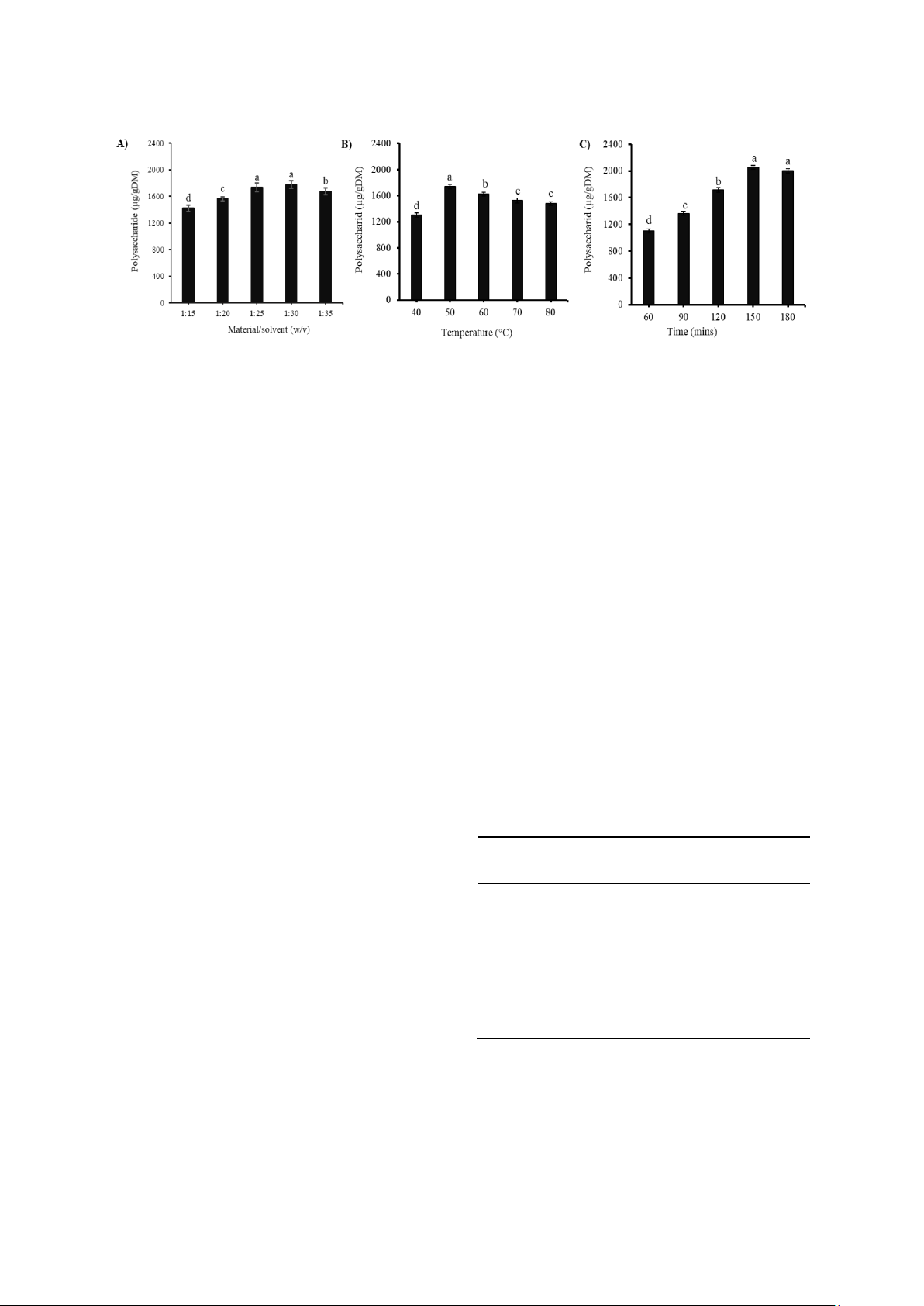
Fig. 1. Effects of material/solvent ratio (A), temperatures (B), and time (C) on polysaccharide extraction
Note: In each graph, different characters on the bar indicate different statistical significance at 5%.
The results indicate that the more used solvent
resulted in higher polysaccharide concentration in the
extract. In fact, the polysaccharide content increased
from 1322.35 µg/gDM (1:10 w/v) to 1728.65 µg/gDM
(1:25 w/v) (Fig. 1A). When this ratio was increased,
the raw materials were in full contact with the
enzymes, leading to the release of many
polysaccharides. As the ratio of materials/solvents
increases, the extraction rate of polysaccharides
decreases because the excess solvents completely
dissolve them.
The effectiveness of polysaccharide extraction
showed an upward trend with the increase in
temperature extraction. At 50 °C, the polysaccharide
content was the highest with 1736.81 μg/gDM
(Fig. 1B); this is because the Viscozyme works well at
this temperature [19]. Enzyme activity is significantly
influenced by the fact that different enzymes have
different optimum temperature conditions for their best
hydrolysis efficiency. Furthermore, a much higher
temperature could result in the thermal breakdown of
polysaccharides, increase energy expenditure, speed
up solvent evaporation, and release impurities in the
extraction process [11]. Besides, 150 mins were cited
as the highest polysaccharide content with
2055.49 μg/gDM (Fig. 1C). The amount of
polysaccharides decreases slightly as extraction time
increases past this point. Enzymes hydrolyzing the
polysaccharide at a specific temperature and extraction
time may be a factor in this phenomenon.
In short, Fig. 1 shows that the highest
polysaccharide content was the material/solvent
1/25 (w/v), temperature 50 °C, and extraction time was
150 minutes were the most appropriate conditions.
3.2. Polysaccharide Purification from S. Oligocystum
Algae
Proteins and polysaccharides are biopolymers
that have a diverse structure. The separation of proteins
from crude polysaccharides is an important step in the
process of splitting and purifying polysaccharides. The
Sevag method is a simple method based on the
principle that reagents denature and precipitate
proteins instead of polysaccharides [20]. After the
extraction, the polysaccharide extract was precipitated
with 96% ethanol, then mixed with water, centrifuged
to collect the supernatant and added the Sevag reagent
to deproteinize for 20 minutes oscillation. Next,
samples were left to stand for 30 minutes so that the
mixtures split into 3 phases; polysaccharides were in
the phase on the top. After three times of this stage, the
obtained polysaccharide content was 1659.34 μg/gDM.
The obtained polysaccharide was then fractioned via
Gel filtration chromatography of Sephadex G75, eluted
by 0.2M NaCl with the rate of 2 mL/min and collected
5 fractions (Table 1).
Based on Table 1, the highest polysaccharide
content was fraction 2 with 2101.28 μg/gDM with a
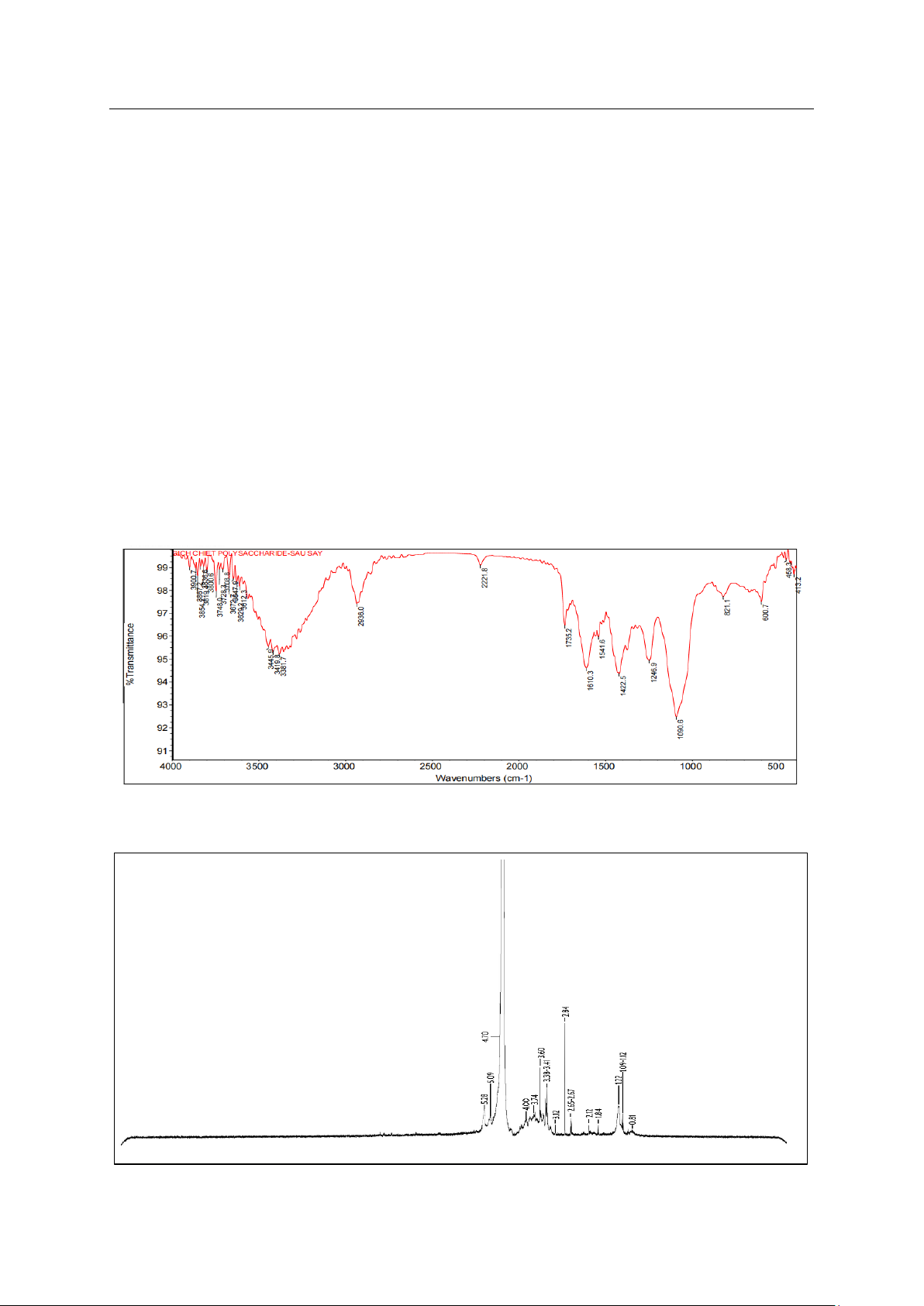
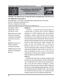
purity of 66.28%. The FT-IR spectrum of this fraction
is shown in Fig. 2.
Table 1. Polysaccharide content and purity during the
purification stage
Fractions Polysaccharide
content (μg/g DM) Purity (%)
1 1156.23 ± 38.59 8.04 ± 0.45
2 2101.28 ± 30.26 66.28 ± 7.41
3 1560.99 ± 32.97 24.47 ± 1.88
4 297.62 ± 33.12 1.84 ± 0.37
5 53.66 ± 35.75 0.15 ± 0.10
The infrared spectrum (Fig. 2) showed a strong
absorption range at 3500-3200 cm−1 with observed
peaks of 3381.7 cm−1, 3419 cm−1, and 3445.9 cm-1
characterizing the elongated oscillations of O-H. The
peak near 2936.0 cm−1 was due to prolonged
oscillations of C-H, peaks appearing at
1730 cm−1 demonstrated that polysaccharides
JST: Engineering and Technology for Sustainable Development
Volume 35, Issue 1, March 2025,001-008
5
contained carboxylic groups and peaks near
1610.3 cm−1 indicated the presence of O-C-O [21]. The
peak at 1246.9 cm-1 was in the extended band at about
1120-1270 cm−1, indicating an elongated S=O sulfate
group branching from the amount of fucoidan or
alginic acid [22]. The peak at 1090.6 cm−1 was
attributed to prolonged oscillations of C-O-C. The
peak at 821.1 cm−1 was considered the signal for the
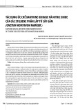
presence of α-mannopyranose [21]. In addition,
1H-NMR spectrum expressed peak of 5.28 ppm
(Fig. 3), which was respond the bond of α -1,2-linked
mannopyranose unit [23].
3.3. Antioxidant Activity of Polysaccharide from
S. Oligocystum Algae
The antioxidant activity of the obtained
polysaccharides was determined via four select assays
of DPPH, ABTS, RP and FRAP. The antioxidant
activity via four selected assays was concentration-
dependent. Regarding antioxidant activity, the IC50
values were 4900 ppm (DPPH assay) and 4010 ppm
(ABTS assay). In addition, higher polysaccharide
concentrations resulted in higher absorbance values,
which revealed better antioxidant properties in both
FRAP and RP assays (Fig. 4).
DPPH free radicals have the ability to absorb
hydrogen molecules of antioxidants. Therefore, DPPH
is widely used to test the antioxidant capacity of
various compounds [22]. As polysaccharide
concentrations increased, antioxidant activity also
increased markedly. The results were similar to those
in the literature [21]. The antioxidant capacity of
polysaccharides of the test sample increased from
1000 ppm to 5000 ppm; the free radical scavenging
increased from 22.38% to 50.96% (Fig. 4A). The
ABTS free radical scavenging increased from 22.66%
(1000 ppm) to 58.23% (5000 ppm) (Fig. 4B). The
ability of polysaccharides from S. oligocystum algae to
scavenge ABTS free radicals was higher than that of
DPPH.
Fig. 2. FT-IR spectrum of polysaccharides
Fig. 3. 1H-NMR spectrum of the isolated polysaccharides

![Giáo trình Sản xuất giống bằng phương pháp nuôi cấy mô tế bào thực vật (Nghề: Kỹ thuật rau, hoa công nghệ cao - TC) - Trường Cao đẳng Đà Lạt [Mới nhất]](https://cdn.tailieu.vn/images/document/thumbnail/2026/20260224/hoacattuong2026/135x160/28171772099038.jpg)


![Giáo trình Đất trồng phân bón (Nghề Bảo vệ thực vật CĐ/TC) - Trường Cao đẳng Gia Lai [Mới Nhất]](https://cdn.tailieu.vn/images/document/thumbnail/2026/20260224/hoacattuong2026/135x160/9591771994804.jpg)
![Giáo trình Kỹ thuật canh tác cây lương thực (Nghề Bảo vệ thực vật - CĐ/TC) - Trường Cao đẳng Gia Lai [Mới nhất]](https://cdn.tailieu.vn/images/document/thumbnail/2026/20260224/hoacattuong2026/135x160/12471771994805.jpg)


![Giáo trình Công nghệ sau thu hoạch (Nghề Bảo vệ thực vật - CĐ) - Trường Cao đẳng Đà Lạt [Mới nhất]](https://cdn.tailieu.vn/images/document/thumbnail/2026/20260224/hoacattuong2026/135x160/32161772096417.jpg)
