
RESEA R C H Open Access
Involvement of aryl hydrocarbon receptor
signaling in the development of small cell lung
cancer induced by HPV E6/E7 oncoproteins
Tonia Buonomo
1
, Laura Carraresi
2
, Mara Rossini
3
, Rosanna Martinelli
1,4*
Abstract
Background: Lung cancers consist of four major types that and for clinical-pathological reasons are often divided
into two broad categories: small cell lung cancer (SCLC) and non-small cell lung cancer (NSCLC). All major
histological types of lung cancer are associated with smoking, although the association is stronger for SCLC and
squamous cell carcinoma than adenocarcinoma. To date, epidemiological studies have identified several
environmental, genetic, hormonal and viral factors associated with lung cancer risk. It has been estimated that
15-25% of human cancers may have a viral etiology. The human papillomavirus (HPV) is a proven cause of most
human cervical cancers, and might have a role in other malignancies including vulva, skin, oesophagus, head and
neck cancer. HPV has also been speculated to have a role in the pathogenesis of lung cancer. To validate the
hypothesis of HPV involvement in small cell lung cancer pathogenesis we performed a gene expression profile of
transgenic mouse model of SCLC induced by HPV-16 E6/E7 oncoproteins.
Methods: Gene expression profile of SCLC has been performed using Agilent whole mouse genome (4 × 44k)
representing ~ 41000 genes and mouse transcripts. Samples were obtained from two HPV16-E6/E7 transgenic
mouse models and from littermate’s normal lung. Data analyses were performed using GeneSpring 10 and the
functional classification of deregulated genes was performed using Ingenuity Pathway Analysis (Ingenuity
®
Systems, http://www.ingenuity.com).
Results: Analysis of deregulated genes induced by the expression of E6/E7 oncoproteins supports the hypothesis
of a linkage between HPV infection and SCLC development. As a matter of fact, comparison of deregulated genes
in our system and those in human SCLC showed that many of them are located in the Aryl Hydrocarbon Receptor
Signal transduction pathway.
Conclusions: In this study, the global gene expression of transgenic mouse model of SCLC induced by HPV-16
E6/E7 oncoproteins led us to identification of several genes involved in SCLC tumor development. Furthermore,
our study reveled that the Aryl Hydrocarbon Receptor Signaling is the primarily affected pathway by the E6/E7
oncoproteins expression and that this pathway is also deregulated in human SCLC. Our results provide the basis
for the development of new therapeutic approaches against human SCLC.
Background
Human papillomaviruses (HPVs) are a collection of
over 200 viruses that can infect humans. HPV is most
often spread through skin-to-skin contact, usually
sexually. Genital HPV infections are very common and
are sexually transmitted. Most HPV infections occur
without any symptoms and go away without any treat-
ment over the course of a few years. However, HPVs
infection sometimes persists for many years in the
host, either through the establishment of latent or
chronic infections, which can ultimately lead to cellular
transformation [1]. It is now well-established that high-
risk HPVs play a role in most cases of cervical cancer,
as well as many cases of vulvar, penile, and anal can-
cers [2,3]. HPV 16 and 18 have been identified not
only in gynecological carcinomas but also in tumors of
* Correspondence: rosanna.martinelli@unina.it
1
CEINGE Biotecnologie Avanzate, Via Comunale Margherita 482, 80145
Napoli, Italy
Full list of author information is available at the end of the article
Buonomo et al.Journal of Translational Medicine 2011, 9:2
http://www.translational-medicine.com/content/9/1/2
© 2011 Buonomo et al; licensee BioMed Central Ltd. This is an Open Access article distributed under the terms of the Creative
Commons Attribution License (http://creativecommons.org/licenses/by/2.0), which permits unrestricted use, distribution, and
reproduction in any medium, provided the original work is properly cited.

other organs, like the upper aerodigestive tract and
oropharynx especially those occurring in young, non-
smoking women. Only a few of these viruses are con-
sidered the “cancer-causing”strains, most notably,
HPV16andHPV18[4-6].
The possibility that HPV may play a role in the devel-
opment of lung cancer was first suggested by Syrjanen
in 1979 who described epithelial changes in bronchial
carcinomas closely resembling those of established HPV
lesions in the genital tract [7]. Since then, several studies
provided evidence of HPV 16 and 18 DNA in lung can-
cers, but there were inconsistency in the reported preva-
lence of infection by HPVs in patients with lung cancer
in different countries, with racial and geographic varia-
tions. In the United States, HPVs DNA is found in
about 20-25% of lung cancers [8]. The most common
strains found are HPV 16 and HPV 18, the same strains
that are commonly found in cervical cancer. More than
90% of lung cancer in Taiwanese females is not related
to cigarette smoking and 55% had HPV16/18 DNA
compared with 11% of non cancer control subjects.
Additionally HPV 16/18 DNA has been uniformly
detected in lung tumor cells but not in the adjacent
noninvolved lung tissue [9]. HPV 16/18 have been
detected in the blood of women with cervical infection
suggesting that HPV 16/18 can infect the lung through
hematic spread from infected sites [10].
A recent review summarizes the studies conducted to
establish the association between the presence of HPVs
and lung cancer [11]. HPVs detection rates in lung can-
cer are highly variable in the different studies published
from several countries, ranging from 0% to 79%. The
mean incidence of HPVs in lung cancer considering all
reviewed articles is 24.5%. While in Europe and in the
USA the average reported incidences is 17% and 15%,
respectively, the mean incidence of HPVs in Asian lung
cancer is 35.7%. The authors concluded that HPV may
be the second leading cause of lung cancer after cigar-
ette smoking.
Although studies of viral-related lung cancer have
been reported, the molecular mechanisms of this disease
remain unclear [12-14]. Therefore, an increase in
knowledge of factors promoting lung carcinogenesis, as
the infection with human papillomavirus, gains in
importance.
In this study we examined the gene expression profile
of previously described transgenic mouse model (CK5-
PAP-2303) of SCLC induced by HPV-16 E6/E7 onco-
proteins [15] and compared data with those obtained
from human tissue with SCLC.
The aim of our study was to identify molecular
mechanisms associated to SCLC development induced
by HPV 16 oncoproteins and in patients affected by
SCLC to validate our “in vivo”model and the derived
cell lines for the development and evaluation of new
anticancer molecules.
Methods
RNA purification, labelling and oligonucleotides
microarray hybridization
Lung tissues from 9-month-old wild-type and transgenic
mice, were homogenised in Qiazol solution (Qiagen) by
rotor-stator and RNA was extracted using RNeasy mini
kit from Qiagen according to manufacturer’sprotocol.
RNA samples were analyzed quantitatively and qualita-
tively by NanoDrop ND-1000 UV-Vis Spectrophot-
ometer (NanoDrop Technologies, Wilmington, DE) and
by Bioanalyzer (Agilent Technologies, Palo Alto, CA).
Only samples with R.I.N. (RNA Integrity Number) >8.0,
260/280 nm absorbance >1.8 and 260/230 absorbance
>2, were used for RNA labelling. Total RNA from lung
tumor and controls, was amplified in the presence of
cyanine-3/cyanine-5 labelled CTP using Agilent low
RNA Input Fluorescent Linear Amplification kit (Agilent
Technologies, Palo Alto, CA) according to manufac-
turer’s protocol. After labelling, targets were purified
using Qiagen’sRNeasyminispincolumntoremove
unincorporated dye-labelled nucleotides. The quality of
labelled targets was determined by calculating the
amount of cDNA produced, the pmoles of dye incorpo-
rated and the frequency of incorporation by NanoDrop.
Equal amounts of cRNAs (825 ng) from control
(labelled with Cy3) and from transgenic mouse (labelled
with Cy5) were mixed together and hybridized to the
microarray in a hybridization oven at 65°C for 17 hours
with rotation at 10 rpm. Gene expression profile of
transgenic SCLC has been performed using Agilent
whole mouse genome (4 × 44k) representing ~ 41000
genes and mouse transcripts. Samples were obtained
from two HPV16-E6/E7 transgenic mice and from 2 lit-
termate’s normal lung. For each sample were performed
the technical replicates.
After hybridization slides were washed with Gene
Expression Wash buffer 1 for 1 minute at room
temperature and Gene Expression Wash buffer 2 for
1 minute at 37°C. Finally to dry the slides and prevent
ozone degradation arrays were treated with the Stabili-
zation and Drying Solution (Agilent Technologies, Palo
Alto, CA) for 30 seconds at room temperature. After
wash the slides were scanned with the Agilent’sdual-
laser microarray scanner (G2565AA) and image data
were processed using Agilent Feature extraction soft-
ware (FE) (Agilent Technologies). This software calcu-
lates log ratios and p-values for valid features on each
array and provides a confidence measure of gene differ-
ential expression performing outlier removal and back-
ground subtraction. Furthermore, FE filters features that
are not positive and significant respect to background
Buonomo et al.Journal of Translational Medicine 2011, 9:2
http://www.translational-medicine.com/content/9/1/2
Page 2 of 11

and/or saturated. FE was also used to perform linear
and LOWESS dye normalization to correct dye bias.
Microarray data analysis
The raw data and associated sample information were
loaded and processed by GeneSpring
®
10 (Agilent Tech-
nologies). Statistical analysis was performed using back-
ground-corrected mean signal intensities from each dye
channel. Microarray data were normalized using inten-
sity-dependent global normalization (LOWESS). Differ-
entially expressed RNAs were identified using a filtering
by the Benjamini and Hochberg False Discovery Rate
(p-Value < 0.05) to minimize selection of false positives.
Of the significantly differentially expressed RNA, only
those with greater than 2-fold increase or 2-fold
decrease in expression compared to the controls were
used for further analysis. All microarray data presented
in this manuscript are in accordance with MIAME
guidelines and have been deposited in the NCBI GEO
database (The Accession Number it is available by
referees).
Functional and network analyses of statistically signifi-
cant gene expression changes were performed using
Ingenuity Pathways Analysis (IPA) 8.0 (Ingenuity
®
Sys-
tems, http://www.ingenuity.com). Analysis considered all
genes from the data set that met the 2-fold (p-value <
0.05) change cut-off and that were associated with biolo-
gical functions in the Ingenuity Pathways Knowledge
Base. For all analyses, Fisher’sexacttestwasusedto
determine the probability that each biological function
assigned to the genes within each data set was due to
chance alone.
Histopathology
Transgenic and control lungs were removed, washed in
PBS and fixed with 4% buffered formaldehyde. Samples
were processed and paraffin-embedded. Sections were
stained with hematoxylin/eosin and observed with a
Zeiss light microscope.
Semiquantitative reverse transcriptase-PCR
Semiquantitative reverse transcriptase-PCR (RT-PCR)
was done essentially as previously described [16]. RNA
(2 μg/reaction) was used to generate cDNA and the
appropriate individual pairs of oligonucleotides (40
pmol/reaction) for the test genes were used to amplify
DNA from the cDNA. Semiquantitative PCR was done
by using 100 μL reaction volumes and taking 33 μL
aliquots at 25, 30, and 35 cycles. The expression of 18
S mRNA, which is ubiquitously expressed, was deter-
mined for each RNA sample to control for variations
in RNA quantity. Ten microliters of each reaction
were electrophoresed in a 1% agarose gel containing
ethidium bromide. The gel was then developed using
theGelDocXRsystem(Bio-Rad) and quantified using
Quantity One (Bio-Rad).
The Bioethics Committee of the University of Siena
approved all the experiments conducted on live animals.
All the experiments were performed in accordance with
guidelines and regulations.
Results
Gene expression profiles
To identify mechanisms associated with SCLC develop-
ment and its neuroendocrine differentiation associated
to E6/E7 oncoproteins expression, we analyzed the gene
expression profile of transgenic lung tumor through
microarrays. Experiments were performed on lung sam-
ples from two different transgenic animals compared to
normal lung tissue. To identify the differentially
expressed genes, using the criteria described in Materials
and Methods for Microarray data analysis, we found
5307 significantly deregulated genes. Among these 2242
genes were up-regulated and 3065 were downregulated.
Up and down regulated genes are reported in the addi-
tional file 1. For each gene the probe ID, fold change, p-
value, gene symbol, Gene Bank and description are
reported. Interestingly, among all the genes deregulated
by the E6/E7 co-expression, 116 genes are associated to
neurogenesis. The list of these genes is reported in addi-
tional file 2. These results support the hypothesis of a
possible role of E6 and E7 in the induction of neuroen-
docrine differentiation of SCLC. To confirm gene array
analysis data and to validate some genes involved in this
process, we performed semiquantitative RT-PCR using
RNA purified from transgenic lung tumour, from litter-
mate normal lung and from the PPAP9 cell line, estab-
lished from the transgenic lung tumour [16]. As shown
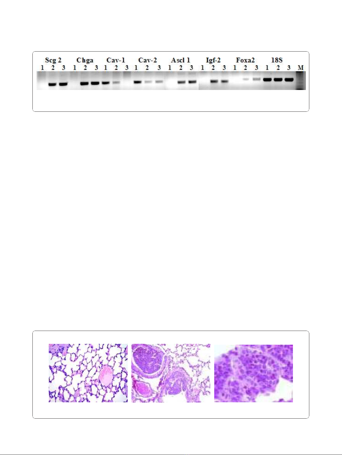
in Figure 1 Ascl1, Igf2, Scg2, Chga and Foxa2, consid-
ered reliable markers of neuroendocrine differentiation,
areup-regulatedintissueandcellsfromtumour
induced by E6/E7 compared to normal lung. Further-
more Cav1 and Cav2 are down regulated, according to
previously published results showing a tumor suppressor
activity of Caveolin-1 and its down-regulation during
lung cancer development [17]. The relative direction of
expression was the same for both the RT-PCR and
microarray results. The primers used for RT-PCR are
reported in additional file 3.
The transgenic mice develop brochiogenic lung cancer
around 6 months of age, and the E6/E7 genes were effi-
ciently expressed in pre-neoplastic and neoplastic cells.
The transgenic mice lung tumors showed a progression
from in situ to invasive carcinoma and in a minor per-
centage brain, liver and pancreas metastases were
observed. Inactivation of p53 and pRB occurs in the
majority of neuroendocrine lung carcinomas in humans
and these observations strongly suggest that E6/E7
Buonomo et al.Journal of Translational Medicine 2011, 9:2
http://www.translational-medicine.com/content/9/1/2
Page 3 of 11
expression most probably causes lung cancer in our
transgenic model, through the inactivation of p53 and
pRB. The transgenic lung carcinoma progress to multi-
ple and bilateral tumors with histopathology and immu-
nophenotype closely mirroring human SCLC constituted
by small cells with a very high nucleus/cytoplasm ratio.
Histological examination of mice lungs showed the
occurrence of multiple dysplastic foci of clustering small
cells in the bronchial and bronchiolar mucosa as shown
in Figure 2. Furthermore intrapulmonary tumors aggres-
sively invade lung parenchyma and vessels and readily
metastasized to extra pulmonary sites, again very similar
to human SCLC. Moreover a subcutaneous injection of
two murine cell lines (PPAP9 and PPAP10), established
from the transgenic SCLC, form primary tumors as well
metastasis typical of the pattern seen in human SCLC
patients [16]. The histological and biological properties
of our model are overlapping to those of two other
described murine SCLC models, obtained using different
experimental approaches [18,19].
Human SCLC metastasizes early and widely and
usuallyitisnottreatablebysurgerymakingtissue
retrieval particularly difficult. Therefore the results
obtained in our experimental system were compared
with those available for human SCLC in Gene Expres-
sion Omnibus (GEO), a public functional genomics data
repository. We imported data files from GSE6044,
selecting five sets derived from human normal lung,
(GSM140185-GSM140189), and five sets from human
SCLC, (GSM140176-GSM140180) [20] and analyzed
data sets with GeneSpring 10. Of the 8793 genes exam-
ined, 561 were differentially expressed to a significant
degree (ANOVA, p < 0.05). Among these, 289 genes
were up-regulated and 272 were down-regulated. Genes
are listed in the additional file 4 reporting for each gene,
probeID,thefoldchange,p-value,genesymbol,Gene
Bank and description. The significant difference in the
number of deregulated genes from the two analyses is
associated with the different number of genes present in
the arrays, 44000 for the Agilent system and 8793 for
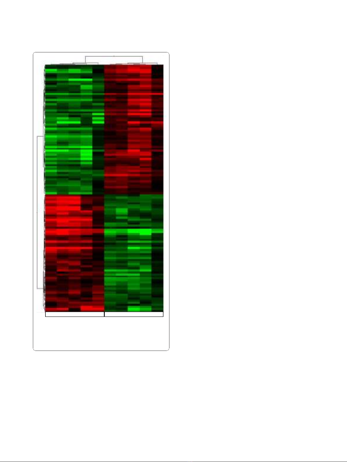
the Affymetrix platform. Hierarchical clustering of the
human differentially expressed genes according to their
expression patterns is reported in Figure 3. Genes up-
regulated in human SCLC are shown in red, down-regu-
lated genes are shown in green, while black bars indicate
genes that are expressed at similar levels in both. To
highlight the molecular mechanisms common to tumor
induced by E6/E7 oncoproteins and human SCLC, we
compared the results obtained in the two systems. We
identified 130 up- and 72-down regulated genes com-
mon to human SCLC and the E6/E7 induced lung
tumour. The list of genes is reported in the additional
file 5 showing for each gene, description, gene symbol,
family name, probe ID for transgenic mouse (Agilent),
probe ID for human SCLC (Affymetrix), the fold change
and relative p-values. To underline similarities among
samples and among genes, we overlaid the unsupervised
two-dimensional hierarchical clustering, obtained from
Figure 1 Confirmation of microarray data. RT-PCR was done using total RNA from wild-type mouse lung (line 1), transgenic mouse lung (line
2) and PPAP9 cells (line 3). Scg2: secretogranin 2, Chga: chromogranin, Cav-1: caveolin1, Cav-2: caveolin 2, Ascl1: achete-scute complex
homologue 1, Igf-2: insulin-like growth factor 2, FoxA2: forkhead box A2, A, 18S: 18 S ribosomal RNA M: marker.
ABC
Figure 2 Normal lung tissue and transgenic lung tumor histology. (A) Normal lung; original magnification 150×; (B) Transgenic SCLC;
original magnification 150×; (C) Transgenic SCLC; original magnification 400×.
Buonomo et al.Journal of Translational Medicine 2011, 9:2
http://www.translational-medicine.com/content/9/1/2
Page 4 of 11
expression profile of human SCLC with results obtained
from the transgenic tumour. Figure 4 and 5 highlight
the expansions of the hierarchical tree containing com-
monly deregulated genes.
Gene network and pathway analysis
We then used Ingenuity Pathways Analysis to highlight
the cellular functions and signaling pathways affected by
the E6/E7 co-expression.
Theanalysisof5307differentiallyexpressedgenesof
SCLC transgenic mouse showed that the molecular and
cellular functions primarily affected by the E6/E7 coex-
pression are associated to cellular development, cell
cycle, cellular growth and proliferation. Interestingly, the
analysis of 561 genes differentially expressed in human
SCLC showed the involvementofthesamemolecular
and cellular functions. Furthermore, the top five canoni-
cal pathways affected by the E6/E7 expression based on
their significance, p-value < 0.01, included the Aryl
Hydrocarbon Receptor Signaling, role of BRCA1 in
DNA Damage Response, LPS/IL-1 Mediated Inhibition
of RXR Function, role of CHK Proteins in Cell Cycle
Checkpoint Control and Pyrimidine Metabolism.
Canonical pathway analysis of transgenic SCLC
revealed the Aryl Hydrocarbon Receptor Signaling as
the most significant signaling pathway modulated by E6/
E7 expression (p-value 1.89 × 10
-7
). Fifty-one genes in
this pathway were deregulated with 20 of them up-regu-
lated and 31 down-regulated. We used these genes to
assemble the pathway depicted in Figure 6. Fifty-one
deregulated genes out of one hundred fifty-four total
genes that map the canonical pathway Aryl Hydrocar-
bon Receptor Signaling are positioned according to sub-
cellular localization. The genes in this pathway have
ascribed not only to detoxification mechanism, but also
to functions such as cell cycle progression, cancer and
cell proliferation. Cyclin dependent kinase inhibitor 2A
(CDKN2A) occupies a focal position in this pathway;
up-regulation of this gene has been previously suggested
to be a specific marker for dysplastic and neoplastic
epithelial cells of the cervix uteri [21].
In addition, canonical pathways were also evaluated
within the human SCLC. The top five canonical path-
ways modulated in human tumour, based on their sig-
nificance, pvalue < 0.01, included the Metabolism of
Xenobiotics by Cytochrome P450, Pyrimidine Metabo-
lism, Bile Acid Biosynthesis, Aryl Hydrocarbon Receptor
Signaling and Mitotic Roles of Polo-Like Kinase. Evalua-
tion of the results obtained in the two systems showed
deregulation of the same pathways in human SCLC and
in that induced experimentally by the E6/E7 oncopro-
teins of HPV16.
To further highlight the similarity of the two systems,
the comparison analysis is shown in Figure 7 where the
first ten canonical pathways based on their significance
(p-value < 0.01) are reported.
Discussion
Human papillomaviruses (HPVs) are small non-envel-
oped DNA viruses that infect squamous epithelial cells.
HPVs give rise to a large spectrum of epithelial lesions,
mainly benign hyperplasia with low malignant potential.
A subgroup of HPVs, the “high-risk”HPV, is associated
Normal Lung Small Cell Lung Cancer
Figure 3 Human SCLC hierarchical clustering of the significantly
deregulated genes. Analysis show human normal control lung
tissues and SCLC samples. Up-regulated genes are shown in red,
down-regulated genes are shown in green and black bars indicate
not significantly changed genes.
Buonomo et al.Journal of Translational Medicine 2011, 9:2
http://www.translational-medicine.com/content/9/1/2
Page 5 of 11


![PET/CT trong ung thư phổi: Báo cáo [Năm]](https://cdn.tailieu.vn/images/document/thumbnail/2024/20240705/sanhobien01/135x160/8121720150427.jpg)


%20--%3e%3cdefs%3e%3cstyle%3e%20.st0%20{%20fill:%20%23fff;%20}%20.st1%20{%20fill:%20%237800fa;%20}%20%3c/style%3e%3c/defs%3e%3cpath%20class='st1'%20d='M117.78,12.18H43.11c2.9,3.47,4.65,7.94,4.65,12.82,0,5.6-2.3,10.66-6.01,14.29h76.02l7.22-13.56-7.22-13.56Z'/%3e%3cg%3e%3cpath%20class='st0'%20d='M53.58,26.17h-.59v-1.46h.59v-4.96h2.83c1.78,0,2.67.94,2.67,2.82v5.76c0,1.87-.89,2.81-2.67,2.81h-2.83v-4.96ZM55.36,21.37v3.34h1.1v1.46h-1.1v3.34h1.01c.61,0,.91-.37.91-1.1v-5.93c0-.74-.3-1.1-.91-1.1h-1.01Z'/%3e%3cpath%20class='st0'%20d='M65.99,31.14h-1.8l-.31-2.07h-2.19l-.31,2.07h-1.64l1.82-11.39h2.62l1.82,11.39ZM65.28,18.04c-.25.46-.51.77-.75.94-.21.15-.47.22-.79.22-.26,0-.57-.07-.92-.22l-.38-.15c-.14-.05-.26-.07-.37-.07-.3,0-.53.18-.71.54l-.91-.68c.25-.46.51-.77.75-.94.21-.14.48-.21.79-.21.26,0,.57.07.92.21l.38.15c.14.05.26.07.37.07.3,0,.53-.18.71-.54l.91.68ZM61.91,27.52h1.73l-.87-5.76-.87,5.76Z'/%3e%3cpath%20class='st0'%20d='M74.53,26.89v1.52c0,1.91-.89,2.86-2.67,2.86s-2.67-.95-2.67-2.86v-5.93c0-1.91.89-2.86,2.67-2.86s2.67.95,2.67,2.86v1.11h-1.69v-1.22c0-.75-.31-1.12-.93-1.12s-.93.37-.93,1.12v6.15c0,.74.31,1.11.93,1.11s.93-.37.93-1.11v-1.63h1.69Z'/%3e%3cpath%20class='st0'%20d='M81.4,31.14h-1.8l-.31-2.07h-2.19l-.31,2.07h-1.64l1.82-11.39h2.62l1.82,11.39ZM75.9,19.2l1.52-1.91h1.71l1.51,1.91h-1.61l-.76-.95-.75.95h-1.61ZM77.32,27.52h1.73l-.87-5.76-.87,5.76ZM83.1,15.99l-1.76,1.91h-1.26l1.17-1.91h1.86Z'/%3e%3cpath%20class='st0'%20d='M84.86,19.75c1.78,0,2.67.94,2.67,2.82v1.48c0,1.87-.89,2.81-2.67,2.81h-.85v4.28h-1.79v-11.39h2.64ZM84.01,21.37v3.86h.85c.58,0,.87-.36.87-1.08v-1.71c0-.71-.29-1.07-.87-1.07h-.85Z'/%3e%3cpath%20class='st0'%20d='M93.51,19.75c1.78,0,2.67.94,2.67,2.82v1.48c0,1.87-.89,2.81-2.67,2.81h-.85v4.28h-1.79v-11.39h2.64ZM92.66,21.37v3.86h.85c.58,0,.87-.36.87-1.08v-1.71c0-.71-.29-1.07-.87-1.07h-.85Z'/%3e%3cpath%20class='st0'%20d='M98.8,31.14h-1.79v-11.39h1.79v4.88h2.03v-4.88h1.83v11.39h-1.83v-4.88h-2.03v4.88Z'/%3e%3cpath%20class='st0'%20d='M105.36,24.55h2.46v1.62h-2.46v3.34h3.09v1.63h-4.88v-11.39h4.88v1.63h-3.09v3.18ZM108.17,17.29l-1.76,1.91h-1.26l1.17-1.91h1.86Z'/%3e%3cpath%20class='st0'%20d='M112.2,19.75c1.78,0,2.67.94,2.67,2.82v1.48c0,1.87-.89,2.81-2.67,2.81h-.85v4.28h-1.79v-11.39h2.64ZM111.35,21.37v3.86h.85c.58,0,.87-.36.87-1.08v-1.71c0-.71-.29-1.07-.87-1.07h-.85Z'/%3e%3c/g%3e%3ccircle%20class='st1'%20cx='25'%20cy='25'%20r='20'/%3e%3cpath%20class='st0'%20d='M32.78,19.27c2.92,0,4.43,2.55,5.28,5.33l.71,2.17c.14.38-.33.75-.71.75h-5.61c.19-.33.24-.71.09-1.08l-.75-2.45c-.43-1.32-.99-2.64-1.79-3.77.75-.57,1.65-.94,2.78-.94h0ZM25,18.38c3.25,0,4.9,2.78,5.89,5.89l.76,2.45c.14.42-.33.8-.8.8h-11.69c-.42,0-.94-.38-.8-.8l.75-2.45c.99-3.11,2.64-5.89,5.89-5.89h0ZM25,11.35c1.74,0,3.11,1.37,3.11,3.11s-1.37,3.11-3.11,3.11-3.11-1.41-3.11-3.11,1.41-3.11,3.11-3.11h0ZM17.27,19.27c1.08,0,1.98.38,2.73.94-.8,1.13-1.37,2.45-1.74,3.77l-.8,2.45c-.14.38-.05.75.09,1.08h-5.56c-.42,0-.9-.38-.75-.75l.71-2.17c.9-2.78,2.41-5.33,5.33-5.33h0ZM17.27,12.91c1.51,0,2.78,1.27,2.78,2.83s-1.27,2.83-2.78,2.83-2.83-1.27-2.83-2.83,1.27-2.83,2.83-2.83h0ZM32.78,12.91c1.56,0,2.78,1.27,2.78,2.83s-1.23,2.83-2.78,2.83-2.83-1.27-2.83-2.83,1.27-2.83,2.83-2.83h0ZM27.07,28.56v.09c0,.57-.24,1.08-.61,1.46h0v.05c-.38.33-.9.57-1.46.57s-1.08-.24-1.46-.61h0c-.38-.38-.61-.9-.61-1.46v-.09h1.41v.09c0,.19.05.38.19.47v.05c.09.09.28.19.47.19s.38-.09.47-.19v-.05c.14-.09.24-.28.24-.47t-.05-.09h1.41ZM30.99,28.56v.09c0,1.65-.66,3.16-1.74,4.24-1.08,1.08-2.59,1.79-4.24,1.79s-3.16-.71-4.24-1.79l-.05-.05c-1.04-1.08-1.7-2.55-1.7-4.2v-.09h1.41v.09c0,1.27.47,2.4,1.27,3.25h.05c.85.85,1.98,1.37,3.25,1.37s2.4-.52,3.25-1.37c.85-.8,1.37-1.98,1.37-3.25v-.09h1.37ZM34.99,28.56v.09c0,2.78-1.13,5.28-2.92,7.07-1.79,1.79-4.29,2.92-7.07,2.92s-5.23-1.13-7.07-2.92c-1.79-1.79-2.92-4.29-2.92-7.07v-.09h1.41v.09c0,2.4.94,4.53,2.5,6.08,1.56,1.56,3.72,2.5,6.08,2.5s4.52-.94,6.08-2.5c1.56-1.56,2.5-3.68,2.5-6.08v-.09h1.41Z'/%3e%3c/svg%3e)