
commentary review reports primary research
CB = carotid body; HPV = hypoxic pulmonary vasoconstriction; K2P channel = tandem P-domain K+channel; NEB = neuroepithelial body; pO2=
partial pressure of oxygen; PKC = protein kinase C; ROS = reactive oxygen species; TASK = TWIK-related, acid-sensitive K2P channel; TWIK =
tandem of P-domains, weakly inward rectifying K2P channel.
Available online http://respiratory-research.com/content/2/3/145
Introduction
Aerobic metabolism requires an adequate supply of O2,
and rapid adaptation to changes in the partial pressures
of inspired atmospheric gases is crucial to survival.
During episodes of compromised O2availability, numer-
ous chemosensory systems, acting in concert, rapidly
modulate pulmonary ventilation and perfusion to optimise
the supply of O2from alveolus to metabolising tissues.
This review focuses on two key systems involved in this
homeostatic response: the carotid bodies (CBs) and
neuroepithelial bodies (NEBs), representative chemo-
receptors of the arterial circulation and the airway,
respectively [1,2]. So far, CBs and NEBs, together with
pulmonary smooth muscle (which will not be examined in
great depth here), have been the most extensively studied
of O2-sensitive tissues, and recent investigations have
provided major new insights into the expression and inter-
actions of molecular components that link a decreased
partial pressure of oxygen (pO2) to appropriate cellular
responses in the circulation and respiratory systems.
CBs are highly vascularised organs, located at the bifurca-
tions of the common carotid arteries, that rapidly initiate
increased activity in afferent chemosensory fibres of the
carotid sinus nerve in response to systemic hypoxaemia.
There is widespread agreement that the sensory elements
of the CB are the type I (glomus) cells, which contain
numerous transmitters and lie in synaptic contact with affer-
ent sensory neurones [1,3]. Type I cells release cate-
cholamines, acetylcholine and ATP in response to hypoxia
to initiate afferent discharge [4]. Commonly located at
airway bifurcations are NEBs, tight clusters of neurone-
derived, transmitter-containing cells that synapse with
branches of both afferent and efferent neurones. They
evoke appropriate responses to airway hypoxia (as opposed
to hypoxaemia) by initiating afferent information to the respi-
ratory centres [5] and releasing peptides and amine modu-
lators [particularly 5-hydroxytryptamine (serotonin)] [6] into
the local pulmonary circulation [2]. The prominence of NEBs
in neonatal lungs and the association of pathological condi-
tions, such as apnoea of prematurity and sudden infant
Review
Acute oxygen sensing: diverse but convergent mechanisms in
airway and arterial chemoreceptors
Chris Peers and Paul J Kemp
University of Leeds, Leeds, UK
Correspondence: Chris Peers, Academic Unit of Cardiovascular Medicine, Worsley Building, University of Leeds, Leeds LS2 9JT, UK.
Tel: +44 113 233 4174; fax: +44 113 233 4803; e-mail: c.s.peers@leeds.ac.uk
Abstract
Airway neuroepithelial bodies sense changes in inspired O2, whereas arterial O2levels are monitored
primarily by the carotid body. Both respond to hypoxia by initiating corrective cardiorespiratory reflexes,
thereby optimising gas exchange in the face of a potentially deleterious O2supply. One unifying theme
underpinning chemotransduction in these tissues is K+channel inhibition. However, the transduction
components, from O2sensor to K+channel, display considerable tissue specificity yet result in
analogous end points. Here we highlight how emerging data are contributing to a more complete
understanding of O2chemosensing at the molecular level.
Keywords: carotid body, chemoreceptor, hypoxia, neuroepithelial body, O2sensing
Received: 20 February 2001
Revisions requested: 27 February 2001
Revisions received: 28 February 2001
Accepted: 1 March 2001
Published: 22 March 2001
Respir Res 2001, 2:145–149
This article may contain supplementary data which can only be found
online at http://respiratory-research.com/content/2/3/145
© 2001 BioMed Central Ltd
(Print ISSN 1465-9921; Online ISSN 1465-993X)

Respiratory Research Vol 2 No 3 Peers and Kemp
death syndrome, with NEB cell hyperplasia strongly suggest
that NEBs are involved in both the initiation of breathing at
birth and cardiorespiratory control postnatally [7].
Although the specific details of the signal transduction
mechanisms that link a decreased pO2to transmitter
release in CBs and NEBs exhibit significant differences,
the unifying response elements in both are pO2-sensitive K+
channels [8]. Thus, decreasing pO2causes, sequentially, K+
channel inhibition [9,10], membrane depolarization [11,12],
activation of voltage-gated Ca2+ channels and Ca2+-
dependent transmitter release [13]. This is not generally
agreed to be so in pulmonary arterioles; there is still contro-
versy about the relative roles of capacitative/voltage-inde-
pendent Ca2+ entry [14] and O2-sensitive K+channels in
hypoxic pulmonary vasoconstriction (HPV) [15,16].
Investigations into the nature of O2sensing in CBs and
NEBs, from sensor to effector, have had surprisingly similar
aetiologies. As more detailed dissection of the signal trans-
duction pathways was required, the use of isolated, cul-
tured and cellular models of CBs and NEBs emerged.
Thus, the precise mechanistic perspectives that are now
available have been derived from the whole gamut of tech-
niques ranging from human studies through intact
CB/sinus nerve and lung slice preparations to cellular and
molecular studies in PC12 cells (a rat phaeochromocytoma
cell line, a model for CBs), H146 cells (a human small cell
carcinoma of the lung cell line, a model for NEBs) and,
most recently, knockout and recombinant experiments.
O2sensor and signal transduction
It has been clear for some time that putative O2sensors
would be drawn from a pool of proteins that naturally under-
went oxido-reductive transitions. Candidates included
plasma membrane bound enzymes, cytosolic enzymes and
mitochondrial complexes that contained, as key elements in
the proposed redox mechanism, one or more transition
metals. Thus, iron-containing haem proteins, including
cytochromes and NADPH oxidases, were proposed some
time ago as potential O2sensors in a variety of cellular
systems. In NEBs, a number of lines of evidence point
towards a significant, if not exclusive, involvement of
NADPH oxidase in airway O2sensing [17–19]. The
NADPH oxidase model for O2chemoreception suggests
that, under normoxic conditions, the oxidase tonically gener-
ates superoxide (O2–•) from O2which is rapidly converted to
H2O2by several enzymes including superoxide dismutase
and catalase. This H2O2is believed to promote channel
activity. Thus, native, isolated and cultured NEB cells
express a number of important proteins that together consti-
tute the multimeric functional NADPH oxidase enzyme
complex, including gp91phox and p22phox [17]. Hypoxia
caused decreased fluorescence of rhodamine 123 (indica-
tive of decreased free radical formation) and K+channel
inhibition, effects that were suppressed by the relatively
non-selective NADPH oxidase inhibitor, diphenylene iodo-
nium (‘DPI’) [17]. Furthermore, H2O2(a product of the
oxidase activity) was able to stimulate K+channels [17].
The suggestion that NADPH oxidase acted as a O2
sensor and transduced the signal via changes in the intra-
cellular redox potential was tested in the human NEB
model, H146 cells [12], by exploiting the fact that NADPH
oxidase activity can be regulated by the protein kinase C
(PKC)-dependent phosphorylation of two components of
the complex, p67phox and p47phox [20]. H146 cells
express these proteins, hypoxia suppresses H2O2levels,
H2O2activates 4-aminopyridine-insensitive K+currents,
and hypoxic K+channel inhibition is suppressed by PKC
activation [19]. These results provide direct functional evi-
dence to support a role for NADPH oxidase in this impor-
tant process and also suggest that PKC might modulate
chemoreception by altering the affinity of the oxidase for
O2. Recently, the involvement of this oxidase has received
further reinforcement by the observation that NEB cell K+
currents recorded from gp91phox knockout mouse lung
slices were acutely insensitive to acute hypoxia [18].
In contrast, the idea that NADPH oxidase provides the
upstream signal for K+channel inhibition has been thor-
oughly investigated and largely discounted by most inves-
tigators in the CB field; the haem hypothesis has gained
greater credence since the observation that hypoxic inhibi-
tion of K+channels can be completely reversed upon the
application of carbon monoxide [21]. Similarly, the involve-
ment of NADPH oxidase as an O2sensor in the pulmonary
circulation has essentially been discounted by the recent
report that HPV is maintained in pulmonary arterioles iso-
lated from gp91phox knockout mice [22].
The generation of reactive oxygen species (ROS) from
mitochondria, as demonstrated in a number of cell types,
has been suggested as one mechanism by which hypoxia
can induce a cellular response [23]. However, results from
most of these studies are inconsistent with mitochondrial
ROS production being the major mechanism for rapid O2
sensing, such as that seen in CBs and NEBs, because
ROS are not significantly elevated during the first 10 min
of the hypoxic challenge and do not become maximal for
up to 2 h [24]. Mitochondrial ROS production is therefore
more likely to underlie responses to chronic hypoxia,
which exerts effects at the level of the gene. This does not
in itself discount mitochondrial involvement in rapid O2
sensing, because specific inhibitors of mitochondrial com-
plexes mimic the actions of hypoxia in isolated type I CB
cells [25], suggesting a potential interaction of different
ROS-generating systems acting on different timescales.
Identity of the O2-sensing K+channels
An interesting parallel has arisen in CB and NEB studies
relating to the specific identity of the K+channels involved
commentary review reports primary research
in the hypoxic response downstream of the sensor. In both
tissues, voltage-dependent and voltage-independent
channels have been implicated, and controversy still exists
about the physiological contribution of each in the overall
cellular response to hypoxia. Studies on CB have been
further complicated by genuine species variation [26] (a
factor that has not yet been thoroughly investigated for
NEBs). In the rat CB, iberiotoxin-sensitive, high-conduc-
tance, Ca2+-activated K+(maxi-K) channels were first pro-
posed as being the O2-sensitive channel [27], but several
years later this was brought into question with the identifi-
cation of a low-conductance, acid-sensitive background
K+channel that was proposed to be TWIK-related, acid-
sensitive K2P channel-1 (TASK1; TWIK refers to ‘tandem
of P-domains, weakly inward rectifying K2P channel’) — a
member of the newly emerging gene family of voltage-
insensitive tandem P-domain K+(K2P) channels [28].
The importance of maxi-K in transducing hypoxic stimuli
into CB transmitter release had been contested until the
recent observation that iberiotoxin (the selective maxi-K
channel inhibitor) could, like acute hypoxia, evoke cate-
cholamine secretion from type I cells in a novel thin slice
preparation of CBs [29]. However, the contribution of
TASK1 to the overall hypoxic response cannot be dis-
counted, and awaits clarification in a preparation in situ.
Similarly, a number of K+channels have been implicated in
HPV but recent recombinant studies point toward a
voltage-activated shaw K+channel (KCNC1), Kv3.1b, as
the primary pulmonary arteriolar effector [16].
In NEBs, a similar controversy has arisen, in part owing to
the vexed nature of consistently isolating native NEB cells
from airway. At present, hypoxic inhibition of both Ca2+-
sensitive and Ca2+-insensitive K+currents has been
demonstrated in NEBs, both isolated [10] and in situ [30],
but there has been a paucity of further information on the
channels that underlie these currents, because of the
unsuitability of primary cultured cells and lung slices for
detailed molecular characterisation. A recent approach to
this problem has been to establish the H146 cell as an
appropriate model in which to study O2sensing in human
Available online http://respiratory-research.com/content/2/3/145
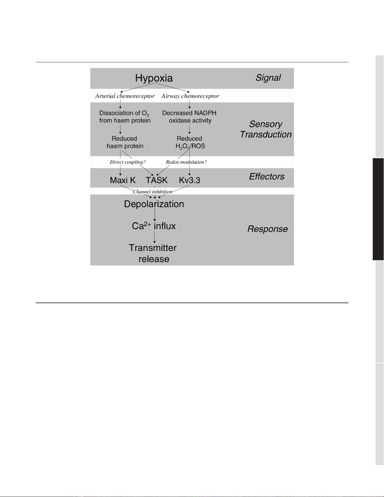
Figure 1
Schematic flow diagram illustrating the diverse but converging transduction pathways linking hypoxia to transmitter release from arterial (carotid
body) and airway (neuroepithelial body) chemoreceptors. Kv3.3 channel, voltage-activated shaw K+channel (KCNC3); Maxi K, high-conductance,
Ca2+-activated K+channel (KCMA1); ROS, reactive oxygen species; TASK, TWIK-related, acid-sensitive K2P channel; TWIK, tandem of P-domains,
weakly inward rectifying K2P channel.

NEB-derived cells [12,19,31]. Employing this model, it has
been possible to verify that O2-sensitive channels are
insensitive to Ca2+ [12], but the contribution of Ca2+-acti-
vated channels still remains to be investigated robustly in
native human cells and lung slices. Notwithstanding that
H146 cells and native cells show some differences, it is
clear that the Ca2+-insensitive components in the two
species are almost certainly identical because their phar-
macologies and biophysical natures are essentially indistin-
guishable. On the basis of these observations, debate still
rages about the molecular identification of the Ca2+-insen-
sitive K+channel: a voltage-activated shaw K+channel
(KCNC3), Kv3.3, is proposed in native NEBs [17] and a
TASK-like conductance is suggested in H146 cells [31].
Screening, by reverse-transcriptase-mediated polymerase
chain reaction, for all the known human homologues of the
K2P gene family has indicated that only TWIK1 and TWIK-
related, arachidonic acid-sensitive K2P channel (‘TRAAK’)
are not expressed in H146 cells [32]. Importantly,
however, in situ hybridisation and immunohistochemical
studies have now exclusively localised TASK to mouse
NEB cells in lung, and recent antisense knock-down
experiments in the H146 cell model have shown a high
correlation between quantitative TASK expression and
functional hypoxic sensitivity [33]. This antisense
approach could not distinguish between TASK1 and
TASK3 because they share such high identity in their
open reading frame sequences; of considerable import,
however, is the recent demonstration that recombinant
TASK1 and TASK3 are exquisitely sensitive to decreased
pO2when expressed in HEK 293 cells [34]. Further phar-
macological dissection (using Zn2+ as a discriminating
blocker) has now lent support to the notion that the O2-
sensitive channel is TASK3, although heterodimerism in
H146 cells cannot at present be excluded (PJ Kemp, GJ
Searle and C Peers, unpublished data).
Conclusion
O2sensing in NEBs and CBs therefore exhibits diverse
yet convergent mechanistic features; these are sum-
marised in Figure 1. Upstream, the main O2sensors in the
two tissues are clearly different, although a contribution by
mitochondrial ROS generation might be shared. Transduc-
tion of the hypoxic signal almost certainly converges, as a
unifying theme, on a K2P channel, but how different K+
channels interact to evoke transmitter release and a full
physiological response to hypoxia in CBs and NEBs is still
debated fiercely and integrative approaches might again
be crucial in resolving this important issue.
Acknowledgements
The authors’ own studies are supported by The Wellcome Trust and
the British Heart Foundation.
References
1. Gonzalez C, Almarez L, Obeso A, Rigual R: Carotid body
chemoreceptors: from natural stimuli to sensory discharges.
Physiol Rev 1994, 74:829–898.
2. Cutz E, Jackson A: Neuroepithelial bodies as airway oxygen
sensors. Respir Physiol 1999, 115:201–214.
3. Peers C, Buckler KJ: Transduction of chemostimuli by the type
I carotid body cell. J Membr Biol 1995, 144:1–9.
4. Zhang M, Zhong H, Vollmer C, Nurse CA: Co-release of ATP
and ACh mediates hypoxic signalling at rat carotid body
chemoreceptors. J Physiol 2000, 525:143–158.
5. Lauweryns JM, VanLommel A: Ultrastructure of nerve endings
and synaptic junctions in rabbit intrapulmonary neuroepithe-
lial bodies. J Anat 1987, 151:65–65.
6. Lauweryns JM, Cokeleare M: Hypoxia sensitive neuroepithelial
bodies intrapulmonary secretory neuroreceptors, modulated
by CNS. Z Zellforsch Mikrosk Anat 1973, 145:521–540.
7. Gillan JE, Curran C, O’Rielly E, Cahalane SF, Unwin AR: Abnor-
mal patterns of pulmonary neuroendocrine cells in victims of
Sudden Infant Death Syndrome. Paediatrics 1989, 84:828–834.
8. Peers C: Oxygen-sensitive ion channels. Trends Pharmacol Sci
1997, 18:405–408.
9. Lopez-Barneo J, Lopez-Lopez JR, Urena J, Gonzalez C: Chemo-
transduction in the carotid body: K+current modulated by pO2
in type I chemoreceptor cells. Science 1988, 241:580–582.
10. Youngson C, Nurse C, Yeger H, Cutz E: Oxygen sensing in
airway chemoreceptors. Nature 1993, 365:153–155.
11. Wyatt CN, Wright C, Bee D, Peers C: O2-sensitive K+currents
in carotid-body chemoreceptor cells from normoxic and
chronically hypoxic rats and their roles in hypoxic chemo-
transduction. Proc Natl Acad Sci USA 1995, 92:295–299.
12. O’Kelly I, Peers C, Kemp PJ: Oxygen-sensitive K+channels in
neuroepithelial body-derived small cell carcinoma cells of the
human lung. Am J Physiol 1998, 275:L709–L716.
13. Urena J, Fernandez-Chacon R, Benot AR, Alvarez de Toledo G,
Lopez-Barneo J: Hypoxia induces voltage-dependent Ca2+
entry and quantal dopamine secretion in carotid body glomus
cells. Proc Natl Acad Sci USA 1994, 91:10208–10211.
14. Robertson TP, Hague D, Aaronson PI, Ward JP: Voltage-indepen-
dent calcium entry in hypoxic pulmonary vasoconstriction of
intrapulmonary arteries of the rat. J Physiol 2000, 525:669–680.
15. Archer SL, Weir EK, Reeve HL, Michelakis E: Molecular identifi-
cation of O2sensors and O2-sensitive potassium channels in
the pulmonary circulation. Adv Exp Med Biol 2000, 475:219–
240.
16. Osipenko ON, Tate RJ, Gurney AM: Potential role for kv3.1b
channels as oxygen sensors. Circ Res 2000, 86:534–540.
17. Wang D, Youngson C, Wong V, Yeger H, Dinauer MC, Vega-
Saenz de Miera E, Rudy B, Cutz E: NADPH-oxidase and hydro-
gen peroxide sensitive K+channel may function as an oxygen
sensor complex in airway chemoreceptors and small cell lung
carcinoma cell lines. Proc Natl Acad Sci USA 1996, 93:13182–
13187.
18. Fu XW, Wang D, Nurse C, Dinauer MC, Cutz E: NADPH oxidase
is an O2sensor in airway chemoreceptors: evidence from K+
current modulation in wild type and oxidase-deficient mice.
Proc Natl Acad Sci USA 2000, 97:4374–4379.
19. O’Kelly I, Lewis A, Peers C, Kemp PJ: O2sensing by airway
chemoreceptor-derived cells: protein kinase C activation
reveals functional evidence for involvement of NADPH
oxidase. J Biol Chem 2000, 275:7684–7692.
20. Tauber AI: Protein kinase C and the activation of the human
neutrophol NADPH-oxidase. Blood 1987, 69:711–720.
21. Lopez-Lopez JR, Gonzalez C: Time course of K+current inhibi-
tion by low oxygen in chemoreceptor cells of adult rabbit
carotid body. Effects of carbon monoxide. FEBS Lett 1992,
299:251–254.
22. Archer SL, Reeve HL, Michelakis E, Puttagunta L, Waite R,
Nelson DP, Dinauer MC, Weir EK: O2sensing is preserved in
mice lacking the gp91 phox subunit of NADPH oxidase. Proc
Natl Acad Sci USA 1999, 96:7944–7949.
23. Chandel NS, Schumacker PT: Cellular oxygen sensing by mito-
chondria: old questions, new insight. J Appl Physiol 2000, 88:
1880–1889.
24. Duranteau J, Chandel NS, Kulisz A, Shao Z, Schumacker PT: Intra-
cellular signaling by reactive oxygen species during hypoxia in
cardiomyocytes. J Biol Chem 1998, 273:11619–11624.
Respiratory Research Vol 2 No 3 Peers and Kemp

25. Buckler KJ, Vaughan-Jones RD: Effects of mitochondrial uncou-
plers on intracellular calcium, pH and membrane potential in
rat carotid body type I cells. J Physiol 1998, 513:819–833.
26. Lopez-Lopez JR, Peers C: Electrical properties of chemorecep-
tor cells. In The Carotid Body Chemoreceptors. Edited by Gonza-
lez C. Austin: Landes Bioscience; 1997:65–78.
27. Peers C: Hypoxic suppression of K+currents in type I carotid
body cells: selective effect on the Ca2+-activated K+current.
Neurosci Lett 1990, 119:253–256.
28. Buckler KJ, Williams BA, Honore E: An oxygen-, acid- and
anaesthetic-sensitive TASK-like background potassium
channel in rat arterial chemoreceptor cells. J Physiol 2000,
525:135–142.
29. Pardal R, Ludewig U, Garcia-Hirschfeld J, Lopez-Barneo J: Secre-
tory responses of intact glomus cells in thin slices of rat
carotid body to hypoxia and tetraethylammonium. Proc Natl
Acad Sci USA 2000, 97:2361–2366.
30. Fu XW, Nurse C, Wang YT, Cutz E: Selective modulation of
membrane currents by hypoxia in intact airway chemorecep-
tors from neonatal rabbit. J Physiol 1999, 514:139–150.
31. O’Kelly I, Stephens RH, Peers C, Kemp PJ: Potential identifica-
tion of the O2-sensitive K+current in a human neuroepithelial
body-derived cell line. Am J Physiol 1999, 276:L96–L104.
32. Kemp PJ, Lewis A, O’Kelly I, Peers C: Regulation of K+currents
in a human neuroepithelial body derived cell line suggests
that hTASK is an airway O2-sensitive K+channel. FASEB J
2001, 15:A817.
33. Kemp PJ, Hartness M, Lewis A, O’Kelly I, Peers C: Antisense
depletion of a specific potassium channel in H146 cells indi-
cates that hTASK is an airway oxygen sensing channel.
FASEB J 2001, 15:A817.
34. Kemp PJ, Lewis A, Searle GJ, Peers C: Direct demonstration
that hTASK1 is an O2-sensitive K+channel. FASEB J 2001,
15:LB37.
Available online http://respiratory-research.com/content/2/3/145
commentary review reports primary research


