
RESEARC H Open Access
Effects of collagen membranes enriched with in
vitro-differentiated N1E-115 cells on rat sciatic
nerve regeneration after end-to-end repair
Sandra Amado
3†
, Jorge M Rodrigues
1,2†
, Ana L Luís
1,2†
, Paulo AS Armada-da-Silva
3
, Márcia Vieira
1
, Andrea Gartner
1
,
Maria J Simões
1
, António P Veloso
3
, Michele Fornaro
4
, Stefania Raimondo
4
, Artur SP Varejão
5
, Stefano Geuna
4*
,
Ana C Maurício
1,2*
Abstract
Peripheral nerves possess the capacity of self-regeneration after traumatic injury but the extent of regeneration is
often poor and may benefit from exogenous factors that enhance growth. The use of cellular systems is a rational
approach for delivering neurotrophic factors at the nerve lesion site, and in the present study we investigated the
effects of enwrapping the site of end-to-end rat sciatic nerve repair with an equine type III collagen membrane
enriched or not with N1E-115 pre-differentiated neural cells. After neurotmesis, the sciatic nerve was repaired by
end-to-end suture (End-to-End group), end-to-end suture enwrapped with an equine collagen type III membrane
(End-to-EndMemb group); and end-to-end suture enwrapped with an equine collagen type III membrane previously
covered with neural cells pre-differentiated in vitro from N1E-115 cells (End-to-EndMembCell group). Along the post-
operative, motor and sensory functional recovery was evaluated using extensor postural thrust (EPT), withdrawal
reflex latency (WRL) and ankle kinematics. After 20 weeks animals were sacrificed and the repaired sciatic nerves
were processed for histological and stereological analysis. Results showed that enwrapment of the rapair site with
a collagen membrane, with or without neural cell enrichment, did not lead to any significant improvement in most
of functional and stereological predictors of nerve regeneration that we have assessed, with the exception of EPT
which recovered significantly better after neural cell enriched membrane employment. It can thus be concluded
that this particular type of nerve tissue engineering approach has very limited effects on nerve regeneration after
sciatic end-to-end nerve reconstruction in the rat.
Background
Nerve regeneration is a complex biological phenom-
enon. In the peripheral nervous system, nerves can
spontaneously regenerate without any treatment if nerve
continuity is maintained (axonotmesis) whereas more
severe type of injuries must be surgically treated by
direct end-to-end surgical reconnection of the damaged
nerve ends [1-3]. Unfortunately, the functional outcomes
of nerve repair are in many cases unsatisfactory [4] thus
calling for research in order to reveal more effective
strategies for improving nerve regeneration. However,
recent advances in neuroscience, cell culture, genetic
techniques, and biomaterials provide optimism for new
treatments for nerve injuries [5-17].
The use of materials of natural origin has several
advantages in tissue engineering. Natural materials are
more likely to be biocompatible than artificial materials.
Also, they are less toxic and provide a good support to
cell adhesion and migration due to the presence of a
variety of surface molecules. Drawbacks of natural mate-
rials include potential difficulties in their isolation and
controlled scale-up [11]. In addition to the use of intact
natural tissues, a great deal of research has focused on
the use of purified natural extracellular matrix (ECM)
molecules, which can be modified to serve as appropri-
ate scaffolding [11]. ECM molecules, such as laminin,
fibronectin and collagen have also been shown to play a
* Correspondence: stefano.geuna@unito.it; ana.colette@hotmail.com
†Contributed equally
1
Centro de Estudos de Ciência Animal (CECA), Instituto de Ciências e
Tecnologias Agrárias e Agro-Alimentares (ICETA), Universidade do Porto (UP),
Portugal
4
Department of Clinical and Biological Sciences, University of Turin, Italy
Amado et al.Journal of NeuroEngineering and Rehabilitation 2010, 7:7
http://www.jneuroengrehab.com/content/7/1/7 JNERJOURNAL OF NEUROENGINEERING
AND REHABILITATION
© 2010 Amado et al; licensee BioMed Central Ltd. This is an Open Access article distributed under the terms of the Creative Commons
Attribution License (http://creativecommons.org/licenses/by/2.0), which permits unrestricted use, distribution, and reproduction in
any medium, provided the original work is properly cited.

significant role in axonal development and regeneration
[12,18-27]. For example, silicone tubes filled with lami-
nin, fibronectin, and collagen led to a better regenera-
tion over a 10 mm rat sciatic nerve gap compared to
empty silicone controls [9]. Collagen filaments have also
been used to guide regenerating axons across 20-30 mm
defects in rats [23-27]. Further studies have shown that
oriented fibers of collagen within gels, aligned using
magnetic fields, provide an improved template for neur-
ite extension compared to randomly oriented collagen
fibers [28,29]. Finally, rates of regeneration comparable
to those using a nerve autograft have been achieved
using collagen tubes containing a porous collagen-glyco-
saminoglycan matrix [30-32]. Nerve regeneration
requires a complex interplay between cells, ECM, and
growth factors. The local presence of growth factors
plays an important role in controlling survival, migra-
tion, proliferation, and differentiation of the various cell
types involved in nerve regeneration [12-14,33]. There-
fore, therapies with relevant growth factors received
increasing attention in recent years although growth fac-
tor therapy is a difficult task because of the high biologi-
cal activity (in pico- to nanomolar range), pleiotrophic
effects (acting on a variety of targets), and short biologi-
cal half-life (few minutes to hours) [34]. Thus, growth
factors should be administered locally to achieve an ade-
quate therapeutic effect with little adverse reactions and
the short biological half-life of growth factors demands
for a delivery system that slowly releases locally the
molecules over a prolonged period of time. Employment
of biodegradable membranes enriched with a cellular
system producing neurotrophic factors has been sug-
gested to be a rational approach for improving nerve
regeneration after neurotmesis [11].
The aim of this study was thus to verify if rat sciatic
nerve regeneration after end-to-end reconstruction can be
improved by seeding in vitro differentiated N1E-115
neural cells on a type III equine collagen membrane and
enwrap the membrane around the lesion site. The N1E-
115 cell line has been established from a mouse neuroblas-
toma [35] and have already been used with conflicting
results as a cellular system to locally produce and deliver
neurotrophic factors [12-14,36,37]. In vitro, the N1E-115
cells undergo neuronal differentiation in response to
dimethylsulfoxide (DMSO), adenosine 3’,5’-cyclic mono-
phosphate (cAMP), or serum withdrawal
[38-43,36,37,12-14]. Upon induction of differentiation,
proliferation of N1E-115 cells ceases, extensive neurite
outgrowth is observed and the membranes become highly
excitable [38-43,36,37,12-14]. The interval period of 48
hours of differentiation was previously determined by
measurement of the intracellular calcium concentration
([Ca
2+
] i). At this time point, the N1E-115 cells present
already the morphological characteristics of neuronal cells
but cell death due to increased [Ca
2+
] i is not yet occurring
as described elsewhere [38-43,36,37,12-14].
Methods
Cell culture
The N1E-115 cells, clones of cells derived from the
mouse neuroblastoma C-130035 retain numerous bio-
chemical, physiological, and morphological properties of
neuronal cells in culture [38-43,36,37,12-14]. N1E-115
neuronal cells were cultured in Petri dishes (around 2 ×
10
6
cells) over collagen type III membranes (Genta-
fleece®, Resorba Wundversorgung GmbH + Co. KG,
Baxter AG) at 37°C, 5% CO
2
in a humidified atmosphere
with 90% Dulbecco’s Modified Eagle’s Medium (DMEM;
Gibco) supplemented with 10% fetal bovine serum (FBS,
Gibco), 100 U/ml penicillin, and 100 μg/ml streptomy-
cin (Gibco). The culture medium was changed every 48
hours and the Petri dishes were observed daily. The
cells were passed or were supplied with differentiating
medium containing 1.5% of DMSO once they reached
approximately 80% confluence, mostly 48 hours after
plating (and before the rats’surgery). The differentiating
medium was composed of 96% DMEM supplemented
with 2.5% of FBS, 100 U/ml penicillin, 100 μg/ml strep-
tomycin and 1.5% of DMSO [12-14,36,37].
Surgical procedure
Adult male Sasco Sprague Dawley rats (Charles River
Laboratories, Barcelona, Spain) weighing 300-350 g,
were randomly divided in 3 groups of 6 or 7 animals
each. All animals were housed in a temperature and
humidity controlled room with 12-12 hours light/dark
cycles, two animals per cage (Makrolon type 4, Tecni-
plast, VA, Italy), and were allowed normal cage activities
under standard laboratory conditions. The animals were
fed with standard chow and water ad libitum. Adequate
measures were taken to minimize pain and discomfort
taking in account human endpoints for animal suffering
and distress. Animals were housed for two weeks before
entering the experiment. For surgery, rats were placed
prone under sterile conditions and the skin from the
clipped lateral right thigh scrubbed in a routine fashion
with antiseptic solution. The surgeries were performed
under an M-650 operating microscope (Leica Microsys-
tems, Wetzlar, Germany). Under deep anaesthesia (keta-
mine 90 mg/Kg; xylazine 12.5 mg/Kg, atropine 0.25 mg/
Kg i.m.), the right sciatic nerve was exposed through a
skin incision extending from the greater trochanter to
themid-thighdistallyfollowedbyamusclesplitting
incision. After nerve mobilisation, a transection injury
was performed (neurotmesis) immediately above the
terminal nerve ramification using straight microsurgical
scissors. Rats were then randomly assigned to three
experimental groups. In one group (End-to-End),
Amado et al.Journal of NeuroEngineering and Rehabilitation 2010, 7:7
http://www.jneuroengrehab.com/content/7/1/7
Page 2 of 13

immediate cooptation with 7/0 monofilament nylon epi-
neurial sutures of the 2 transected nerve endings was
performed, in a second group (End-to-EndMemb)nerve
transection was reconstructed by end-to-end suture, like
in the first group, and then enveloped by a membrane
of equine collagen type III. In a third group (End-to-
EndMembCell) animals received the same treatment as
the previous group but with equine collagen type III
membranes covered with neural cells differentiated in
vitro. Sciatic nerves from the contralateral site were left
intact in all groups and served as controls. To prevent
autotomy, a deterrent substance was applied to rats’
right foot [44,45]. The animals were intensively exam-
ined for signs of autotomy and contracture during the
postoperative and none presented severe wounds, infec-
tions or contractures. All procedures were performed
with the approval of the Veterinary Authorities of Por-
tugal in accordance with the European Communities
Council Directive of November 1986 (86/609/EEC).
Evaluation of motor performance (EPT) and nociceptive
function (WRL)
Motor performance and nociceptive function were eval-
uated by measuring extensor postural thrust (EPT) and
withdrawal reflex latency (WRL), respectively. The ani-
mals were tested pre-operatively (week-0), at weeks 1, 2,
and every two weeks thereafter until week-20. The ani-
mals were gently handled, and tested in a quiet environ-
ment to minimize stress levels. The EPT was originally
proposed by Thalhammer and collaborators, in 1995
[46] as a part of the neurological recovery evaluation in
the rat after sciatic nerve injury. For this test, the entire
body of the rat, excepting the hind-limbs, was wrapped
in a surgical towel. Supporting the animal by the thorax
and lowering the affected hind-limb towards the plat-
form of a digital balance, elicits the EPT. As the animal
is lowered to the platform, it extends the hind-limb,
anticipating the contact made by the distal metatarsus
and digits. The force in grams (g) applied to the digital
platform balance (model TM 560; Gibertini, Milan,
Italy) was recorded. The same procedure was applied to
the contralateral, unaffected limb. Each EPT test was
repeated 3 times and the average result was considered.
The normal (unaffected limb) EPT (NEPT) and experi-
mental EPT (EEPT) values were incorporated into an
equation (Equation 1) to derive the functional deficit
(varying between 0 and 1), as described by Koka and
Hadlock, in 2001 [47].
Motor Deficit NEPT EEPT NEPT()/ (1)
To assess the nociceptive withdrawal reflex (WRL),
the hotplate test was modified as described by Masters
and collaborators [48]. The rat was wrapped in a
surgical towel above its waist and then positioned to
stand with the affected hind paw on a hot plate at 56°C
(model 35-D, IITC Life Science Instruments, Woodland
Hill, CA). WRL is defined as the time elapsed from the
onset of hotplate contact to withdrawal of the hind paw
and measured with a stopwatch. Normal rats withdraw
their paws from the hotplate within 4.3 s or less [49].
The affected limbs were tested 3 times, with an interval
of 2 min between consecutive tests to prevent sensitiza-
tion, and the three latencies were averaged to obtain a
final result [50,51]. If there was no paw withdrawal after
12 s, the heat stimulus was removed to prevent tissue
damage, and the animal was assigned the maximal WRL
of 12 s [52].
Kinematic Analysis
Ankle kinematics during the stance phase of the rat
walk was recorded prior nerve injury (week-0), at week-
2 and every 4 weeks during the 20-week follow-up time.
Animals walked on a Perspex track with length, width
and height of respectively 120, 12, and 15 cm. In order
to ensure locomotion in a straight direction, the width
of the apparatus was adjusted to the size of the rats
during the experiments, and a darkened cage was placed
at the end of the corridor to attract the animals. The
rats gait was video recorded at a rate of 100 images per
second (JVC GR-DVL9800, New Jersey, USA). The
camera was positioned perpendicular to the mid-point
of the corridor length at a 1-m distance thus obtaining
a visualization field of 14-cm wide. Only walking trials
with stance phases lasting between 150 and 400 ms
were considered for analysis, since this corresponds to
the normal walking velocity of the rat (20-60 cm/s)
[52-54]. The video images were stored in a computer
hard disk for latter analysis using an appropriate soft-
ware APAS® (Ariel Performance Analysis System, Ariel
Dynamics, San Diego, USA). 2-D biomechanical analysis
(sagittal plan) was carried out applying a two-segment
model of the ankle joint, adopted from the model firstly
developed by Varejão and collaborators [52-55]. Skin
landmarks were tattooed at points in the proximal edge
of the tibia, in the lateral malleolus and, in the fifth
metatarsal head. The rats’ankle angle was determined
using the scalar product between a vector representing
the foot and a vector representing the lower leg. With
this model, positive and negative values of position of
the ankle joint indicate dorsiflexion and plantarflexion,
respectively. For each stance phase the following time
points were identified: initial contact (IC), opposite toe-
off (OT), heel-rise (HR) and toe-off (TO) [52-55], and
were time normalized for 100% of the stance phase.
The normalized temporal parameters were averaged
over all recorded trials. Angular velocity of the ankle
joint was also determined where negative values
Amado et al.Journal of NeuroEngineering and Rehabilitation 2010, 7:7
http://www.jneuroengrehab.com/content/7/1/7
Page 3 of 13

correspond to dorsiflexion. Four steps were analysed for
each animal [55].
Histological and Stereological analysis
A 10-mm-long segment of the sciatic nerve distal to the
site of lesion was removed, fixed, and prepared for
quantitative morphometry of myelinated nerve fibers. A
10-mm segment of uninjured sciatic nerve was also
withdrawn from control animals (N = 6). The harvested
nerve segments were immersed immediately in a fixa-
tion solution containing 2.5% purified glutaraldehyde
and 0.5% saccarose in 0.1 M Sorensen phosphate buffer
for 6-8 hours. Specimens were processed for resin
embedding as described in details elsewhere [56,57]. Ser-
ies of 2-μm thick semi-thin transverse sections were cut
using a Leica Ultracut UCT ultramicrotome (Leica
Microsystems, Wetzlar, Germany) and stained by Tolui-
dine blue for stereological analysis of regenerated nerve
fibers. The slides were observed with a DM4000B
microscope equipped with a DFC320 digital camera and
an IM50 image manager system (Leica Microsystems,
Wetzlar, Germany). One semi-thin section from each
nerve was randomly selected and used for the morpho-
quantitative analysis. The total cross-sectional area of
the nerve was measured and sampling fields were then
randomly selected using a protocol previously described
[57-59]. Bias arising from the “edge effect”was coped
with the employment of a two-dimensional disector pro-
cedure which is based on sampling the “tops”of fibers
[60,61]. Mean fiber density in each disector was then
calculated by dividing the number of nerve fibers
counted by the disector’sarea(N/mm
2
). Finally, total
fiber number (N) in the nerve was estimated by multi-
plying the mean fiber density by the total cross-sectional
area of the whole nerve. Two-dimensional disector
probes were also used for the unbiased selection of a
representative sample of myelinated nerve fibers for esti-
mating circle-fitting diameter and myelin thickness. Pre-
cision and accuracy of the estimates were evaluated by
calculating the coefficient of variation (CV = SD/mean)
and coefficient of error (CE = SEM/mean) [57-59].
Statistical analysis
Two-way mixed factorial ANOVA was used to test for
the effect of time in the End-to-End group (within sub-
jects effect; 12 time points) and experimental groups
(between subjects effect, 3 groups). The sphericity
assumption was evaluated by the Mauchly’stestand
when this test could not be computed or when spheri-
city assumption was violated, adjustment of the degrees
of freedom was done with the Greenhouse-Geiser’s epsi-
lon. When time main effect was significant (within sub-
jects factor), simple planned contrasts (General Linear
Model, simple contrasts) were used to compare pooled
data across the three experimental groups along the
recovery with data at week-0. When a significant main
effect of treatment existed (between subjects factor),
pairwise comparisions were carried out using the
Tukey’s HSD test. At week-0, kinematic data was
recorded only from the End-to-End group so the main
effect of time was evaluated only in this group. Evalua-
tion of the main effect of treatment on ankle motion
variables used only data after nerve injury. In this case,
and when appropriate, pairwise comparisons were made
using the Tukey’s HSD test. Statistical comparisons of
stereological morpho-quantitative data on nerve fibers
were accomplished with one-way ANOVA test. Statisti-
cal significance was established as p < 0.05. All statistical
procedures were performed by using the statistical pack-
age SPSS (version 14.0, SPSS, Inc) except stereological
data that were analysed using the software “Statistica
per discipline bio-mediche“(McGraw-Hill, Milan, Italy).
All data in this study is presented as mean ± standard
error of the mean (SEM).
Results
Motor deficit and Nociception function
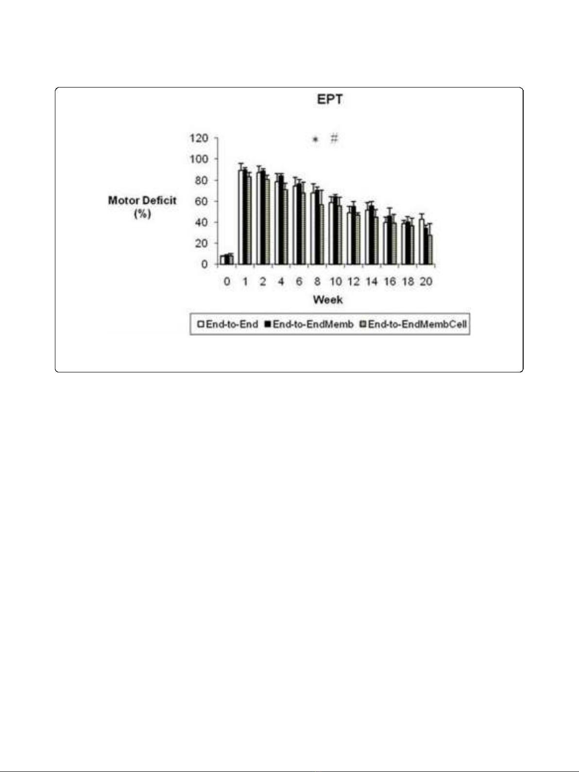
Motor deficit (EPT)
Before sciatic injury, EPT was similar in both hindlimbs
in all experimental groups (figure 1). In the first week
after sciatic nerve transection, near total EPT loss was
observed in the operated hindlimb, leading to a motor
deficit ranging between 83 to 90%. The EPT response
steadily improved during recovery but at week-20 the
EPT values of the injured side were still significantly
lower compared to values at week-0 (p < 0.05). A signif-
icant main effect for treatment was found [F
(2,17)
=
14.202; p = 0.000], with pairwise comparisons showing
significantly better recovery of the EPT response in the
End-to-EndMembCell groupwhencomparedtothe
other two experimental groups (p < 0.05). At week-20,
motor deficit decreased to 27% in the End-to-EndMemb-
Cell and to 34% and 42% in the End-to-End and End-to-
EndMemb groups, respectively (figure 1).
Nociception function (WRL)
In the week after sciatic transaction, all the animals pre-
sented a severe loss of sensory and nociception function
acutely after sciatic nerve transection and the WRL test
has to be interrupted at the 12 s-cutoff time (figure 2).
During the following weeks there was recovery in paw
nociception which was more clearly seen between weeks
6 and 8 post-surgery. At week-6, half of the animals still
had no withdrawal response to the noxious thermal sti-
mulus in the operated side, which is in contrast with
week-8, when all animals presented a consistent,
although delayed, response. Despite such improvement
in WRL response, contrast analysis showed persistence
of sensory deficit in all groups by the end of the 20-
Amado et al.Journal of NeuroEngineering and Rehabilitation 2010, 7:7
http://www.jneuroengrehab.com/content/7/1/7
Page 4 of 13
weeks recovery time (p < 0.05). No differences between
the groups was observed in the level of WRL impair-
ment after the sciatic nerve transection [F
(2,17)
=1.563;
p = 0.238].
Kinematics Analysis
Figures 3 and 4 display the mean plots, respectively for
ankle joint angle and ankle joint velocity during the
stance phase of the rat walk. Comparisons to the normal
ankle motion can only be draw for the End-to-End
group for reasons explained in the Methods section. In
the weeks following sciatic nerve transection, ankle joint
motion became severely abnormal, particularly through-
out the second half of stance corresponding to the
push-off sub-phase. In clear contrast to the normal pat-
tern of ankle movement, at week-2 post-injury animals
were unable to extend this joint and dorsiflexion contin-
ued increasing during the entire stance, which is
explained by the paralysis of plantarflexor muscles. The
pattern of the ankle joint motion seemed to have
improved only slightly during recovery. Contrast analysis
was performed for each of the kinematic parameters
(tables 1 and 2) with somewhat different results. For OT
velocity and HR angle no differences existed before and
after sciatic nerve transection, whereas for OT angle dif-
ferences from pre-injury values were significant only at
weeks 2 and 16 of recovery (p < 0.05). The angle at IC
showed a unique pattern of changes, being unaffected at
week-2 post-injury and altered from normal in the fol-
lowing weeks of recovery. Probably the most consistent
results are those of HR velocity, TO angle and TO velo-
city. These parameters were affected immediately after
the nerve injury and remained abnormal along the
entire 20-weeks recovery period (p < 0.05). The effect of
the different tissue engineering strategies was assessed
comparing the kinematic data of the experimental
groups only during the recovery period (see Methods).
Statistical analysis demonstrated that the collagen mem-
brane and the cells had no or little effect on ankle
motion pattern recovery. Generally, no differences in
the kinematic parameters were found between the
groups. Exceptions were IC velocity in the End-to-End-
MembCell group, which was different from the other
two groups (p < 0.05), and OT angle in the End-to-End-
Memb group that was also different from the other two
groups (p < 0.05).
Histological and Stereological Analysis
Figure 5 shows representative light micrographs of the
regenerated sciatic nerves of the three groups (figure
5A-C) and control sciatic normal nerves (figure 5D). As
expected, regeneration of axons was organized in many
smaller fascicles in comparison to controls. The results
of the stereological analysis of myelinated nerve fibers
are reported in Table 3. Statistical analysis by ANOVA
test revealed no significant (p > 0.05) difference
Figure 1 Weekly values of the percentage of motor deficit obtained by the Extensor Postural Thrust (EPT) test. * Significantly different
from week-0 all groups pooled together (p < 0.05). # Group End-to-EndMembCell significantly different from the other groups (p < 0.05). Results
are presented as mean and standard error of the mean (SEM).
Amado et al.Journal of NeuroEngineering and Rehabilitation 2010, 7:7
http://www.jneuroengrehab.com/content/7/1/7
Page 5 of 13


