
Structure and activity of the atypical serine kinase Rio1
Nicole LaRonde-LeBlanc
1
, Tad Guszczynski
2
, Terry Copeland
2
and Alexander Wlodawer
1
1 Protein Structure Section, Macromolecular Crystallography Laboratory, National Cancer Institute, NCI-Frederick, MD, USA
2 Laboratory of Protein Dynamics and Signaling, National Cancer Institute, NCI-Frederick, MD, USA
Ribosome biogenesis is fundamental to cell growth
and proliferation and thereby to tumorigenesis. It has
been shown that ribosome biogenesis and cell cycle
progression are tightly linked through a number of
mechanisms [1,2]. Not surprisingly, several oncogenes
have been shown to deregulate ribosome biogenesis,
in order to meet the demand for cell growth and
increased protein production [3]. For example,
increased levels of ribosome biogenesis have been
reported for human breast cancer cells with decreased
pRb and p53 activity [4]. Ongoing studies in yeast have
identified many of the nonribosomal factors necessary
for the proper processing of ribosomal RNA (rRNA)
[5]. More recent efforts using proteomics methods have
begun to pinpoint the protein factors required for this
critical process. Although many of the factors have
been identified, the specific roles they play in rRNA
processing or ribosomal subunit assembly have not
been clarified. Understanding these basic pathways on
a molecular level is important for providing insight
into how the connection between ribosome biogenesis
and cell cycle control might be used to our advantage,
such as design of new classes of drugs.
Protein kinases are known players in the regulation
of cell cycle control, in addition to their role in a wide
variety of cellular processes including transcription,
DNA replication, and metabolic functions. This large
protein superfamily contains over 500 members in the
human genome [6] and represents one of the largest
protein superfamilies in eukaryotes [7]. One major class
of eukaryotic protein kinases (ePKs) catalyzes phos-
phorylation of serine or threonine, while another one
phosphorylates tyrosine residues [8–10]. All these
enzymes contain catalytic domains composed of con-
served secondary structure elements and catalytically
important sequences referred to as ‘subdomains’ that
create two globular ‘lobes’ linked by a flexible ‘hinge’
[7,8,10]. Twelve subdomains are recognized in ePKs: I
to IV comprising the N-terminal lobe, V producing
the hinge, and VIa, VIb, and VII to XI forming the
C-terminal lobe. The three-dimensional structure of
the ePK kinase domain is well established and the
Keywords
autophosphorylation; nucleotide complex;
protein kinase; ribosome biogenesis; Rio1
Correspondence
A. Wlodawer, National Cancer Institute,
MCL, Bldg. 536, Rm. 5, Frederick,
MD 21702–1201, USA
Fax: +1 301 8466322
Tel: +1 301 8465036
E-mail: wlodawer@ncifcrf.gov
(Received 21 April 2005, revised 24 May
2005, accepted 27 May 2005)
doi:10.1111/j.1742-4658.2005.04796.x
Rio1 is the founding member of the RIO family of atypical serine kinases
that are universally present in all organisms from archaea to mammals.
Activity of Rio1 was shown to be absolutely essential in Saccharomyces
cerevisiae for the processing of 18S ribosomal RNA, as well as for proper
cell cycle progression and chromosome maintenance. We determined high-
resolution crystal structures of Archaeoglobus fulgidus Rio1 in the presence
and absence of bound nucleotides. Crystallization of Rio1 in the presence
of ATP or ADP and manganese ions demonstrated major conformational
changes in the active site, compared with the uncomplexed protein. Com-
parisons of the structure of Rio1 with the previously determined structure
of the Rio2 kinase defined the minimal RIO domain and the distinct fea-
tures of the RIO subfamilies. We report here that Ser108 represents the
sole autophosphorylation site of A. fulgidus Rio1 and have therefore estab-
lished its putative peptide substrate. In addition, we show that a mutant
enzyme that cannot be autophosphorylated can still phosphorylate an
inactive form of Rio1, as well as a number of typical kinase substrates.
Abbreviations
aPK, atypical protein kinase; ePK, eukaryotic protein kinase; MAD, multiwavelength anomalous diffraction; N-lobe, N-terminal kinase lobe;
RMSD, root mean square deviation.
3698 FEBS Journal 272 (2005) 3698–3713 ª2005 FEBS

conserved subdomain residues have been shown to be
involved in phosphotransfer, as well as in recognition
and binding of ATP or substrate peptides [8,9,11,12].
Several protein subfamilies have been identified that
are not significantly related to ePKs in sequence but
contain a ‘kinase signature’ [6]. Based on the presence
of these limited sequence motifs and ⁄or demonstrated
kinase activity, these proteins have been collectively
named atypical protein kinases (aPKs) [6]. Unlike
ePKs, aPK families are small, typically containing only
a few (1–6) members per organism [6]. The RIO pro-
tein family has been classified as aPK based on dem-
onstrated kinase activity of the yeast Rio1p and Rio2p
and on the identification of a conserved kinase signa-
ture, although these enzymes exhibit no significant
homology to ePKs [6]. The RIO family is the only
aPK family conserved in archaea, and it has been sug-
gested that this family represents an evolutionary link
between prokaryotic lipid kinases and ePKs [13].
The founding member of the RIO kinase family is
Rio1p, an essential gene product in Saccharomyces
cerevisiae that functions as a nonribosomal factor
necessary for late 18S rRNA processing [14,15]. Deple-
tion of Rio1p results in accumulation of 20S pre-
rRNA, cell cycle arrest, and aberrant chromosome
maintenance [14,16]. Sequence alignments have demon-
strated that members of two RIO subfamilies, Rio1 and
Rio2, are represented in organisms from archaea to
mammals [13,17,18], whereas a third subfamily, Rio3, is
found strictly in higher eukaryotes. The RIO kinase
domain is generally conserved among the three sub-
families, but with distinct differences. In addition, the
Rio2 and Rio3 subfamilies are characterized by con-
served N-terminal domains outside of the RIO domain
that are unique to each of the two subfamilies and are
not present in Rio1. Yeast contains one Rio1 and one
Rio2 protein, but no members of the Rio3 subfamily.
Depletion of yeast Rio2 also affects growth rate and
results in an accumulation of 20S pre-rRNA [18,19].
Therefore, both RIO proteins are critically important
for ribosome biogenesis. Although there is significant
sequence similarity between Rio1 and Rio2 proteins
(43% similarity between the yeast enzymes), Rio1 pro-
teins are functionally distinct from Rio2 proteins and
do not complement their activity, as deletion of Rio2 in
yeast is also lethal, despite functional Rio1 [19].
Yeast RIO proteins are capable of serine phosphory-
lation in vitro, and residues equivalent to the conserved
catalytic residues of ePKs are required for their in vivo
function [15–18]. Our recently reported crystal struc-
ture of Rio2 from Archaeoglobus fulgidus has demon-
strated that the RIO domain resembles a trimmed
version of an ePK kinase domain [20]. It consists of
two lobes which sandwich ATP and contains the cata-
lytic loop, the metal-binding loop, and the nucleotide-
binding loop (P-loop, glycine-rich loop), but lacks the
classical substrate-binding and activation loops (subdo-
mains VIII, X and XI) present in ePKs. The structure
also revealed that the conserved Rio2-specific domain
contains a winged helix motif, usually found in DNA-
binding proteins, tightly connected through extensive
interdomain contacts to the RIO kinase domain. An
entire 18 amino acid loop in the N-terminal kinase
lobe (N-lobe) of Rio2, containing several subfamily
specific conserved residues, was not observed in the
crystal structure due to its flexibility. Differences
between the sequences of the Rio1 and Rio2 kinases in
several key regions of the RIO domain have led us
to the conclusion that structural differences may exist
between them which could explain their distinct func-
tionality and separate conservation.
To investigate the functional distinction of Rio1 and
its relationship to Rio2, we have solved several X-ray
crystal structures of Rio1 from A. fulgidus (AfRio1),
with and without bound nucleotides. Crystallization of
Rio1 protein in the presence of ATP and manganese
demonstrated partial hydrolysis of ATP, consistent
with data that indicate much higher autophosphoryla-
tion activity of Rio1 than Rio2. We have also shown
that Rio1 is active in phosphorylating several kinase
substrates and characterized its autophosphorylation
site. Analysis of the data reported here allowed us to
identify the key differences between Rio1 and Rio2
proteins and highlighted the unique features of RIO
proteins in general.
Results
Structure determination and the overall fold
of AfRio1
Full-length Rio1 from the thermophilic organism
A. fulgidus was expressed in Escherichia coli in the pres-
ence of selenomethionine (Se-Met). The enzyme was
purified using heat denaturation (in order to denature
E. coli proteins while leaving the thermostable Rio1
protein intact), affinity chromatography, and size-
exclusion chromatography. Mass spectrometry con-
firmed that the purified protein contained all the
expected residues (1–258). We obtained two substan-
tially different crystal forms of AfRio1. Crystals grown
without explicit addition of ATP or its analogs belong
to the space group P2
1
, contain one molecule per asym-
metric unit, and diffract to the resolution better than
2.0 A
˚. The structure was solved using the multiwave-
length anomalous diffraction (MAD) phasing technique
N. LaRonde-LeBlanc et al. Structure and activity of the Rio1 kinase
FEBS Journal 272 (2005) 3698–3713 ª2005 FEBS 3699

with Se-Met substituted protein at 1.9 A
˚. The model
contains residues 6–257 of the 258 residues of AfRio1,
with both termini being flexible. Crystals grown in the
presence of adenosine-5¢-triphosphate (ATP) or adeno-
sine-5¢-diphosphate (ADP) and Mn
2+
ions also belong
to the space group P2
1
, but are quite distinct, contain-
ing four molecules in the asymmetric unit. Manganese
ions were used in the place of magnesium ions for
better detection in electron density maps, and have
been shown to support catalysis in vitro with this
enzyme (data not shown). The nucleotide-complex
structures were solved by molecular replacement, using
the coordinates described above as the search model.
Data collection and crystallographic refinement statis-
tics for both crystal forms are shown in Table 1.
The determination of the structure of Rio1 and the
availability of the previously determined structure of
Rio2, has enabled us to define the minimal consensus
RIO domain (Fig. 1A). Similar to ePKs, it consists of
an N-lobe comprised of a twisted b-sheet (b1–b6) and
a long a-helix (aC) that closes the back of the ATP-
binding pocket, a hinge region, and a C-lobe which
forms the platform for the metal-binding loop and the
catalytic loop. However, the RIO kinase domain con-
tains only three of the canonical ePK a-helices (aE,
aF, and aI) in the C-lobe. In both Rio1 and Rio2,
an additional a-helix (aR), located N-terminal to the
canonical N-lobe b-sheet, extends the RIO domain
(Figs 1A and 2A). All RIO domains also contain an
insertion of 18–27 amino acids between aC and b3. In
the Rio1 structure solved from data obtained using
crystals grown in the absence of ATP (APO-Rio1), we
were able to trace that part of the chain in its entirety
(Figs 1A and 2A). In the structures of AfRio2, how-
ever, no electron density was observed for most of this
region, and thus we have called it the ‘flexible loop’
(Fig. 1B). The overall fold of the kinase domain of
Rio1 is very homologous to that of the Rio2, but sig-
nificant local differences between the two proteins
result in root mean square deviation (RMSD) of
1.39 A
˚(for 217 Capairs of complexes with ATP and
Mn
2+
ions). Comparison of the Rio1 structure with
that of c-AMP-dependent protein kinase (PKA)
showed that like Rio2, Rio1 lacks the activation or
‘APE’ loop (subdomain VIII) and subdomains X and
XI seen in canonical ePKs (Fig. 1C). In addition to
the N-terminal a-helix specific to RIO domain, Rio1
contains another two a-helices N-terminal to the RIO
domain, as opposed to the complete winged helix
domain present in Rio2 (Fig. 1A,B).
Although no nucleotide was added to the protein
used for the determination of the APO-Rio1 structure,
electron density in which we could model an adenosine
molecule was observed in the active site. However, no
density which would correspond to any part of a tri-
phosphate group was seen. The bound molecule
(Fig. 1A) must have remained complexed to the
enzyme through all steps of purification of the Rio1
protein, which is quite remarkable as two affinity col-
umn purification steps and one size-exclusion column
purification step were performed. As such, this mole-
cule must bind to Rio1 with extremely high affinity,
Table 1. Data collection and refinement statistics for the APO, ATP- and ADP-bound Rio1.
Crystal data
Space group P2
1
Se-Met MAD
ATP-Mn ADP-Mn
Peak Edge Remote
a(A
˚) 42.99 53.31 53.41
b(A
˚) 52.70 80.37 80.08
c(A
˚) 63.78 121.32 121.06
b() 108.89 90.02 90.17
k(A
˚) 0.97947 0.97934 0.99997 0.96860 0.96860
Resolution (A
˚) 40–1.90 40–2.10 40–1.99 30–2.00 30–1.89
R
sym
(last shell) 0.075 (0.259) 0.085 (0.283) 0.066 (0.233) 0.106 (0.286) 0.147 (0.350)
Reflections 40612 (3463) 30503 (2899) 35301 (3339) 65296 (5149) 82669 (6470)
Redundancy 3.6 (2.8) 3.6 (2.7) 3.7 (3.1) 3.8 (3.7) 7.5 (6.8)
Completeness (%) 97.6 (83.5) 98.8 (93.0) 98.5 (83.5) 90.1 (71.4) 94.2 (74.1)
R⁄R
free
(%)
(Last shell)
15.9 ⁄24.7 17.7 ⁄24.9 18.9 ⁄26.3
Mean B factor (A
˚
2
) 31.20 23.42 30.51
Residues 253 980 980
Waters 275 921 831
RMS Deviations
Lengths (A
˚) 0.032 0.018 0.016
Angles () 2.29 1.68 1.60
Structure and activity of the Rio1 kinase N. LaRonde-LeBlanc et al.
3700 FEBS Journal 272 (2005) 3698–3713 ª2005 FEBS
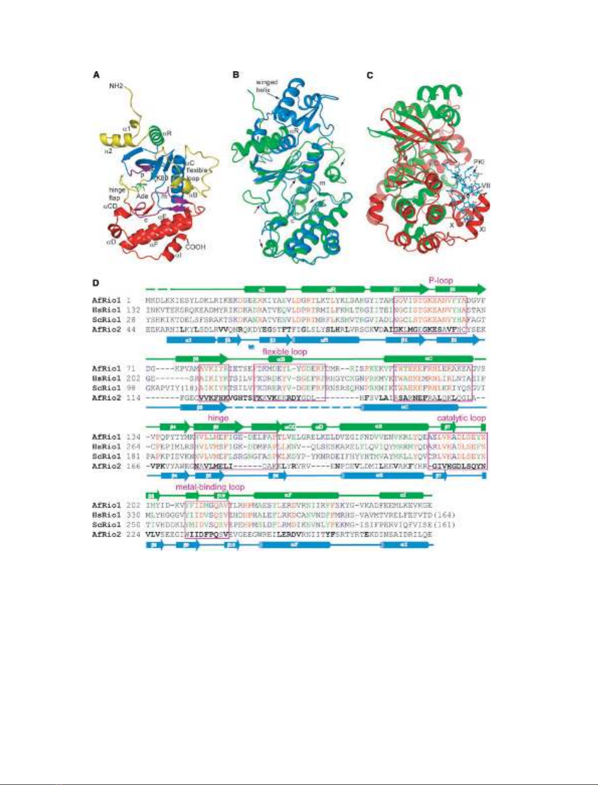
Fig. 1. The structure and conservation of Rio1. (A) The structure of APO-Rio1 showing the important kinase domain features and the Rio1-
specific loops (yellow). The P-loop, metal-binding loop and catalytic loop are indicated in all figures by p, m and c, respectively (purple). (B)
An alignment of the polypeptide chains of the ATP-Mn complexes AfRio1 (green) with AfRio2 (blue; PDB code 1ZAO). Arrows indicate signi-
ficant differences in structure between the two molecules. The position of aR and the winged helix of Rio2 is also indicated. (C) An align-
ment of the Rio2–ATP–Mn complex (green) with the PKA-ATP-Mn-peptide inhibitor complex (red; PDB code 1ATP). The peptide inhibitor is
shown in cyan stick representation (PKI), and the subdomains of PKA molecule absent in Rio1 are labeled. (D) Sequence alignment of AfRio1
with the enzymes from (H). sapiens and S. cerevesiae, as well as with AfRio2. Rio1 sequences are colored red for identical, green for highly
similar, and blue for weakly similar residues as calculated by CLUSTALW using the sequences shown as well as those from Caenorhabditis ele-
gans,Drosophila melanogaster and Xenopus laevis. The AfRio2 sequence is structurally aligned to the AfRio1 and is bolded for residues that
are identical or highly similar among Rio2 proteins. The elements of secondary structure of the Archaeoglobus enzymes are shown above
and below the alignments, with colors corresponding to (B).
N. LaRonde-LeBlanc et al. Structure and activity of the Rio1 kinase
FEBS Journal 272 (2005) 3698–3713 ª2005 FEBS 3701
and the model presented here does not represent a true
APO form. However, the structure in the absence of
added nucleotide does not represent an ATP-bound
form either. When nucleotide is added, Rio1 undergoes
conformational changes that result in a new crystal
form. Comparison of the APO and nucleotide-bound
structures indicates that in the presence of ATP or
ADP, two portions of the flexible loop become disor-
dered, and that the part that remains ordered changes
conformation and position relative to the rest of Rio1
molecule (Fig. 3A,B). In addition, the catalytic loop
and the metal-binding loop both move significantly
when ATP is added (Fig. 3A,B). The overall RMSD
between the two states is 0.91 A
˚for 228 Capairs. The
c-phosphate is modeled with partial occupancy, as
high temperature factors suggested that a fraction of
the molecules were hydrolyzed. Comparisons of the
four crystallographically independent molecules in the
Rio1-ATP complex showed that the N-terminal Rio1-
specific helices and aD adopt different positions, and
two of the molecules show a slightly different position-
ing of the ATP c-phosphate relative to the other two
(Fig. 3C). The structures of the Rio1-ATP-Mn and the
Rio1-ADP-Mn complexes are virtually identical, indi-
cating that the conformational changes which occur
require neither the presence of the c-phosphate nor
autophosphorylation (Fig. 4A).
The flexible loop and hinge region of the Rio1
kinases
The loop between aC and b3 of the RIO kinase
domain shows distinct conservation in each RIO
subfamily (Fig. 1D). In the case of Rio2, the electron
density for that region was not observed in any crys-
tals that have been studied to date. However, the
sequence in this region is highly conserved, suggesting
that it plays an important role in the function of Rio2
kinases. Similarly, Rio1 kinases also exhibit significant
conservation of residues in this loop (Fig. 1D). Align-
ment of A. fulgidus and S. cerevisiae Rio1 with human,
zebrafish, dog, plant, fly, and worm homologs yields
60% similarity and 20% identity in the sequence in
this region (data not shown). This increases to 87.5%
similarity and 66% identity when the yeast and archaeal
sequences are omitted from the alignment. In the
structure of APO-Rio1 presented here, this loop con-
sists of 27 amino acids (Arg83 of b3 through Glu111
of aC) and is significantly longer than the 18 amino
acids long disordered loop of Rio2 (Fig. 1D). In the
APO-Rio1 structure, this loop starts with a poorly
ordered chain between residues 84 and 90. This region
is characterized by weak density and high temperature
factors and makes no direct contact with other parts
of the protein, thus none of the side chains were mode-
led (Fig. 2A). Residues 90–96 form a small a-helix, fol-
lowed by a b-turn between Leu96 and Asp99. Three
more b-turns follow between Asp99 and Phe102,
Met104 and Ile107, and Ser108 and Glu111, which
marks the start of aC. The entire flexible loop packs
between the N-terminal portion of aC and part of the
C-lobe (Figs 2A and 3A).
The interactions between the flexible loop and the
rest of the protein include several hydrogen bonds
between conserved residues (Fig. 2A). The side chain
of Asp93 makes a hydrogen bond to Lys112, which is
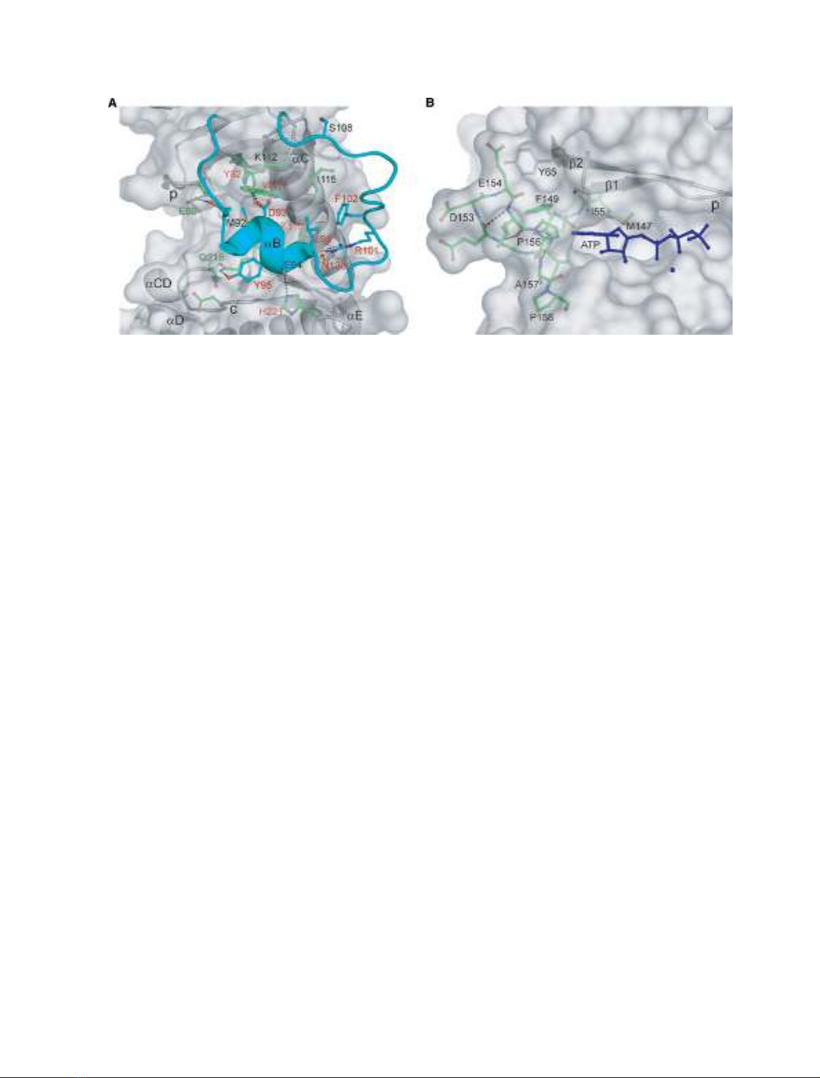
Fig. 2. The flexible loop and flap of Rio1. (A) The flexible loop of Rio1 showing the interactions between the loop and the rest of the pro-
teins. The loop is colored in cyan, residues that are involved in the interaction are shown in stick representation. Rio1-conserved residues
are labeled in red text. Those residues that are also conserved in Rio2 proteins are indicated in green text. (B) The structure of the flap in
the hinge region. Residues of the hinge region are shown in green stick representation.
Structure and activity of the Rio1 kinase N. LaRonde-LeBlanc et al.
3702 FEBS Journal 272 (2005) 3698–3713 ª2005 FEBS


