
REVIEW ARTICLE
Structure and function of plant aspartic proteinases
Isaura Simo
˜es and Carlos Faro
Departamento de Biologia Molecular e Biotecnologia, Centro de Neurocie
ˆncias e Biologia Celular, Universidade de Coimbra and
Departamento de Bioquı´mica, Faculdade de Cie
ˆncias e Tecnologia, Universidade de Coimbra, Portugal
Aspartic proteinases of the A1 family are widely distributed
among plant species and have been purified from a variety
of tissues. They are most active at acidic pH, are specifically
inhibited by pepstatin A and contain two aspartic residues
indispensible for catalytic activity. The three-dimensional
structure of two plant aspartic proteinases has been deter-
mined, sharing significant structural similarity with other
known structures of mammalian aspartic proteinases. With
a few exceptions, the majority of plant aspartic proteinases
identified so far are synthesized with a prepro-domain and
subsequently converted to mature two-chain enzymes. A
characteristic feature of the majority of plant aspartic pro-
teinase precursors is the presence of an extra protein domain
of about 100 amino acids known as the plant-specific insert,
which is highly similar both in sequence and structure to
saposin-like proteins. This insert is usually removed during
processing and is absent from the mature form of the
enzyme. Its functions are still unclear but a role in the vac-
uolar targeting of the precursors has been proposed. The
biological role of plant aspartic proteinases is also not
completely established. Nevertheless, their involvement in
protein processing or degradation under different conditions
and in different stages of plant development suggests some
functional specialization. Based on the recent findings on the
diversity of A1 family members in Arabidopsis thaliana,new
questions concerning novel structure–function relationships
among plant aspartic proteinases are now starting to be
addressed.
Keywords: aspartic proteinases; cardosin; phytepsin;
programmed cell death; stress response.
Introduction
Aspartic proteinases (APs; EC 3.4.23) have been extensively
studied and characterized and are widely distributed among
vertebrates, plants, yeast, nematodes, parasites, fungi and
viruses [1,2]. AP activity has also been detected in recom-
binant proteins from bacterial origin [3]. According to the
MEROPS database (http://www.merops.ac.uk), created by
Rawlings & Barrett [4], APs are now grouped into 14
different families, on the basis of their amino acid sequence
homology, which in turn are assembled into six different
clans based on their evolutionary relationship and tertiary
structure. Plant APs have been distributed among families
A1, A3, A11 and A12 of clan AA, and family A22 of clan
AD. The majority of plant APs belongs to the A1 family,
together with pepsin-like enzymes from many different
origins.
In common with other members of the A1 family, plant
APs are active at acidic pH, are specifically inhibited by
pepstatin and have two aspartic acid residues responsible for
the catalytic activity [2,5]. However, there are several
structural and functional features that make plant APs
unique among aspartic proteinases. These aspects will be
highlighted throughout the present review article which
aims to provide an overview of the current knowledge about
plant aspartic proteinases in terms of their structure,
processing, inactivation, localization, proposed biological
functions and genomic diversity.
Primary structure organization
The majority of plant APs identified so far are synthesized
as single-chain preproenzymes and subsequently converted
to mature enzymes that can be either single- or two-chain
enzymes. The cDNA derived amino acid sequences of
several plant APs revealed that the primary structures of
their precursors are quite similar [6–15]. These precursors
are characterized by the presence of a hydrophobic
N-terminal signal sequence, responsible for translocation
into the ER, followed by a prosegment of about 40 amino
acids, and a N-terminal domain and a C-terminal domain
separated by an insertion comprising approximately 100
amino acids, named as plant-specific insert (PSI) (Fig. 1).
While the prosegment is present in all APs and is involved
either in the inactivation or in the correct folding, stability
and intracellular sorting of several zymogens [16], the PSI is
an insertion only identified in plant APs, which is highly
similar to saposins and saposin-like proteins and whose
biological function has not been completely established
[8,13,17–21].
Correspondence to C. Faro, Departamento de Bioquı
´mica,
Universidade de Coimbra, Apt. 3126, 3000 Coimbra, Portugal.
Fax: + 351 239 480208, Tel.: + 351 239 480210,
E-mail: cfaro@imagem.ibili.uc.pt
Abbreviations: AP, aspartic proteinase; PSI, plant specific insert;
PCD, programmed cell death; PR, pathogenesis-related;
SAPLIP, saposin-like protein.
Enzymes: aspartic proteinases (EC 3.4.23).
(Received 19 February 2004, revised 25 March 2004,
accepted 31 March 2004)
Eur. J. Biochem. 271, 2067–2075 (2004) ÓFEBS 2004 doi:10.1111/j.1432-1033.2004.04136.x
To date, the only exceptions to this primary structure
organization are nucellin, specifically expressed in barley
ovule nucellar cells [22], an AP-like protein from tobacco
chloroplasts [23] and an AP encoded by the cdr-1
1gene
involved in disease resistance [24].
In general, plant APs share high amino acid sequence
similarity in their N- and C-terminal domains (over 60%
identity), and about 45% identity with cathepsin D, the
closest AP of nonplant origin. The two catalytic sequence
motifs are Asp-Thr-Gly (DTG) and Asp-Ser-Gly (DSG) in
all plant APs belonging to the A1 family with the exception
of chlapsin, the AP from Chlamydomonas reinhardtii that
contains DTG/DTG (accession number: AJ579366) (C. M.
Almeida
2and C. Faro, unpublished results). In some APs
from fungi and from protozoa, the catalytic Asp residues
also occur within the DTG/DSG motifs (http://www.
merops.ac.uk). The evolutionary or biological significance
of this variation observed in APs of different kingdoms has
not been established.
Three-dimensional structure
The three-dimensional structures of several members of the
A1 family have been determined and they share significant
structural similarity [2]. Regarding plant APs, only two
crystal structures have been determined – mature cardosin
A (PDB code: 1B5F) [17] and prophytepsin, the precursor
form of barley AP containing the prosegment and the PSI
(PDB code: 1QDM) [25] (Fig. 2). Both APs are two-chain
polypeptides in their mature forms and present a very
similar fold to what was found for other APs. The overall
secondary structure consists essentially of b-strands with
very little a-helix. The molecules are bilobal with the active
site located in a large cleft between the two similar b-barrel-
like domains, each contributing one of the catalytic
sequence motifs (DTG/DSG). The catalytic aspartic resi-
dues are located at the base of this large cleft. Three
conserved disulfide bridges stabilize the structure and both
polypeptide chains are held together by hydrophobic
interactions and hydrogen bonds. As in the other AP
structures, there is a flexible region known as the flap which
projects out over the cleft and encloses substrates and
inhibitors in the active site [5].
Besides the common pepsin-like topology for the main
body of mature phytepsin, the structural characterization
of the enzyme precursor also gave new insights about
the prosegment and the PSI [25]. Although part of the
prosegment was not traced due to a disordered structure,
it was shown that its N-terminal part is involved in the
formation of the six-stranded b-sheet, while the helical
portion of this prosegment approaches the active site and
partially covers it. As will be discussed below, the authors
propose an inactivation mechanism based on the inter-
actions found between the prosegment and the active site.
The PSI forms an independent subunit in the prophy-
tepsin structure that is inserted into the C-terminal domain.
Structurally, the PSI comprises five amphipathic a-helices
folded into a compact globular domain and linked with
each other by three disulfide bridges. A quite similar
structure was described for NK-lysin, which is also a
saposin-like protein [26].
Processing and inactivation mechanism
Plant AP precursors undergo several proteolytic cleavages
to produce mature single-chain or two-chain form of the
enzymes. Proteolytic processing of plant APs starts with
removal of the signal sequence upon translocation to the ER
lumen. The following conversion steps include cleavage of
the prosegment and total or partial removal of the internal
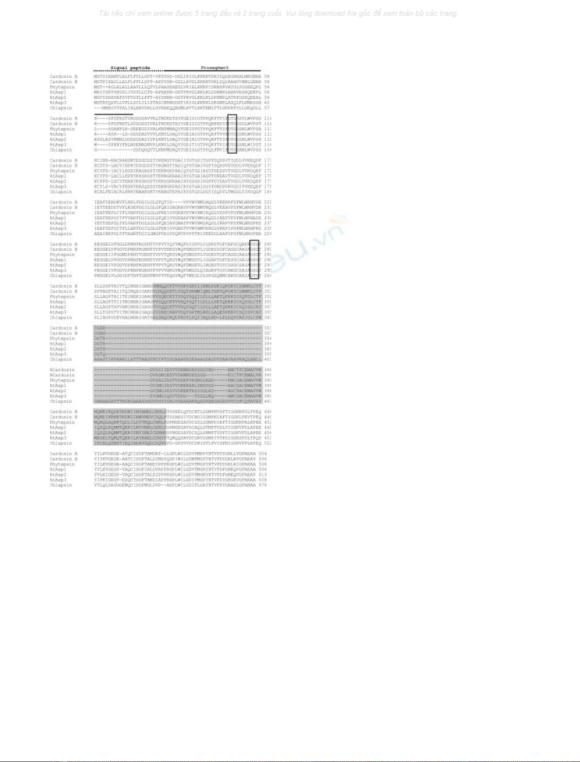
Fig. 1. Plant aspartic proteinase precursors. Comparison of the amino
acid sequences of representative members of the A1 family of plant
aspartic proteinases. The regions corresponding to the signal peptide
(dotted line), the prosegment (solid line) and the plant specific insert
(shaded grey) are highlighted. The catalytic aspartic acid residues are
boxed. Cardosin A and cardosin B were purified from C. carduncu-
lus L. (accession numbers
25 : AJ132884 and AJ237674, respectively – EBI
Data Bank); phytepsin was purified from barley (H. vulgare)(acces-
sion number: X56136); AtAsp1, AtAsp2 and AtAsp3 are A. thaliana
aspartic proteinases (accession numbers: U51036, AY070453 and
AF076243, respectively); chlapsin was purified from Chlamydomonas
reinhardtii (accession number: AJ579366).
2068 I. Simo
˜es and C. Faro (Eur. J. Biochem. 271)ÓFEBS 2004
PSI. Proteolytic removal of the prosegment is an important
step in generation of active protease from inactive zymogen
[1]. Zymogen conversion generally occurs by limited
proteolysis and removal of the Ôactivation segmentÕ.It
may involve accessory molecules that trigger activation or
the process may be autocatalytic requiring only a drop in
pH [27] as is described for the gastric APs [28].
In general, processing of plant aspartic proteinase
precursors involves removal of the prosegment and the
PSI domain [18,20,21,29–33]. Nevertheless there are some
variations on the mechanism and order by which each
segment is removed from the precursor.
Procardosin A, the precursor of cardosin A, undergoes
proteolytic processing as the flower matures and during this
process the PSI is totally removed, probably by an aspartic
proteinase, before the prosegment. Its conversion into an
active form is likely to occur inside the vacuoles where the
protein is accumulated [20]. Processing by a similar auto-
catalytic mechanism has also been proposed for cenprosin,
the AP from Centaurea calcitrapa [30] and for recombinant
oryzasin 1, the rice AP [29].
A slightly different picture has emerged for prophytepsin.
Using metabolic labeling and immunoprecipitation it was
shown that prophytepsin in barley roots is sequentially
processed into two different two-chain forms by cleavage of
the prosegment and partial removal of the PSI (and not
completely like in procardosin A) [18]. Although it was not
clearly established which is removed first, whether the
prosegment or the PSI, a recent paper proposed a model in
which the prosegment is removed prior to the PSI [33]. As
the intermediate forms and final products obtained in vitro
are slightly different from those detected in vivo,itwas
suggested that complete maturation of the protein probably
requires the presence of other proteinases/exopeptidases
besides the autoactivation mechanism [18].
The activation of recombinant cyprosin produced in
Pichia pastoris has given us a third processing scheme. Like
prophytepsin, the precursor form of cyprosin was processed
in different isoforms by the excision of the prosegment and
of most of the PSI [21]. Conversely to what has been found
in vivo [31], heavy and light chains of the processed forms of
recombinant cyprosin are held together by disulfide bonds.
It has been suggested that this different processing is caused
by the action of host cell proteinases and not by auto-
activation [21]. A similar processing mechanism has been
suggested for the sunflower seed AP. The precursor is
sequentially cleaved into different intermediate forms,
whose chains remain associated by disulfide bridges.
However, and in contrast to recombinant cyprosin, the
PSIisfinallyremovedtocompletioninordertogenerate
the mature form of the sunflower AP in which the chains
are no longer held together by disulfide bridges [32].
In any case, processing of plant AP precursors leads
ultimately to the formation of a two-chain enzyme, without
the prosegment and the PSI domain, with a domain
organization similar to that of mammalian or microbial APs.
An inactivation mechanism for plant APs has been
proposed by Kervinen et al. based on the three-dimensional
structure of phytepsin precursor [25]. The inactivation
mechanism proposed for prophytepsin resembles the mech-
anism accepted for mammalian gastric APs zymogens,
progastricsin and pepsinogen, with a preformed active site
blocked by the prosegment [34,35]. In prophytepsin the
active site is blocked not only by the prosegment, but also by
the 13 residues of the N-terminal of the mature enzyme and
by the ÔflapÕ. The anchorage of the prosegment and of part
of the N-terminus in the active site cleft is made by ionic
interactions established between Lys11/Tyr13 of the mature
enzyme sequence and the catalytic aspartic acids at the
bottom of the cleft. In fact, these two residues replace the
characteristic Lys36p/Tyr37p (where p stands for proseg-
ment) found in mammalian APs zymogens and known to be
responsible for the ionic interactions with the Asp residues
oftheactivesite.
Most plant APs contains a Lys/Tyr sequence in a position
equivalent to Lys11/Tyr13 of prophytepsin suggesting a
similar inactivation mechanism. However, cardosin A,
cardosin B and two rice APs do not contain this sequence
either in the prosegment or in the N-terminus of the mature
enzyme. Biochemical studies with recombinant precursors
of cardosins revealed that, conversely to other zymogens,
procardosins are active (M. Vieira
3& C. Faro, unpublished
results). These evidences suggest that procardosins probably
do not share the inactivation mechanism described above.
Most likely, the interactions between the prosegment and
the active site render the prosegment more flexible and
enable the substrate to enter the catalytic cleft. Nevertheless,
only the structural characterization of procardosins and
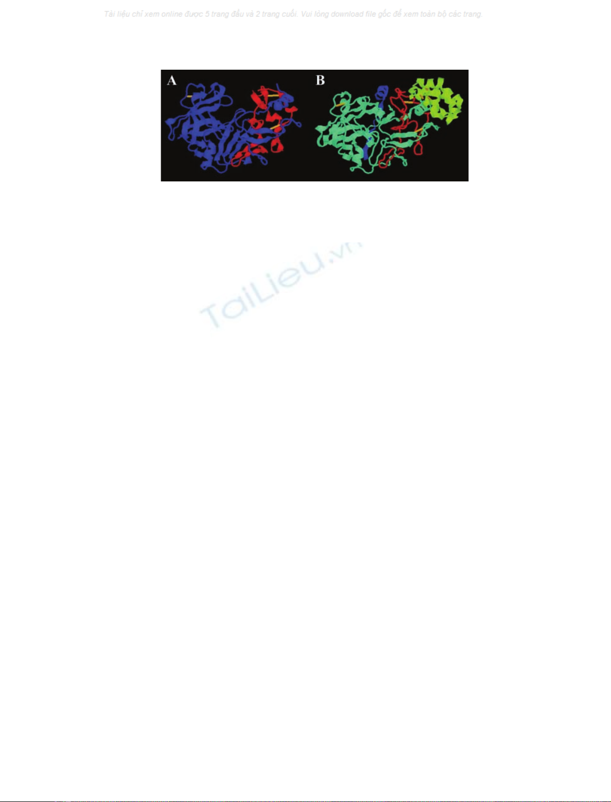
Fig. 2. Ribbon representation of the crystal structures of cardosin A (A) and prophytepsin (B). (A) Structure of mature cardosin A from C. car-
dunculus L. (PDB code: 1B5F) [17]. The heavy chain is shown in blue, the light chain in red and disulfide bridges in yellow. (B) Structure
of prophytepsin from H. vulgare L. (PDB code: 1QDM) [25]. The propeptide is shown in blue, the mature protein is shown in cyan
26 (heavy chain)
and red (light chain), the plant specific insert (PSI) in green and disulfide bridges in yellow. Prepared with the program
PROTEIN EXPLORER
http://www.proteinexplorer.org.
27
ÓFEBS 2004 Plant aspartic proteinases (Eur. J. Biochem. 271) 2069

other precursors will give new clues about the different
modes of inactivation in plant APs.
The plant-specific insert
Except for the barley nucellin [22], an AP-like protein from
tobacco chloroplasts [23] and the product of cdr-1 gene from
Arabidopsis [24], all plant APs identified so far are charac-
terized by the presence of an extra protein domain of
approximately 100 amino acids known as the plant specific
insert (PSI). This segment, inserted into the C-terminal
domain of the plant APs precursors, is usually removed
during the proteolytic maturation of the proteinases. The
PSI sequence shows no homology with mammalian or
microbial APs, but is highly similar to that of saposin-like
proteins (SAPLIPs) [36]. This protein family includes
saposins, which are lysosomal sphingolipid-activator pro-
teins [37], NK-lysin, granulysin, surfactant protein B,
amoebapores and domains of acid sphingomyelinase and
acyloxyacyl hydrolase [38–40]. Like other members of this
family, the PSI contains six conserved cysteines, several
hydrophobic residues and a consensus glycosylation site. In
the particular case of Chlamydomonas reinhardtii AP, and
besides these common features, the PSI domain comprises
an extra region of approximately 80 amino acids rich in
alanine triplets whose function is still unknown (C. M.
Almeida
4& C. Faro, unpublished results) (Fig. 1).
The structural characterization of prophytepsin’s PSI
revealed the same Ôsaposin foldÕ[25] as first determined for
NK-lysin [26] and recently for granulysin [41]. In fact, the
proteins belonging to this SAPLIPs family all share a closely
related compact globular structure comprising five amphi-
pathic a-helices linked with each other by three disulfide
bridges. A unique feature of the PSI is the swap of the
N- and C-terminal portions of the saposin-like domain,
where the C-terminal portion of one saposin is linked to the
N-terminal portion of the other saposin. Hence, the PSI is
not a true saposin but a swaposin [25,38,42] (Fig. 3).
The functions of the PSI are still unclear, however, an
important role in vacuolar targeting of plant AP precursors
has been proposed. Besides its possible direct interaction
with lipid bilayers, as described for other SAPLIPs members
[38], the structural characterization of phytepsin PSI
revealed a putative membrane-binding region comprising
the PSI and an adjacent area of the mature enzyme [25].
Thus, the authors suggest that this saposin-like domain in
plant APs may be responsible for bringing AP precursors
into contact with membranes or membrane-bound receptor
proteins mediating the sorting of enzyme precursors during
Golgi-mediated intracellular transport to the vacuoles. In
fact, the role of the PSI in protein sorting to vacuoles has
also been demonstrated in transient expression experiments
in tobacco protoplasts [33] where it was shown that deletion
of the PSI from phytepsin results in secretion of the
truncated phytepsin, whereas the wild-type phytepsin still
accumulates inside the vacuoles. In addition to this role of
the PSI as a vacuolar sorting signal it is also suggested that
this domain may have a strong influence on how phytepsin
leaves the ER, implying that the vacuolar sorting may not
be restricted to the Golgi apparatus but can start as early as
the ER [33]. The proposed role of the PSI in the targeting of
plant APs to the vacuole resembles what has been described
for mammalian saposin C and cathepsin D. It has been
suggested that the association of saposin C with cathep-
sin D may be responsible for the mannose-6-phosphate
independent targeting of the latter to the lysosome [43,44].
An important difference between both targeting mecha-
nisms is that in plants, APs and the PSI sorting domain
5
are encoded in the same precursor molecule, whereas in
mammalian cells different genes encode cathepsin D and
saposin C. However, and similarly to what has been
described for saposin C [38], intracellular protein targeting
may not be the only function of the PSI. In fact, Egas et al.
demonstrated that besides its ability to interact with
membranes, the PSI of cardosin A is a potent inducer of
vesicle leakage [45]. The results described either with
procardosin A or with recombinant PSI support the idea
that plant AP precursors are bifunctional molecules con-
taining a membrane-destabilizing domain in addition to
their protease domain. Thus, the authors suggest that the
PSI may take part in defensive mechanisms against
pathogens and/or as an effector of cell death. Based on
these results it was also suggested that the PSI from
carnivorous plants may contribute to prey digestion by
destroying prey cell membranes [6].
Distribution and localization
Plant APs are widely distributed in the plant kingdom and
have been detected or purified from monocotyledonous and
dicotyledonous species as well as gymnosperms. Recently,
the cDNA of an AP was cloned from Chlamydomonas
Fig. 3. The ‘saposin fold’. (A) Ribbon representation of the structure of NK-lysin, a saposin-like protein [26]. The N-terminal domain is shown in
blue and C-terminal domain in red. (B) Ribbon representation of the structure of the PSI domain of barley prophytepsin [25] (N-terminal domain,
blue; C-terminal domain, red). (C) Model structure of the PSI domain of cardosin A based on the crystal structure of prophytepsin PSI (N-terminal
domain, blue; C-terminal domain, red). Prepared with the program
PROTEIN EXPLORER
http://www.proteinexplorer.org.
28
2070 I. Simo
˜es and C. Faro (Eur. J. Biochem. 271)ÓFEBS 2004

reinhardtii indicating therefore that the A1 family of AP
is also represented in the unicellular green algae which
are the closest ancestral precursors of vascular plants
(C. M. Almeida
6& C. Faro, unpublished results).
In gymnosperms, AP activity has been detected in the
seeds of two pine species [46], whereas in angiosperms APs
have been detected or purified in monocotyledonous plants
such as barley, rice, wheat, sorghum and maize [7,47–54]
and in dicotyledonous plants
7like cucumber, squash, figleaf
gourd, castor bean, sunflower, cacao, Arabidopsis,Brassica,
spinach, potato, tobacco, tomato, cardoon, Centaurea
calcitrapa and carnivorous plants such as Nepenthes
[12,30,31,55–69].
Plant APs are either single-chain (cucumber, squash,
spinach, potato, sorghum, Brassica, rice, wheat, tomato and
tobacco) or two-chain (barley, figleaf gourd, castor bean,
sunflower, cacao, Centaurea, cardoon, Arabidopsis and
maize) enzymes. However, it has not been established what
determines the additional processing step of converting a
single-chain inactive enzyme into a two-chain active form.
Some authors suggest that these processing differences may
be caused by the presence or absence of protein-processing
enzymes responsible for the conversion because, in terms of
primary structure organization, plant APs precursors are, in
general, very similar.
Like for monocotyledonous plants, AP expression or
activity in some dicotyledonous plants has been detected
in other tissues besides those where the protein was first
purified [6,8,10,14,70–75]. However, tissue-specific localiza-
tion has been described for some plant APs and revealed
that these enzymes are not randomly distributed throughout
the organs. Moreover, it is now clear that some plant species
have multiple genes for APs. In fact, the differential
expression observed for these AP homologs in Cynara
cardunculus L., Arabidopsis,barleyandNepenthes clearly
suggests some functional specialization and imply the
potential involvement of the different APs in a wide variety
of cellular processes [6,8,15,22,54,76,77].
In barley, two independent studies demonstrated that in
developing grains and during seed germination the local-
ization of the AP (phytepsin) was very specific. Immuno-
histochemical studies in barley roots have also revealed that
phytepsin is specifically expressed in developing tracheary
elements and sieve cells [77]. Castor bean AP was localized
in the endosperm of maturing seeds [56] and in Nepenthes
alata, transcripts of two of the five AP homologues were
detected, by in situ hybridization, in the digestive glands of
the pitchers, the trapping organs of the plant [6]. Using
immunohistochemistry and immunogold transmission EM,
APs purified from the flowers of the cardoon Cynara
cardunculus L. have been specifically localized in the floral
transmitting tissue (cardosin B) [15], in the stigmatic
papillae (cardosin A) [76] and in the epidermal cells of the
style (cardosin A and cyprosins) [76,78]. In a recently
published report, Chen et al. demonstrated, by in situ
hybridization studies, the differential expression of the three
typical aspartic proteinases of Arabidopsis [8] and confirmed
previously published results on the AP localization in seed
tissues [79]. In the recently published paper, the authors
showed that transcripts of these three APs are detected in all
seed cell types, in the outer cell layers of the anthers early in
flower development and in the guard cells of the sepals. The
mRNA of one of the APs (AtPaspA2) was also weakly
detected in the transmitting tract of the flowers [8].
The great majority of the purified plant APs are
intracellular, and subcellular localization studies revealed
that they accumulate essentially inside protein storage
vacuoles. Biochemical and immunocytochemistry analysis
of barley roots and leaves showed that phytepsin was
localized to the vacuoles of these cells [80] and, in a different
study, phytepsin was also shown to accumulate in protein
bodies and large vacuoles of barley seeds [81]. The same
vacuolar localization was found for the APs present in the
seeds of castor bean [56], buckwheat [72] and Arabidopsis
[79]. Cardosin A, one of the APs purified from the flowers
of C. cardunculus L. also accumulates in protein storage
vacuoles in the stigmatic papillae [76].
The exceptions to this intracellular location are the
secreted APs found in the extracellular matrix of tobacco
[64] and tomato leaves [63], cardosin B found in the
extracellular matrix of the floral transmitting tissue in
C. cardunculus L. [15], the APs from Nepenthes that are
secreted into the pitchers [66] and the AP encoded by the
Arabidopsis cdr-1 gene [24]. The AP purified from maize
pollen is believed to be in the cell wall [51] and, surprisingly,
the AP from spinach has been localized to the plastids [62].
Biological functions
Plant APs have been detected and purified from many
different plant species. However, their biological functions
are not as well assigned or characterized as those of their
mammalian, microbial or viral counterparts that were
shown to perform many different and diverse functions,
including specific protein processing (e.g. rennin, cathep-
sin D and yapsins), protein degradation (e.g. gastric
enzymes such as chymosin, pepsin and gastricsin) or viral
polyprotein processing (human immunodeficiency virus
AP) [1,5,19]. For the great majority of plant APs no
definitive role has been assigned and the biological functions
are still hypothetical. Actually, much of our knowledge
about plant AP functions arises from colocalization studies
with putative protein substrates, experimental evidences for
the processing or degradation of those substrates in vitro
and/or specific expression in certain tissues or under specific
conditions. In general, plant APs have been implicated
in protein processing and/or degradation in different plant
organs, as well as in plant senescence, stress responses,
programmed cell death and reproduction.
Protein processing and/or degradation
as nitrogen source
In citrus leaf extracts, an AP has been implicated in the
proteolysis of the photosynthetic enzyme ribulose-1,5-
bisphosphate carboxylase/oxygenase which plays a signi-
ficant role as a nitrogen source during the growth of new
organs [70]. In carnivorous plants like Nepenthes or Drosera,
APs secreted into the pitchers may participate in the
degradation of insect proteins suggesting that these plants
may use insect proteins as nitrogen sources [6,66]. Partici-
pation of plant APs in storage protein degradation during
the mobilization of reserve proteins in seed germination has
been proposed for rice and wheat. In rice seeds it was
ÓFEBS 2004 Plant aspartic proteinases (Eur. J. Biochem. 271) 2071


