
Open Access
Available online http://arthritis-research.com/content/11/3/R84
Page 1 of 12
(page number not for citation purposes)
Vol 11 No 3
Research article
Alterations in peripheral blood memory B cells in patients with
active rheumatoid arthritis are dependent on the action of tumour
necrosis factor
M Margarida Souto-Carneiro1*, Vijayabhanu Mahadevan2*, Kazuki Takada3, Ruth Fritsch-Stork4,
Toshihiro Nanki3, Margaret Brown5, Thomas A Fleisher5, Mildred Wilson2, Raphaela Goldbach-
Mansky2 and Peter E Lipsky2
1Centro de Neurociências e Biologia Celular, Department of Zoology, University of Coimbra, 3004-517 Coimbra, Portugal
2National Institute of Arthritis and Musculoskeletal and Skin Diseases, NIH, 9000 Rockville Pike, Bethesda, MD 20892, USA
3Department of Medicine and Rheumatology, Graduate School, Tokyo Medical and Dental University, 1-5-45, Yushima, Bunkyo-ku, Tokyo 113-8519,
Japan
4Department of Rheumatology, UMC Utrecht, Heidelberglaan 100, 3584 CX Utrecht, The Netherlands
5Department of Laboratory Medicine, Warren Magnuson Center, NIH, 9000 Rockville Pike, Bethesda, MD 20892, USA
* Contributed equally
Corresponding author: Peter E Lipsky, peterlipsky@comcast.net
Received: 23 Jan 2009 Revisions requested: 6 Mar 2009 Revisions received: 20 Apr 2009 Accepted: 5 Jun 2009 Published: 5 Jun 2009
Arthritis Research & Therapy 2009, 11:R84 (doi:10.1186/ar2718)
This article is online at: http://arthritis-research.com/content/11/3/R84
© 2009 Souto-Carneiro et al.; licensee BioMed Central Ltd.
This is an open access article distributed under the terms of the Creative Commons Attribution License (http://creativecommons.org/licenses/by/2.0),
which permits unrestricted use, distribution, and reproduction in any medium, provided the original work is properly cited.
Abstract
Introduction Disturbances in peripheral blood memory B cell
subpopulations have been observed in various autoimmune
diseases, but have not been fully delineated in rheumatoid
arthritis (RA). Additionally, the possible role of tumour necrosis
factor (TNF) in regulating changes in specific peripheral blood
memory B cell subsets in RA is still unclear.
Methods The frequency and distribution of B cell subsets in the
peripheral blood and synovial membrane of active RA patients
with long-standing disease have been analysed. Additionally, the
possible role of TNF in causing disturbances in memory B cell
subsets in RA patients was assessed in a clinical trial with the
specific TNF-neutralising antibody, infliximab.
Results RA patients, independent of disease duration, have a
significantly lower frequency of peripheral blood pre-switch
IgD+CD27+ memory B cells than healthy individuals, whereas
post-switch IgD-CD27+ accumulate with increased disease
duration. Notably, both pre-switch IgD+CD27+ and post-switch
IgD-CD27+ memory B cells accumulate in the synovial
membrane of RA patients. Finally, anti-TNF therapy increased
the frequency of pre-switch IgD+CD27 memory B cells in the
peripheral blood.
Conclusions The data suggest that decreases in peripheral
blood IgD+CD27+ pre-switch memory B cells in RA reflect their
accumulation in the synovial tissue. Moreover, the significant
increase in the peripheral blood pre-switch memory B cells in
patients who underwent specific TNF-blockade with infliximab
indicates that trafficking of memory B cells into inflamed tissue
in RA patients is regulated by TNF and can be corrected by
neutralising TNF.
Introduction
Rheumatoid arthritis (RA) is a chronic systemic autoimmune
disease, characterised by inflammatory polyarthritis and joint
damage resulting in progressive disability [1]. The inflamma-
tory infiltrate in RA includes T cells, B cells and dendritic cells
[2-4], and in approximately 20% of patients lymphoid neogen-
APC: allophycocyanin; BSA: bovine serum albumin; CRP: C-reactive protein; DMARD: disease-modifying anti-rheumatic drugs; DMEM: Dulbecco's
Modified Eagle's Medium; ELISA: enzyme-linked immunosorbent assay; ESR: erythrocyte sedimentation rate; FCS: fetal calf serum; FDC: follicular
dendritic cell; FITC: fluorescein isothiocyanate; ICAM: intercellular adhesion molecule; IgVH: immunoglobulin heavy chain variable region; IL: inter-
leukin; LT: lymphotoxin; MAb: monoclonal antibodies; MTX: methotrexate; PBMC: peripheral blood mononuclear cells; PBS: phosphate-buffered
saline; PCR: polymerase chain reaction; PE: phycoerythrin; PerCpCy5.5: peridinin-chlorophyll-protein Cy5.5; RA: rheumatoid arthritis; SLE: systemic
lupus erythematosus; SS: Sjögren's syndrome; TNF: tumour necrosis factor; VCAM: vascular cell adhesion molecule; VEGF: vascular endothelial
growth factor.

Arthritis Research & Therapy Vol 11 No 3 Souto-Carneiro et al.
Page 2 of 12
(page number not for citation purposes)
esis develops with the formation of ectopic germinal centres
[5-8].
The importance of B cells in RA has been emphasised by the
success of therapeutic approaches using anti-CD20 mono-
clonal antibodies (mAbs) [9]. It is currently unknown whether
this approach to treatment is successful because of the pro-
duction of early plasma cells due to the loss of rheumatoid fac-
tor or because of other functions of B cells.
Functionally distinct B cell subsets can be defined by the sur-
face expression of immunoglobulin (Ig) D and CD27. These
include naïve IgD+CD27-; pre-switch memory IgD+CD27+;
and post-switch memory IgD-CD27+ [10-12]. Importantly,
CD27 expression by B cells has been considered a hallmark
for cells that have undergone somatic hypermutation [13],
although recently a CD27- population of memory B cells with
mutated Ig genes has been described [14-16], which is ele-
vated in patients with systemic lupus erythematosus (SLE)
[15]. Abnormalities in the frequencies of peripheral blood
memory B cells have been reported in SLE [17], and Sjögren's
syndrome (SS) [18]. However, in RA the data on possible dis-
turbances of peripheral blood B cell distributions have not
been delineated as well. Part of this could relate to differences
in disease duration and therapy of the cohorts studied [19-21].
Treatment with TNF blockers ameliorates the signs and symp-
toms of RA and disease progression [22-25]. Recently, a
study of peripheral blood and tonsilar biopsies from RA
patients undergoing treatment with the combined TNF and
lymphotoxin α (LTα) antagonist, etanercept, suggested that
part of the success of this therapy in RA could be linked to a
disruption of follicular dendritic cell (FDC) networks in second-
ary lymphoid organs, thus impairing germinal centre formation,
and decreasing the number of CD27+memory B cells in the
blood [19]. However, this effect was noted in the tonsil, mak-
ing it uncertain whether etanercept would have a similar
impact on germinal centres in the spleen and lymph nodes.
Etanercept neutralises both TNF and LTα, so it is difficult to
determine the possible contribution of each cytokine to the
effects noted. TNF and LTα have many non-overlapping func-
tions and, therefore, distinct effects of blocking each of these
two cytokines on memory B cell homeostasis are possible. For
example, TNF is involved in the regulation of the expression of
adhesion molecules, such as vascular cell adhesion molecule
(VCAM-1), intercellular adhesion molecule (ICAM-1), P-selec-
tin, E-selectin, and L-selectin (reviewed in [26]) and also vas-
cular endothelial growth factor (VEGF)-C [27], suggesting
that it may play a crucial role in the neovascularisation of rheu-
matoid synovium and also recruitment of lymphocytes into the
inflamed synovium.
In order to study the changes in peripheral memory B cell sub-
populations in RA patients, and to understand the possible
role of TNF in regulating changes in specific memory B cells,
we analysed the frequency and distribution of B cell subsets
in the peripheral blood and synovial membrane of active RA
patients with long-standing disease. Subsequently, we
assessed whether treatment with the specific TNF-blocker, inf-
liximab, normalised the distribution of these peripheral B cell
subsets. Our results show, for the first time, that RA patients,
independent of disease duration, have a much lower fre-
quency of peripheral blood pre-switch IgD+CD27+ memory B
cells than healthy individuals, whereas post-switch IgD-CD27+
memory B cells accumulate with increased disease duration.
Additionally, we present evidence that pre-switch IgD+CD27+
memory B cells accumulate in the synovial membrane of RA
patients, and that this accumulation might be related to the
influence of TNF, because anti-TNF therapy increased the fre-
quency of pre-switch IgD+CD27 memory B cells in the periph-
eral blood. These results document disease-related and TNF-
dependent abnormalities in memory B cell subsets in RA and
suggest that part of the success of TNF neutralising therapy
could relate to normalisation of memory B cell abnormalities.
Materials and methods
Patients and controls
Peripheral blood samples from 40 healthy donors (26 females,
14 males; mean age 44 years) were obtained from the
National Institutes of Health blood bank, and from 33 patients
(28 females, five males; mean age 57 years) with long-stand-
ing RA (median disease length, 13 years) enrolled in a natural
history protocol (00-AR-0222) at the Warren G. Magnuson
Clinical Center (National Institutes of Health, Bethesda, Mary-
land, USA).
In addition, blood samples were obtained from 23 patients (20
females, 3 males; mean age 48.5 years) with active RA
(defined as having greater than four tender and swollen joints,
erythrocyte sedimentation rate (ESR) greater than 20 mm/
hour or C-reactive protein (CRP) greater than 0.8 mg/dl) who
failed treatment with methotrexate (MTX; 12.5 to 15 mg/week)
and were entering a clinical trial of infliximab therapy (00-AR-
0220). For this trial, patients on prednisone had to be on 7.5
mg or less per day to be eligible to participate. Patients were
randomised to receive either monthly infliximab infusions (3
mg/kg infliximab with MTX 15 mg/week), or monthly control
infusions and weekly MTX alone (<25 mg/week). All patients
fulfilled the revised American College of Rheumatology criteria
for RA [28]. MB, carrying out the flow cytometric analysis, was
blinded to the measurements of clinical response and disease
activity scores.
The group of patients enrolled in the natural history protocol,
with a median disease length of 13 years, were considered as
the long-standing disease group. The group of patients
enrolled in the clinical trial for infliximab, with a median disease
duration of 4.4 years, were considered as the group with
shorter disease duration.

Available online http://arthritis-research.com/content/11/3/R84
Page 3 of 12
(page number not for citation purposes)
Synovial specimens and peripheral blood samples were col-
lected at the Department of Rheumatology, Tokyo Medical and
Dental University from 10 RA subjects with long-standing dis-
ease (median disease length of 13.5 years).
The characteristics of all patients studied are shown in Table
1.
The local institutional review board or the ethics committees
(National Institutes of Health and Tokyo Medical and Dental
University) approved the studies and all patients signed an
informed consent before participating in this study. Patient's
management was performed in accordance with the local
standard practice and the study was conducted in accord-
ance with the regulations governing clinical trials, such as the
Declaration of Helsinki as amended in Edinburgh (2000).
Lymphocyte phenotyping
Peripheral Blood
Peripheral blood samples from the controls and natural history
patients were obtained during a single scheduled outpatient
visit. Peripheral blood mononuclear cells (PBMCs) were iso-
lated by Ficoll gradient centrifugation and re-suspended in 1.5
ml PBS and 1% BSA (1 × 10⁶ cells/100 μL). Isolated PBMCs
were stained by standard methods with fluorescein isothiocy-
anate (FITC), phycoerythrin (PE), peridinin-chlorophyll-protein
Cy5.5 (PerCpCy5.5) or allophycocyanin (APC) conjugated
mAb specific for the following human cell surface markers:
anti-CD19 PerCpCy5.5, anti-CD27 PE, anti-IgD FITC and
anti-IgM FITC (all mAb were obtained from BD Pharmingen,
Franklin Lakes, NJ, USA). Data were acquired on a FACSCal-
ibur (BD Biosciences, Franklin Lakes, NJ, USA).
Peripheral blood samples from the RA patients treated with
MTX and infliximab or MTX alone were obtained before and
after treatment. Anticoagulated samples were stained for
three-colour flow cytometry using a whole blood staining
method at the National Institutes of Health Clinical Center lab-
oratory. B cells were identified by staining with anti-CD20
APC and anti-CD27 PE (BD Biosciences, San Jose, CA,
USA) and anti-IgD FITC (Caltag, Burlingame, CA, USA). T
cells were identified by anti-CD3 APC or PE, anti-CD4 PE,
anti-CD8 FITC or APC, anti-CD45RA FITC and anti-CD45R0
APC (BD Biosciences, San Jose, CA, USA).
To calculate absolute numbers of each lymphocyte subset, the
percentage of cells staining positively was multiplied by the
absolute peripheral blood lymphocyte count, which was deter-
mined by cell counting with a Celldyne 3500 (Abbott, Santa
Clara, CA, USA) blood cell counting machine. With all experi-
ments, peripheral blood from healthy adult patients was
stained and analysed as controls.
To determine the chemokine receptor expression by B cells
and their subsets, the following APC-conjugated anti-human
mAbs were used: anti-CXCR1, anti CXCR2 and anti-CCR2
(R&D Systems, Minneapolis, MN, USA); and anti-CXCR4 (BD
Biosciences, San Jose, CA, USA).
Irrelevant, directly conjugated, murine IgG1 (BD Biosciences,
San Jose, CA, USA) was used to ascertain background stain-
Table 1
Clinical and demographic characteristics of the RA patients
Treatment trial
Healthy controls Long-standing RA RA patients for synovium
collection
MTX only Infliximab and MTX
Number of subjects 40 33 10 8 15
Age (years) 44 ± 9 57 ± 12 62 ± 10 52 ± 16 45 ± 11
Female/male ratio 26:14 28:5 9:1 7:1 13:2
Disease duration (years) -- 13 ± 12 13.5 ± 11.0 5.7 ± 1.5 3.0 ± 0.5
% RF positive patients -- 79% 90% 50% 73%
ESR (mm/hour) -- 36 ± 27 -- 36.9 ± 14.3 60.2 ± 30.3
CRP (mg/dl) -- <0.4 ± 9.75 2.8 ± 2.3 0.8 ± 1.1 1.8 ± 1.9
% Patients on MTX (dose) -- 85% (15 mg/wk) 60% (4 mg/wk) 100% (14 mg/wk) 100% (14 mg/wk)
% Patients on GC (dose) -- 49% (5 mg/day) 80% (5 mg/day) 50% (6 mg/day) 64% (6 mg/day)
% Patients on other DMARD -- 82% 50% -- --
Data are means ± standard deviation.
CRP = C-reactive protein; DMARD = disease-modifying anti-rheumatic drug; ESR = erythrocyte sedimentation rate; GC = glucocorticoids; MTX
= methotrexate; RA = rheumatoid arthritis; RF = rheumatoid factor.

Arthritis Research & Therapy Vol 11 No 3 Souto-Carneiro et al.
Page 4 of 12
(page number not for citation purposes)
ing. Samples were run on a FACScan or a FACSCalibur (BD
Biosciences, San Jose, CA, USA). Data were analysed using
the WinList software, version 5.0, and FloJo software (TreeS-
tar, Stanford University, CA, USA). B cells (CD20+ or CD19+)
were gated and the percentages of CD27+ (total memory),
IgD+CD27- (naïve), IgD+CD27+ (pre-switch memory) and IgD-
CD27+ (post-switch memory) populations in the gated B cells
were calculated. Although anti-CD20 mAb do not identify all
plasmablasts, most of which are CD19+CD27++IgD-, the
results from both staining protocols were pooled together,
because no significant differences in total or post-switch
memory B cells were observed when analysing the results
separately.
T cells (CD3+) were gated, and the precentages of CD4+
(total helper), CD8+ (total cytotoxic), CD4+CD45RA+ (total
naïve helper) and CD4+CD45R0+ (total memory helper) pop-
ulations within the T cell population were calculated.
Synovial specimens
Synovial tissues were obtained during joint replacement sur-
gery from 10 RA patients. Specimens were minced and incu-
bated with 0.3 mg/ml of collagenase (Sigma, St. Louis, MO,
USA) for one hour at 37°C in Dulbecco's Modified Eagle's
Medium (DMEM; Sigma, St. Louis, MO, USA). Partially
digested pieces of the tissue were pressed through a metal
screen to obtain single cell suspensions. Cells were stained
with anti-CD19 PECy5 (Beckman Coulter, Fullerton, CA,
USA), anti-CD27 FITC, anti-IgM PE and anti-IgD PE (all from
Becton Dickinson, Fullerton, CA, USA), anti-CXCR1 PE, anti-
CXCR2 PE, anti-CXCR4 PE and anti-CCR2 PE (all from R&D
Systems Inc., Minneapolis, MN. USA). Synovial tissue cells
were adjusted to 1 × 10⁵ cells, and incubated with the above
mAbs for 30 minutes, rinsed with PBS-3% FCS, and analysed
with a FACSCalibur (Becton Dickinson, Fullerton, CA, USA).
Amplification of the IgV heavy chain by single-cell PCR
CD19+IgD+CD27- naïve B cells and CD19+IgD+CD27+ mem-
ory B cells from four patients with RA were sorted using a
Beckton Dickinson FACS DIVA (Fullerton, CA, USA) or a
Dako Cytomation MoFlo (Dako Cytomation, Ft Collins, CO,
USA) and 1 to 1.5 cells/5 μL PBS, and then plated into 96-
well PCR plates containing 10 μL lysis buffer (2 × PCR buffer
+ 0.4 mg/ml proteinase K (Sigma, St. Louis, MO, USA)), sub-
jected to primer extension pre-amplification and then VH3 and
VH4 genes were amplified by nested PCR, as previously
described [29]. PCR products were purified using the Per-
forma® 96-Well Standard Plate kit (Edge BioSystems, Gaith-
ersburg, MD, USA) and sequenced on a model 3100 capillary
sequencer (Applied Biosystems, Foster City, CA, USA) using
the Big Dye® Terminator v1.1 Cycle Sequencing Kit (Applied
Biosystems, Foster City, CA, USA). Ig variable heavy chain
rearrangements were analysed for somatic mutations using
the web-based algorithm JOINSOLVER® (NIAMS/CIT, Mary-
land, USA) [30].
Soluble CD27 ELISA
The level of soluble CD27 was determined in serum samples
from RA patients in the natural history protocol and healthy
controls using the PeliKline Compact human soluble CD27
ELISA kit (CLB, Central Laboratory of the Netherlands Red
Cross, Amsterdam, The Netherlands) according to the manu-
facturer's instructions.
Statistical analyses
Data were checked for a normal distribution in order to decide
whether to use parametric or non-parametric tests. Median
group values (with standard error of the mean) for percentage
and absolute numbers of the different B cell populations were
compared in patients and healthy controls using the nonpara-
metric unpaired Mann-Whitney test.
Mean values (with standard deviation) of the CD27+ memory
B cell population were compared between the synovium and
peripheral blood of 10 RA patients undergoing synovectomy
using a paired Student's t-test.
Median group values (with standard error of the mean) of the
different B cell populations compared pre- and post-treatment
in the 23 RA patients who were treated with infliximab plus
MTX or MTX monotherapy using the nonparametric paired
Wilcoxon Signed Rank test. A P < 0.05 was considered sta-
tistically significant.
Results
Characteristics of the RA patients
The demographic and clinical characteristics of the RA patient
groups evaluated in this study are shown in Table 1. Most of
the 33 patients with long-standing RA were women with
chronic (median disease duration of 13 years), rheumatoid
factor-positive erosive disease. All patients were receiving
MTX alone or in combination with other disease-modifying
anti-rheumatic drugs (DMARDs). Most of the subjects from
whom synovial specimens were obtained were also older
women with chronic rheumatoid factor-positive RA.
The 23 RA patients enrolled in a clinical trial comparing MTX
plus infliximab with MTX alone had disease of shorter duration
(median 4.4, infliximab + MTX: 3.0 and MTX: 5.7 years).
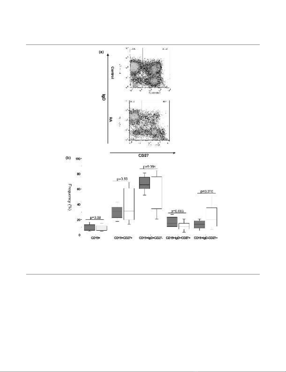
RA patients have a reduced peripheral blood pre-switch
IgD+CD27+ memory B cell population
The frequencies of B cell subsets defined by the expression of
IgD and CD27 in the peripheral blood of patients with long-
standing RA were compared with healthy donors (Figures 1a,
b). One striking finding was that the subjects with long-stand-
ing RA had a significantly (P = 0.0031) lower frequency of
IgD+CD27+ pre-switch memory B cells than the healthy
donors (median RA 10.4 ± 1.3% vs control 15.1 ± 1.1%). This
significant difference (P = 0.0036) was maintained when ana-
lysing the absolute number of pre-switch memory B cells
Available online http://arthritis-research.com/content/11/3/R84
Page 5 of 12
(page number not for citation purposes)
(median RA: 13.8 ± 4.7 cells/μl vs control: 21.3 ± 3.9 cells/
μl). On the other hand, the frequency – but not the absolute
numbers – of the IgD-CD27+ post-switch memory population
was significantly (P = 0.0101) increased in subjects with long-
standing RA when compared with the control individuals
(median RA 19.6 ± 2.9% vs control 13.2 ± 1.0%). Interest-
ingly, no significant difference could be seen between RA
patients and controls in the frequency or absolute number of
the total CD27+ memory B cell pool (median RA 31.3 ± 3.8%
vs control 30.3 ± 1.6%, P = 0.6258; median RA 41.0 ± 11.3
cells/μl vs control: 44.6 ± 5.0 cells/μl, P = 0.7022). Finally, the
frequency of IgD+CD27- naïve B cell population in the periph-
eral blood of subjects with long-standing RA was comparable
with the healthy donors (median RA 57.3 ± 4.1% vs control
65.6 ± 1.7%). However, the absolute number of naïve B cells
was significantly (P = 0.0231) lower in the long-standing RA
patients when compared with the control individuals (median
RA: 61.4 ± 28.6 cells/μl vs control: 100.5 ± 10.7 cells/μl).
These differences could not be the result of B cell lymphope-
nia, because the absolute number of the total B cell pool in the
long-standing RA patients was comparable to the healthy
Figure 1
RA patients, irrespective of disease duration show marked shifts in the frequency of the peripheral blood B cell subsetsRA patients, irrespective of disease duration show marked shifts in the frequency of the peripheral blood B cell subsets. (a) Dot-plots of IgD versus
CD27 of peripheral blood CD19+ B cells from representative healthy control and a long-standing rheumatoid arthritis (RA) patients illustrating the
differences in the frequency of each B cell subset. (b) Box-plots representing the 10th, 25th, 50th (median), 75th and 90th percentiles of the fre-
quencies of the total B cells (as a percentage of lymphocytes), total CD27+ memory B cells, naïve IgD+ CD27- B cells, pre-switch IgD+ CD27+ mem-
ory B cells and post-switch IgD-CD27+ memory B cells (each as a percentage of B cells) in the peripheral blood of healthy donors (n = 40, white
bars) and long-standing RA patients (n = 33, grey bars). *Significant (P < 0.01) difference from control donors.


