
Hallgren et al. Respiratory Research 2010, 11:55
http://respiratory-research.com/content/11/1/55
Open Access
RESEARCH
BioMed Central
© 2010 Hallgren et al; licensee BioMed Central Ltd. This is an Open Access article distributed under the terms of the Creative Commons
Attribution License (http://creativecommons.org/licenses/by/2.0), which permits unrestricted use, distribution, and reproduction in
any medium, provided the original work is properly cited.
Research
Altered fibroblast proteoglycan production in
COPD
Oskar Hallgren*
1,2
, Kristian Nihlberg
1
, Magnus Dahlbäck
3
, Leif Bjermer
2
, Leif T Eriksson
3
, Jonas S Erjefält
1
, Claes-
Göran Löfdahl
2
and Gunilla Westergren-Thorsson
1
Abstract
Background: Airway remodeling in COPD includes reorganization of the extracellular matrix. Proteoglycans play a
crucial role in this process as regulators of the integrity of the extracellular matrix. Altered proteoglycan
immunostaining has been demonstrated in COPD lungs and this has been suggested to contribute to the
pathogenesis. The major cell type responsible for production and maintenance of ECM constituents, such as
proteoglycans, are fibroblasts. Interestingly, it has been proposed that central airways and alveolar lung parenchyma
contain distinct fibroblast populations. This study explores the hypothesis that altered depositions of proteoglycans in
COPD lungs, and in particular versican and perlecan, is a result of dysregulated fibroblast proteoglycan production.
Methods: Proliferation, proteoglycan production and the response to TGF-β1 were examined in vitro in centrally and
distally derived fibroblasts isolated from COPD patients (GOLD stage IV) and from control subjects.
Results: Phenotypically different fibroblast populations were identified in central airways and in the lung parenchyma.
Versican production was higher in distal fibroblasts from COPD patients than from control subjects (p < 0.01). In
addition, perlecan production was lower in centrally derived fibroblasts from COPD patients than from control subjects
(p < 0.01). TGF-β1 triggered similar increases in proteoglycan production in distally derived fibroblasts from COPD
patients and control subjects. In contrast, centrally derived fibroblasts from COPD patients were less responsive to TGF-
β1 than those from control subjects.
Conclusions: The results show that fibroblasts from COPD patients have alterations in proteoglycan production that
may contribute to disease development. Distally derived fibroblasts from COPD patients have enhanced production of
versican that may have a negative influence on the elastic recoil. In addition, a lower perlecan production in centrally
derived fibroblasts from COPD patients may indicate alterations in bronchial basement membrane integrity in severe
COPD.
Background
Chronic obstructive pulmonary disease (COPD) is a pro-
gressive disease characterized by a reduction in respira-
tory airflow that is not possible to normalize [1]. The
reduced airflow is caused by tissue remodeling, including
reorganization of the extracellular matrix (ECM). In
bronchi, epithelial dysregulation results in impaired
mucocilliary clearance, over-production of mucus, and
squamous cell metaplasia. In parallel with this, subepi-
thelial fibrosis is often observed in bronchi and bronchi-
oles. Degradation of alveolar walls (emphysema) is a
hallmark of COPD, which limits the area of air-blood
exchange and the elastic recoil [2]. Other structural
changes in COPD, such as thickening of the airway wall
and reticular basement membrane, have been implicated
as factors that contribute to reduction in airflow [3,4].
Interestingly, in terms of the turnover of ECM, oppos-
ing pathological processes occur in the COPD lung as the
ECM is degraded in alveoli and there is excessive deposi-
tion of ECM (fibrosis) in bronchi and in bronchioles [5].
The major cell type responsible for production and main-
tenance of the ECM are fibroblasts. Recently, it was sug-
gested that central airways and alveolar lung parenchyma
contain distinct fibroblast populations [6,7]. Fibroblasts
* Correspondence: oskar.hallgren@med.lu.se
1 Department of Experimental Medical Science, BMC D12 Lund, Lund
University, Sweden
Full list of author information is available at the end of the article

Hallgren et al. Respiratory Research 2010, 11:55
http://respiratory-research.com/content/11/1/55
Page 2 of 11
from these anatomical sites were found to have different
morphology, proliferation, and ECM production. This
distinction is important to consider in COPD, as the
ECM turnover is different in bronchi and alveoli. A key
family of molecules for ECM integrity is the proteogly-
cans. The production of proteoglycans and other ECM
molecules have been reported to be modulated by the
profibrotic signal molecule TGF-β [8,9]. Proteoglycans
have been shown to be differentially expressed in COPD
lungs [10,11]. For example, enhanced alveolar immunos-
taining of the large proteoglycan versican has been
reported in COPD patients [10]. As versican may inhibit
the assembly of elastic fibers, it may have a negative effect
on the elastic recoil and thereby possibly contribute to
the pathogenic development of COPD. Moreover, perle-
can, a heparan sulphate proteoglycan, is crucial for base-
ment membrane integrity and reduced perlecan
immunostaining has been demonstrated in the lungs of
COPD patients [11].
In this study, we hypothesized that altered levels of pro-
teoglycans in COPD lungs may be dependent on dysregu-
lated proteoglycan production in fibroblasts and hence
that there are alterations in fibroblast phenotypes in
COPD patients compared to control subjects. In particu-
lar, we wanted to determine whether enhanced alveolar
versican deposition is due to higher versican production
by distal fibroblasts. We also wanted to examine whether
perlecan production was altered in centrally derived
fibroblasts. Thus, we isolated centrally and distally
derived fibroblasts from lung explants from COPD
patients and from biopsies from healthy control subjects
in order to assess proteoglycan production, proliferative
potential, and responsiveness to TGF-β1 in vitro.
Methods
Patients
Patients (n = 8) suffering from very severe COPD (GOLD
stage IV) who were undergoing lung transplantation at
Lund University Hospital were included in the study. The
patients had quitted smoking at least 6 months before the
lung transplantation. Non-smokers (n = 12) with no clini-
cal history of asthma, (reversibility < 12% after adminis-
tration of the β2-agonist salbutamol (400 μg) and did not
respond to methacholine test doses (< 2,000 μg)), or other
lung diseases were included as control subjects. This
study was approved by the Swedish Research Ethical
Committee in Lund (FEK 91/2006 and FEK 213/2005).
Isolation of cells
Fibroblasts were isolated from explants from COPD
patients and from bronchial and transbronchial biopsies
from control subjects as previously described [12].
Briefly, biopsies from control subjects were immediately
after sampling transferred to cell culture medium
(DMEM supplemented with 10% FBS, Gentamicin, PEST,
and Ampotericin B (all from Gibco BRL, Paisley, UK)).
Specimens from the lung explants were dissected directly
after removal from the COPD patients and were immedi-
ately transferred to cell culture medium. Bronchial tissue
was collected from the luminal side from the same locali-
sation as where bronchial biopsies were taken and were
chopped into small pieces. Alveolar parenchymal speci-
mens from explants were collected 2-3 cm from the
pleura in the lower lobes, i.e. from the same location as
where transbronchial biopsies were taken. Vessels and
small airways were removed from the peripheral lung tis-
sues and the remaining tissues were chopped into small
pieces. After rinsing, bronchial and parenchymal pieces
from explants and biopsies were allowed to adhere to the
plastic of cell culture flasks for 4 h and were then kept in
cell culture medium in 37°C cell incubators until out-
growth of fibroblasts were observed. Bronchial and
parenchymal fibroblasts were then referred to as central
and distal fibroblasts, respectively. Experiments were per-
formed at passages 3-6. The cell cultures were continu-
ously stained to verify the mesenchymal identity and to
estimate the purity. In the few cases when the cellular
staining was less clear then the cell morphology was veri-
fied to be representative for the culture as a whole.
Proliferation assay
Proliferation was determined as previously described
[13]. Briefly, cells were plated and fixed after 6, 24, 48, h.
Cells were stained with Crystal Violet and cell numbers
were quantified indirectly by absorbance at 595 nm on a
spectrophotometer plate reader (ELX800; Biotek Instru-
ments, Winooski, VT,). Proliferation was defined as
absorbance at 48 h divided by the absorbance after 6 h.
With this method the amount of adsorbed dye has been
shown to be proportional to the cell number recorded on
a Coulter counter [14].
Immunohistochemistry
Staining of fibroblasts
Fibroblasts (7000/well) grown overnight were fixed in 4%
paraformaldehyde for 15 minutes. Thereafter unspecific
binding sites were blocked by 2% BSA-TBS containing 5%
goat serum (Vector laboratories, Burlingame, CA) and
0,1% Triton X for 30 minutes. Cells were incubated with
primary antibodies: monoclonal mouse antibody against
Prolyl 4-Hydroxylase (Acris antibodies, Hiddenhausen,
Germany), monoclonal mouse IgM antibody against
Vimentin (Santa Cruz Biotechnology, Santa Cruz, CA),
monoclonal mouse IgG2a antibody against α-SMA
(Dako, Glostrup, Denmark), monoclonal IgG and anti-
body against SM22-alpha (Abcam, Cambridge, UK), and
with secondary antibodies: Alexafluor 488-conjugated
goat anti-mouse antibody and Alexafluor 555-conjugated

Hallgren et al. Respiratory Research 2010, 11:55
http://respiratory-research.com/content/11/1/55
Page 3 of 11
goat anti-mouse antibody (both from Molecular Probes
Invitrogen, Eugene, OR). The DNA-binding molecule
DAPI (Molecular Probes Invitrogen, Eugene, OR) was
used to stain cell nuclei before final mounting (Dako fluo-
rescence mounting medium, Dako, Glostrup, Denmark).
Cells were photographed using a TE2000-E fluorescence
microscope (Nikon, Tokyo, Japan) equipped with a
DXM1200C camera (Nikon, Tokyo, Japan).
Tissue staining of proteoglycans
In short, tissue, from same locations as where pieces for
cell isolations were taken, was fixed in 4% paraformalde-
hyde and embedded in paraffin. 5 μm sections were
deparaffinized, rehydrated and then pre-treated over-
night in buffer containing chondrotinase ABC (Seika-
gaku, Tokyo, Japan), in 37°C to make epitopes accessible
for antibodies. Endogenous peroxidase activity was
blocked in 3% hydrogen peroxidase (Merck, Damstadt,
Germany) followed by a 30 minutes block with 2% BSA-
TBS containing 5% serum raised in the same species as
the secondary antibodies used. Furthermore, endogenous
avidin and biotin binding sites were blocked (Vector avi-
din/biotin blocking kit, Vector laboratories, Burlingame,
CA) according to the manufacturer's protocol. Sections
were incubated with primary antibodies: rabbit poly-
clonal antibody against versican (Santa Cruz Biotechnol-
ogy, Santa Cruz, CA), mouse polyclonal antibody against
perlecan (Zymed laboratories, San Francisco, CA), goat
polyclonal antibody against biglycan (Santa Cruz Bio-
technology, Santa Cruz, CA), mouse monoclonal anti-
body against decorin (Abcam, Cambridge, UK). This was
followed by incubation with secondary antibodies: biotin-
conjugated goat anti-rabbit (Vector laboratories, Burl-
ingame, CA), biotin-conjugated horse anti-mouse (Vec-
tor laboratories, Burlingame, CA), and biotin-conjugated
donkey anti-goat (Jackson ImmunoResarch, West Grove,
PA). Sections were incubated with avidin and biotin (Vec-
tor laboratories, Burlingame, CA) according to the manu-
facturer's instructions and were developed with DAB
(Vector laboratories, Burlingame, CA) to visualize bound
antibodies and then counterstained with Mayer's hema-
toxylin. Sections were photographed using a TE2000-E
fluorescence microscope (Nikon, Tokyo, Japan) equipped
with a DXM1200C camera (Nikon, Tokyo, Japan).
Quantification of proteoglycans
Proteoglycan production in fibroblasts was determined
as previously described [15]. Briefly, cells were incubated
in sulfate-poor Dulbecco's MEM (Gibco BRL, Paisley,
UK) supplemented with 0.4% FBS w/wo 10 ng/ml TGF-
β1 (R&D Systems, Minneapolis, MN). COPD fibroblasts
had a very contractile phenotype and experiments were
therefore performed on cell culture plastics coated with
1% collagen-1 (PureCOL; Inamed Biomaterials, Fremont,
CA). This modification did not significantly alter proteo-
glycan production (data not shown). Proteoglycans were
quantified by [35S]-sulfate incorporation into glycosoam-
inoglycan side-chains measured on a scintillation counter
(Wallac; Perkin Ellmer, Boston, MA). Individual proteo-
glycans were separated by SDS-PAGE and quantified
using densitometry. The contribution of individual pro-
teoglycans to the total proteoglycan content was calcu-
lated as the value for each proteoglycan divided by the
sum of all the measured proteoglycans (versican, perle-
can, biglycan and decorin) by densitometry.
Statistics
Data are expressed as mean ± SEM. Statistical differences
between groups were determined by multiple compari-
sons using Kruskal-Wallis test. The Mann-Whitney test
was used to compare statistical differences between
groups. The Wilcoxon signed rank test was used to deter-
mine whether TGF-β1-stimulated proteoglycan produc-
tion was different from basal levels. Differences were
considered significant at p < 0.05. All analyses were per-
formed using GraphPad Prism software version 4.00
(GraphPad Software, San Diego, CA).
Results
Study subjects
Characteristics of COPD patients and control subjects
are shown in Table 1. Predicted FEV1 was 19.7% (14-24%);
(mean and range) in COPD patients. The corresponding
numbers were 106.7% (95-116%) for control subjects.
Mean age was 62 years (53-66) in the COPD group. For
control subjects the mean age was 30 years (24-41). All
the COPD patients were ex-smokers whereas none of the
controls had a history of smoking. Distal fibroblast cul-
tures were obtained from all patients and control sub-
jects, while central fibroblasts were obtained from 6 of 8
COPD patients and 7 of 12 control subjects.
Qualitative evaluation of proteoglycan localization
The localization of versican, perlecan, biglycan, and
decorin staining from one representative COPD subject
is presented in Figure 1. Immunoreactivity for perlecan
was, as expected, identified in the basement membrane of
bronchi, bronchioles and blood vessels. Unexpectedly,
the bronchial and bronchiolar reticular basement mem-
branes showed immunoreactivity for biglycan and deco-
rin. The basement membrane of pulmonary vessels were
positive for decorin but not for biglycan. The lamina pro-
pria tissue between basement membranes and smooth
muscle layers in bronchi and bronchioles was positive for
versican, biglycan, and decorin. Immunoreactivity for
these three proteoglycans was also observed in the tunica
media of pulmonary arteries, as well as in the adventia of
both bronchioles and arterioles. Staining was also present
in alveolar walls. Finally, smooth muscle cell layers cir-

Hallgren et al. Respiratory Research 2010, 11:55
http://respiratory-research.com/content/11/1/55
Page 4 of 11
cumscribing bronchi and bronchioles were slightly posi-
tive for perlecan, decorin and biglycan.
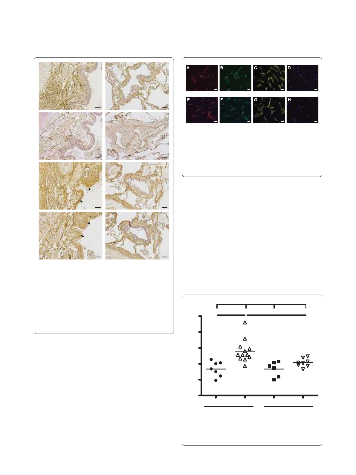
Characterization of fibroblasts
Isolated fibroblasts from COPD patients and control sub-
jects were characterized using antibodies to mesenchy-
mal markers (Figure 2). Both central and distal fibroblasts
were positive for α-smooth muscle actin (α-SMA), as
shown in Figure 2A and 2E. Furthermore, central and dis-
tal fibroblasts showed immunoreactivity for the fibroblast
markers prolyl 4-hydroxylase and vimentin (Figure 2B, C,
F and 2G). The contractile protein Sm22 has been found
to be expressed by smooth muscle cells and myofibro-
blasts but not by fibroblasts [16]. The isolated fibroblasts
were negative for sm22.
Fibroblast proliferation
The proliferative potential of isolated fibroblasts was
quantified using the crystal violet assay (Figure 3). In con-
trol subjects, central fibroblasts had a significantly lower
proliferation potential (1.72 ± 0.46) than distal fibroblasts
(2.80 ± 0.72) (p < 0.01). No such difference was observed
for fibroblasts from COPD patients. Distal fibroblasts
from COPD patients had a significantly lower prolifera-
tion potential (2.07 ± 0.27) than distal fibroblasts from
control subjects (2.80 ± 0.72) (p < 0.01). No difference
was seen between central fibroblasts from COPD patients
and control subjects.
Basal proteoglycan production
The basal proteoglycan production was investigated, as
shown in Figure 4. In control subjects, distal fibroblasts
had a significantly higher production of biglycan than
central fibroblasts (255 ± 43 vs. 81 ± 13) (p < 0.05). No
such difference was observed for fibroblasts from COPD
patients. Distal fibroblasts from COPD patients had a sig-
nificantly higher production of versican than distal fibro-
blasts from control subjects (324 ± 198 vs. 90 ± 47) (p <
0.01). Perlecan production was significantly lower in cen-
tral fibroblasts from COPD patients (111 ± 29) than in
fibroblasts from control subjects (285 ± 55) (p < 0.05).
There were no significant differences between the groups
in the basal production of decorin.
TGF-β1-induced proteoglycan production
Fibroblasts were stimulated with TGF-β1 for 24 hours and
the proteoglycan production was quantified and com-
pared to basal levels (Figure 5). In central fibroblasts from
control subjects TGF-β1 induced significant increases in
the production of versican (2.1-fold) and biglycan (3.6-
fold). In distal fibroblasts from control subjects TGF-β1
induced significant increases of versican (1.8-fold), perle-
can (1.4-fold) and biglycan (3.1-fold). However, TGF-β1
induced a significant decrease of decorin (1.4-fold) but no
change in the production of the other proteoglycans in
central fibroblasts from COPD patients. In distal fibro-
blasts from COPD patients, TGF-β1 induced significant
increases in the production of versican (2.1-fold), perle-
can (1.5-fold) and biglycan (2.9-fold). Distal fibroblasts
from COPD patients had a higher TGF-β1-response in
the production of versican (p < 0.05), perlecan (p < 0.05),
biglycan (p < 0.01), and decorin (p < 0.01) than central
fibroblasts from COPD patients. No such difference was
Table 1: COPD patients and control subjects in the study
Characteristics Controls COPD
No. 12 8
Age (range) 30 (24--41) 62 (53--66)
Pack years (range) 0 41 (25--60)
Gender, M/F in % 42/58 37/63
Lung function
FEV14.0 (3.0--5.4) 0.57 (0.4--0.9)
FEV1 % predicted 106.7 (95--116) 19.7 (14--24)
FVC 4.8 (3.5--6.4) 2.0 (1.3--2.8)
FEV1 % predicted/
FVC
22 (17--28) 31 (20--39)
DLCO m 1.4 (1.4--1.5)†
DLCO % predicted m 24 (14--42)§
† Only values from 4 patients. § Only values from 5 patients. m denotes that data is missing
Data is presented as mean (range)
Hallgren et al. Respiratory Research 2010, 11:55
http://respiratory-research.com/content/11/1/55
Page 5 of 11
observed between distal and central fibroblasts from con-
trol subjects.
Proteoglycan production profiles
We next examined the contribution of each proteoglycan
to the total proteoglycan production defined as the sum
of versican, perlecan, biglycan and decorin (Figure 6).
The contribution of perlecan to the total proteoglycan
production was significantly higher in distal fibroblasts
(0.19 ± 0.02) compared to central fibroblasts (0.13 ± 0.02)
from COPD patients (p < 0.05). There was no such differ-
ence in fibroblasts from control subjects. However, in
control subjects the contribution of biglycan to the total
proteoglycan production was significantly higher in distal
fibroblasts (0.26 ± 0.02) compared to central fibroblasts
(0.13 ± 0.03) (p < 0.05). There was no such difference in
fibroblasts from COPD patients. The contribution of ver-
sican to the total proteoglycan production was signifi-
cantly higher in central fibroblasts from COPD patients
(0.27 ± 0.04) than in central fibroblasts from control sub-
jects (0.12 ± 0.03) (p < 0.05). There was a similar differ-
ence (p < 0.01) between distal fibroblasts from COPD
patients (0.31 ± 0.03) and control subjects (0.15 ± 0.03) in
the contribution of versican to the total proteoglycan
production. The contribution of perlecan to the total pro-
teoglycan production was significantly lower (p < 0.01) in
central fibroblasts from COPD patients (0.13 ± 0.02) than
from control subjects (0.42 ± 0.09). A similar difference in
the contribution of perlecan was recorded between distal
Figure 1 Proteoglycan staining in lung sections from COPD pa-
tients. Representative micrographs of lung sections from one COPD
patient. Antibodies were visualized by DAB staining (shown in brown)
and sections were counterstained with Mayer's hematoxylin, which
stains cell nuclei blue. A, C, E, and G (left panel) are representative mi-
crographs from the central airways (bronchi) and B, D, F, and H (right
panel) show the small airways and parenchyma. A and B show versi-
can, C and D perlecan, E and F biglycan, and G and H decorin. L de-
notes lumen of the bronchi, and A and V denote airway and vessels,
respectively. Black arrowheads show staining in the lamina reticularis.
Scale bars represent 100 μm.
AB
CD
EF
GH
L
L
L
L
A
A
A
A
A
V
V
V
Figure 2 Immunostaining of COPD fibroblasts. Isolated fibroblasts
were immunostained to verify their mesenchymal origin. Cell nuclei
are visualized by DAPI staining, shown in blue. A--D (upper panel): rep-
resentative micrographs of central fibroblasts. E--H (lower panel) show
distal fibroblasts. Antibodies to α-SMA were used in A and E, antibod-
ies to prolyl 4-hydroxylase in B and F, antibodies to vimentin in C and
G, and antibodies to sm22 in D and H. Scale bars represent 50 μm
E
α−SMA
F
prolyl 4-H
G
vimentin
H
sm22
A B C D
Central fibroblasts
Distal fibroblasts
Figure 3 Proliferation potential of isolated fibroblasts. Prolifera-
tion potential was determined by crystal violet assay, as described in
Methods. The data presented are those for 48 hours, as compared to
those for 6 hours. *P < 0.01, +++P < 0.001.
5
4
3
2
1
0
Control COPD
Proliferation rate
******
+++
Central Distal Central Distal


