
Page 1 of 10
(page number not for citation purposes)
Available online http://arthritis-research.com/content/11/1/212
Abstract
The autoinflammatory diseases, also known as periodic fever
syndromes, are disorders of innate immunity which can be
inherited or acquired and which cause recurrent, self-limiting,
seemingly spontaneous episodes of systemic inflammation and
fever in the absence of autoantibody production or infection. There
has been much recent progress in elucidating their aetiologies and
treatment. With the exception of familial Mediterranean fever, which
is common in certain populations, autoinflammatory diseases are
mostly rare but should not be overlooked in the differential
diagnosis of recurrent fevers since DNA diagnosis and effective
therapies are available for many of them.
Introduction
The autoinflammatory conditions are a group of multisystem
disorders of innate immunity characterised by fluctuating or
irregularly recurring episodes of fever and systemic inflam-
mation, affecting the skin, eyes, joints, and serosal surfaces.
They include the hereditary syndromes familial Mediterranean
fever (FMF), tumour necrosis factor (TNF) receptor-asso-
ciated periodic syndrome (TRAPS), the hyper-IgD and
periodic fever syndrome (HIDS), and the cryopyrin-associated
periodic syndrome (CAPS) and acquired diseases of adult-
hood, including urate arthropathy and Schnitzler syndrome.
Despite some similarities in symptoms, there are major
distinctions in the aetiology, inheritance, duration and
frequency of ‘attacks’, and the overall clinical picture of the
various disorders (Table 1). These diseases are generally
compatible with normal life expectancy, bar the significant risk
of developing AA amyloidosis. Recent insights into their
molecular pathogenesis with identification of susceptibility
genes and characterisation of new proteins and pathways
have led to improved diagnosis and development of rational
therapies and have shed fascinating new light on aspects of
the innate immune system.
The inherited fever syndromes
Familial Mediterranean fever
This was first described in New York in 1945 by Sheppard
Siegal, although the term familial Mediterranean fever was not
coined until 1958 [1].
Genetics and pathophysiology
The gene associated with FMF, MEFV on chromosome 16,
encodes a protein called pyrin and was identified through
positional cloning in 1997 [2,3]. MEFV is constitutively
expressed in neutrophils, eosinophils, monocytes, dendritic
cells, and synovial fibroblasts and is upregulated in response
to inflammatory activators such as interferon-γand TNF-α[4].
The more than 40 MEFV mutations associated with FMF
encode either single amino acid substitutions or deletions
(Infevers registry database [5]). Disease-causing mutations
occur mostly in exon 10 but also occur in exons 1, 2, 3, 5,
and 9. Mutations in each of the two MEFV alleles are found in
85% of patients with FMF, whilst the great majority of
individuals with a single mutated allele are healthy carriers [6].
The methionine residue at position 694 may be especially
important for pyrin’s function; three different mutations
involving M694 have been identified, and homozygosity for
M694V is associated with a severe phenotype. Interestingly,
simple heterozygous deletion of this residue has been
associated with autosomal dominant FMF in northern
Review
Developments in the scientific and clinical understanding of
autoinflammatory disorders
Helen J Lachmann and Philip N Hawkins
National Amyloidosis Centre and Centre for Acute Phase Proteins, Department of Medicine, University College London Medical School,
Hampstead Campus, Rowland Hill Street, London NW3 2PF, UK
Corresponding author: Helen J Lachmann, h.lachmann@medsch.ucl.ac.uk
Published: 30 January 2009 Arthritis Research & Therapy 2009, 11:212 (doi:10.1186/ar2579)
This article is online at http://arthritis-research.com/content/11/1/212
© 2009 BioMed Central Ltd
CAPS = cryopyrin-associated periodic syndrome; CB2BP1 = CD2-binding protein-1; CINCA = chronic infantile neurological, cutaneous, and artic-
ular syndrome; CPPD = calcium pyrophosphate dihydrate; FCAS = familial cold autoinflammatory syndrome; FMF = familial Mediterranean fever;
HIDS = hyper-IgD and periodic fever syndrome; IL = interleukin; LRR = leucine-rich repeat; MSU = monosodium urate; MVA = mevalonic aciduria;
MVK = mevalonate kinase; MWS = Muckle-Wells syndrome; NF-κB = nuclear factor-kappa-B; NOMID = neonatal onset multisystem inflammatory
disease; PAMP = pathogen associated molecular patterns; PAPA = pyogenic sterile arthritis, pyoderma gangrenosum, and acne; PYD = pyrin
domain; SAA = serum amyloid A protein; TNF = tumour necrosis factor; TNFR1 = tumour necrosis factor receptor 1; TNFRSF1A = tumour necro-
sis factor receptor superfamily 1A; TRAPS = tumour necrosis factor receptor-associated periodic syndrome.

Page 2 of 10
(page number not for citation purposes)
Arthritis Research & Therapy Vol 11 No 1 Lachmann and Hawkins
Table 1
The autoinflammatory conditions of known genetic aetiology
Periodic Predominant Potential Distinctive Typical Typical Characteristic
fever Mode of ethnic Usual age precipitants clinical duration frequency laboratory
syndrome Gene inheritance groups at onset of attacks features of attacks of attacks abnormalities Treatment
FMF MEFV Autosomal Eastern Childhood/ Usually none Short severe 1 to 3 days Variable Marked acute- Colchicine
Chromosome recessive Mediterranean early adult and occasionally attacks, phase response
16 (dominant in menstruation, colchicine- during attacks
rare families) fasting, stress, responsive, and
and trauma erysipelas-like
erythema
TRAPS TNFRSF1A Autosomal Northern Childhood/ Usually none Prolonged More than a Variable and Marked acute- Etanercept and
Chromosome dominant European early adult symptoms week and may may be phase response high-dose
12 and can be but reported be very continuous during attacks corticosteroids
de novo in many prolonged and low levels
ethnic groups of soluble TNFR1
when well
HIDS MVK Autosomal Northern Infancy Immunisations Diarrhoea and 3 to 7 days 1 to 2 monthly Elevated IgD Anti-TNF and
Chromosome recessive European lymphadenopathy and IgA, acute- anti-IL-1
12 phase response, therapies
and mevalonate
aciduria during
attacks
FCAS NLRP3 Autosomal Northern Childhood Exposure to Cold-induced 24 to 48 hours Depends on Acute-phase Cold avoidance
Chromosome 1 dominant European cold fever, arthralgia, environmental response during and anti-IL-1
environment rash, and factors attacks and to a therapies
conjunctivitis lesser extent
when well
MWS NLRP3 Autosomal Northern Neonatal/ Marked diurnal Urticarial rash, Continuous, Often daily Varying but Anti-IL-1
Chromosome 1 dominant European infancy variation and conjunctivitis, often worse in marked acute- therapies
cold environment and sensorineural the evenings phase response
but less marked deafness most of the time
than in FCAS
CINCA/ NLRP3 Sporadic Northern Infancy None Urticarial rash, Continuous Continuous Varying but Anti-IL-1
NOMID Chromosome 1 European aseptic meningitis, marked acute- therapies
deforming arthropathy, phase response
ensorineural deafness, most of the time
and mental retardation
PAPA PSTPIP1 Autosomal Northern Childhood None Pyogenic arthritis, Intermittent Variable and Acute-phase Anti-TNF
(CD2BP1) dominant European pyoderma attacks with may be response during therapy
Chromosome (only 3 families gangrenosum, migratory arthritis continuous attacks
15 reported) and cystic acne
Blau NOD2 Autosomal None Childhood None Granulomatous Continuous Continuous Sustained Corticosteroids
syndrome (CARD15) dominant polyarthritis, iritis, modest acute-
Chromosome 16 and dermatitis phase response
CINCA, chronic infantile neurological, cutaneous, and articular syndrome; FCAS, familial cold autoinflammatory syndrome; FMF, familial Mediterranean fever; IL, interleukin; MVK, mevalonate
kinase; MWS, Muckle-Wells syndrome; NOMID, neonatal onset multisystem inflammatory disease; PAPA, pyogenic sterile arthritis, pyoderma gangrenosum, and acne; TNF, tumour necrosis
factor; TNFR1, tumour necrosis factor receptor 1; TNFRSF1A, tumour necrosis factor receptor superfamily 1A; TRAPS, tumour necrosis factor receptor-associated periodic syndrome.

Page 3 of 10
(page number not for citation purposes)
Europeans [7]. Greater disruption of a single MEFV allele by
two or more mutations can also cause dominant inheritance,
although FMF affecting more than one generation in typical
populations usually represents pseudodominant inheritance
due to consanguinity or a high prevalence of carriers.
One particular pyrin variant, E148Q encoded in exon 2, has
allele frequencies of 10% to 20% in Asian populations and
up to 1% to 2% in Caucasians. Whilst pyrin E148Q can
cause FMF when coupled with an exon 10 mutation,
homozygosity for E148Q alone is not associated with the
disease in the vast majority of cases. There is some evidence
that FMF carriers, perhaps especially those with pyrin
E148Q, may have an augmented response to some types of
non-FMF inflammation [8,9].
Neither the structure nor the function of pyrin has yet been
fully characterised, although subtle abnormalities of leukocyte
function have been reported in FMF patients and upregulated
MEFV expression has been identified in critically ill children
with multiple organ failure [10]. The putative 781-amino acid
protein has sequence homologies with a number of proteins
of apparently disparate function and cellular localisation. Pyrin
is thought to interact with a variety of proteins within the
cytoplasm and to play a key role in the modulation of
inflammation and apoptosis [11]. Many of its interactions
appear to involve its 90-amino acid N-terminal death domain,
which is now classified generically as a pyrin domain (PYD) in
other proteins that have homology with pyrin’s N-terminal
sequence [12]. Members of the death domain superfamily are
involved in the assembly and activation of apoptotic and
inflammatory complexes through homotypic protein-protein
interactions [13]. Proteins with PYDs play important roles in the
regulation of caspase-1 and thus modulate production of inter-
leukin-1 (IL-1). In this regard, pyrin is thought to interact with
another member of the superfamily, apoptosis-associated
speck-like protein with a caspase recruitment domain (ASC).
Recent work also suggests that pyrin may itself be a substrate
for cleavage by caspase-1 and that pyrin variants may serve as a
more efficient substrate than the wild-type protein [14]. Another
postulated mechanism by which variant pyrin could promote
inflammation is translocation of the resulting N-terminal PYD
cleavage fragments to the nucleus, where they could potentiate
activation of nuclear factor-kappa-B (NF-κB) [15].
Clinical features
FMF is the most common in Middle Eastern populations but
occurs worldwide [16]. The prevalence of FMF is estimated
to be 1/250 to 1/500 among Sephardic Jews and 1/1,000 in
the Turkish population. Carrier frequency exceeds 1 in 4 in
some eastern Mediterranean populations, prompting specu-
lation that the FMF trait may have conferred survival benefit,
possibly through enhanced resistance to microbial infection
mediated via an upregulated innate immune response
[17,18]. Males and females are affected equally and the
disease usually presents in childhood.
Attacks of FMF occur irregularly and apparently sponta-
neously although some may be precipitated by minor physical
or emotional stress, the menstrual cycle, or diet. Attacks
evolve rapidly and symptoms resolve within 72 hours. Fever
with serositis are the cardinal features, and these can vary
from mild to incapacitating. Peritonitis that can mimic an
acute surgical abdomen occurs in 85% of cases, and indeed
40% of patients will undergo exploratory surgery before FMF
is diagnosed. Pleuritic chest pain occurs in 40% of patients,
characteristically unilaterally, either alone or with peritonitis.
Headache with features of meningism has been reported in
children in particular, but the nervous system is not usually
involved. Orchitis occurs in less than 5% of patients, most
commonly in early childhood, and can be confused with
testicular torsion. Joint involvement usually affects the lower
limbs: arthralgia is common in acute attacks and usually
subsides within a couple of days, but a chronic destructive
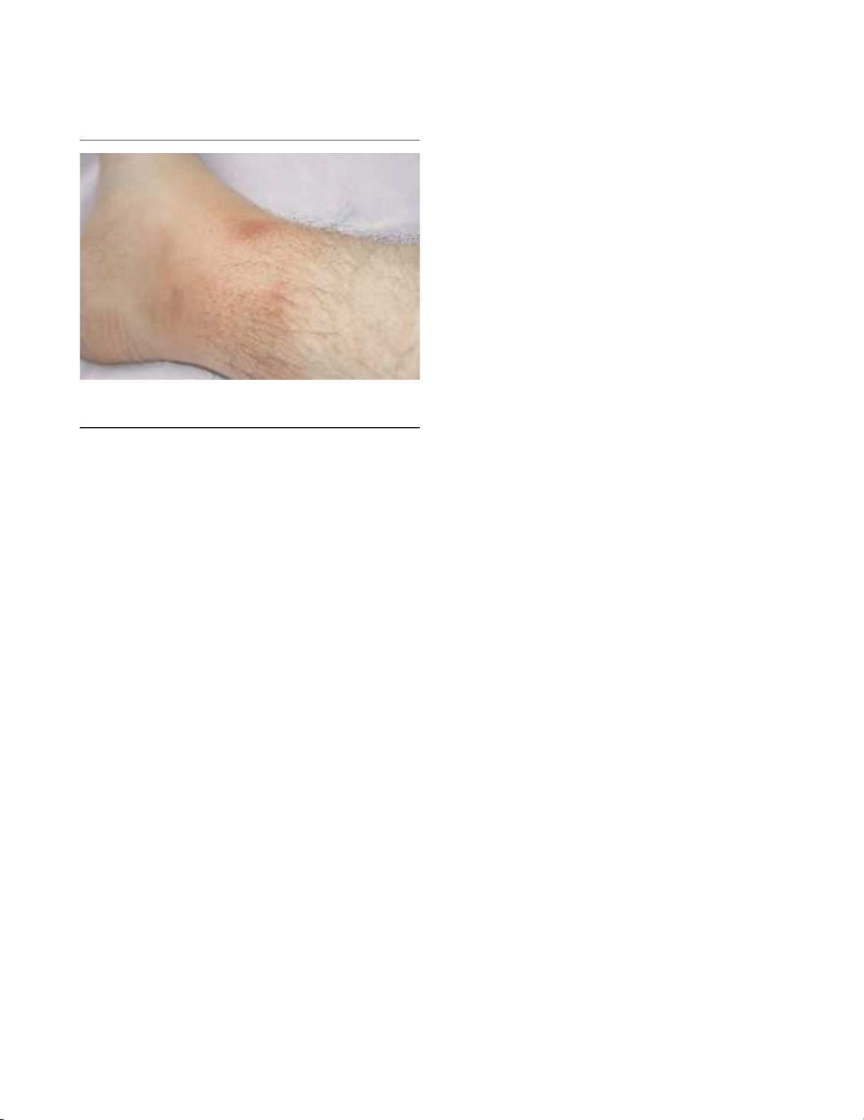
arthritis can rarely occur. A characteristic erysipelas-like rash
occurs in 20% of patients, usually around the ankles
(Figure 1). A degree of myalgia can occur during acute
attacks, but up to a fifth of patients complain of persistent
muscle pain on exertion, usually affecting the calves. Pro-
tracted febrile myalgia is rare and is characterised by severe
pain in the lower limbs or abdominal musculature which may
persist for weeks and can be accompanied by a vasculitic
rash; it usually responds to corticosteroids therapy.
Acute attacks are accompanied by a neutrophilic leuko-
cytosis, raised erythrocyte sedimentation rate, and a dramatic
acute-phase response. Investigations may be required to
exclude other diagnoses but imaging by x-ray, ultrasound, or
echocardiography during attacks is usually unrewarding.
Diagnosis is supported by DNA analysis but essentially
remains clinical and centres on the history of recurrent self-
limiting idiopathic attacks of fever and serositis that can be
prevented by prophylatic colchicine treatment. Genetic
results must be interpreted cautiously given that certain
individuals with paired pathogenic MEFV mutations never
develop FMF and that others with heterozygous carrier status
can do so. Furthermore, most diagnostic laboratories offer
only limited analysis of the large 10-exon MEFV gene.
Treatment
Supportive measures, including analgesia, are often required
during acute attacks, but the mainstay of management is
long-term prophylactic treatment with low-dose colchicine.
This was discovered serendipitously in 1972 by Goldfinger
[19] and has entirely transformed the outlook of this pre-
viously disabling disease. Continuous treatment with
colchicine at a dose of 1 to 2 mg daily in adults prevents or
substantially reduces symptoms of FMF in at least 95% of
cases and almost completely eliminates the risk of AA
amyloidosis (see below). The mechanism of action of
colchicine remains incompletely understood, but colchicine
binds to tubulin and evidently modulates neutrophil adhesion,
Available online http://arthritis-research.com/content/11/1/212
mobility, and cytokine release in a presumably rather specific
manner in patients with defective pyrin variants [20,21].
Long-term colchicine is advisable in every patient with FMF and
mandatory in those who already have AA amyloidosis. Although
colchicine is very toxic in acute overdose, the low daily doses
required for treatment of FMF are generally very well tolerated.
Diarrhoea is the most common side effect and usually can be
avoided by gradual introduction of the drug. Despite theoretical
concerns, there is no evidence that colchicine causes infertility
or birth defects and it can be taken safely by nursing mothers
[22]. Colchicine is a purely prophylactic treatment in FMF, and
introduction or dose escalation during an acute FMF attack is
not generally effective.
Genuine resistance to colchicine is probably very rare, although
issues of compliance are surprisingly common. Anecdotal
reports of benefit from treatment with etanercept or anakinra in
‘refractory’ patients are beginning to emerge [23,24].
Tumour necrosis factor receptor-associated periodic
syndrome
TRAPS is the second most common inherited fever
syndrome, although with an estimated prevalence of about 1
per million in the UK, it is very rare.
Genetics and pathophysiology
TRAPS is an autosomal dominant disease associated with
mutations in the gene for TNF receptor superfamily 1A
(TNFRSF1A), a 10-exon gene located on chromosome
12p13 [25]. TNF is a key mediator of inflammation with pleio-
tropic actions, including pyrexia, cachexia, leukocyte activa-
tion, induction of cytokine secretion, expression of adhesion
molecules, and resistance to intracellular pathogens. TNF
receptor 1 (TNFR1) is a member of the death domain
superfamily and comprises an extracellular motif containing
four cysteine-rich domains, a transmembrane domain, and an
intracellular death domain. Binding of soluble circulating TNF
causes trimerization of the receptor and activation of NF-κB,
with downstream induction of inflammation and inhibition of
apoptosis via production of cellular caspase-8-like inhibitory
protein (cFLIP). Events following endocytosis of the activated
TNFR1 complex result in apoptosis. The mechanism(s) by
which heterozygous TRFRSF1A mutations cause TRAPS
remain unclear and may well differ between mutations. Most
TRAPS-associated mutations lie within exons 2 to 4, of which
about half are missense substitutions affecting highly
conserved cysteine residues that disrupt structurally impor-
tant cysteine-cysteine disulphide bonds in the extracellular
domain. Under normal circumstances, TNF signalling is
terminated by metalloproteinase-dependent cleavage of a
proximal region of the extracellular domain, which releases
soluble TNFR1 that competitively inhibits binding of circu-
lating TNF to cell surface receptors. Whilst cleavage of
certain TNFR1 variants is impaired producing a ‘shedding
defect’, this is not the case with other TRAPS-causing
mutations, which must exert their pathogenic effect by
different means. It is thought that mutant misfolded receptors
may give rise to enhanced or prolonged signalling, possibly
through retention within the endoplasmic reticulum [26-29].
Despite initial hopes to the contrary, the mechanisms and
downstream effects by which TNFR1 mutations result in
TRAPS remain far from clear.
Clinical features
The clinical entity now known as TRAPS was described in
1982 as familial Hibernian fever [30], reflecting the Irish/
Scottish ancestry of patients in early reports, but TRAPS has
subsequently been reported in many ethnic groups, including
Jews, Arabs, and Central Americans. Males and females are
affected equally and presentation is usually before 4 years of
age. Most mutations are associated with high penetrance, but
two variants, P46L and R92Q, that can be associated with
TRAPS are present in approximately 10% of healthy West
Africans [31] and 1% of healthy Caucasians, respectively.
Attacks in TRAPS are far less distinct than in FMF. Febrile
episodes typically last 1 to 4 weeks and symptoms are nearly
continuous in a third of patients. Approximately half of
patients give no clear family history, many of whom have the
P46L or R92Q variants, which are also associated with
milder disease and later onset [32]. The clinical picture
varies: more than 95% of patients experience fever, and 80%
have arthralgia or myalgia that typically follows a centripetal
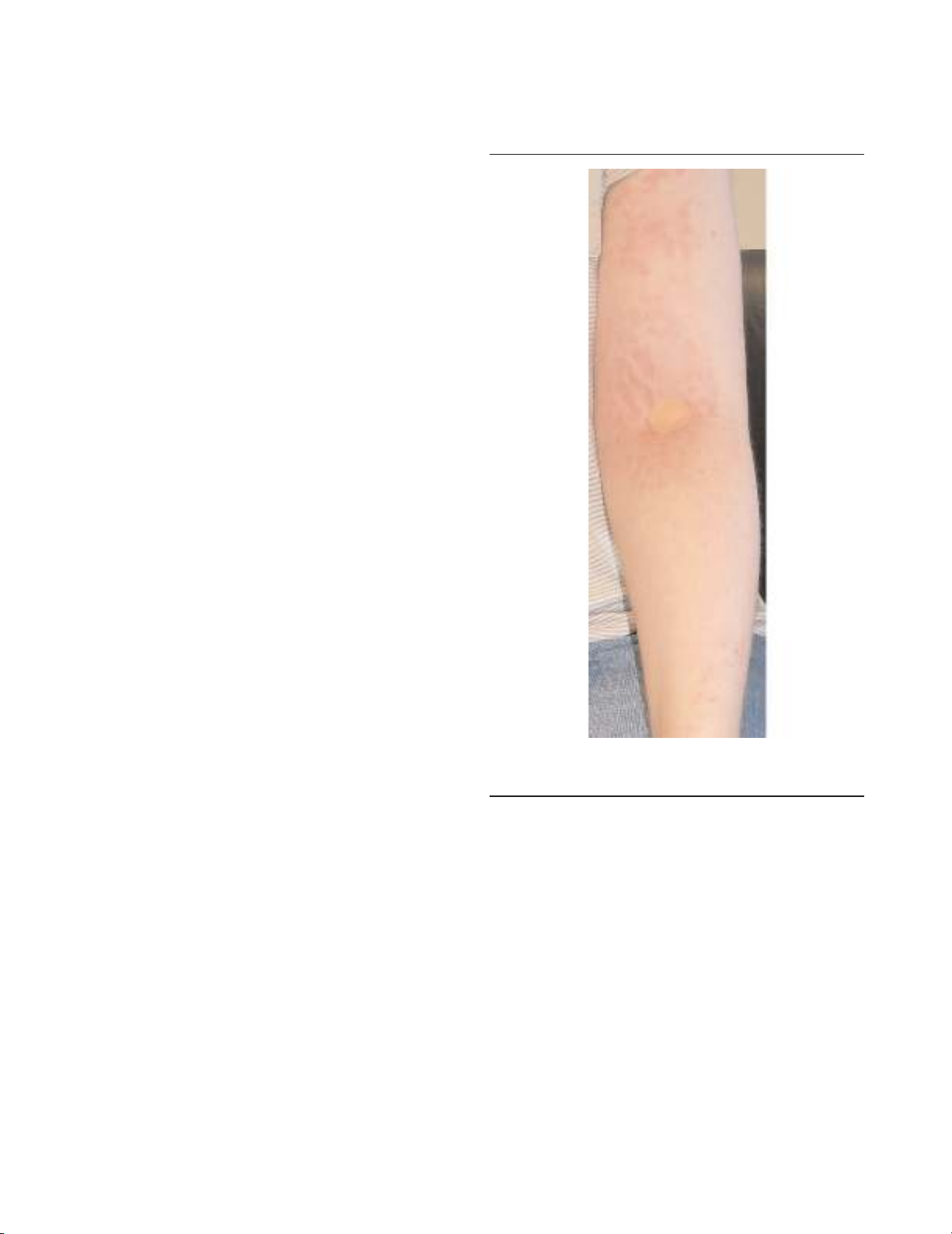
migratory path; abdominal pain occurs in 80%; and skin
manifestations, including erythematous rash (Figure 2),
oedematous plaques (often overlying areas of mylagic pain),
and discrete reticulate or serpiginous lesions, occur in 70%
of patients. Other features include headache, pleuritic pain,
lymphadenopathy, conjunctivitis, and periorbital oedema.
There are also reports of central nervous system manifes-
tations and imaging findings resembling multiple sclerosis
Arthritis Research & Therapy Vol 11 No 1 Lachmann and Hawkins
Page 4 of 10
(page number not for citation purposes)
Figure 1
Erysipelas-like erythema around the ankle, the characteristic painful
rash seen in attacks of familial Mediterranean fever.
[33]. Symptoms are almost universally accompanied by a
marked acute-phase response. During quiescent periods, the
plasma concentration of soluble TNFR1 may be abnormally
low in patients with decreased receptor shedding. Genetic
testing is central to diagnosis.
Treatment
Despite high initial hopes for response to anti-TNF biologics,
treatment of TRAPS often remains disappointing. Acute
attacks do respond to high-dose corticosteroids, and
etanercept (but interestingly not infliximab) is useful in some
patients, although response may gradually decline [34]. A
recent report suggested that IL-1 blockade with anakinra can
be very effective in some patients [35].
The hyper IgD and periodic fever syndrome
Genetics and pathophysiology
Hyper IgD and periodic fever syndrome (HIDS) is an auto-
somal recessive disease caused by mutations in the mevalo-
nate kinase (MVK) gene on the long arm of chromosome 12
[36]. About 60 mutations have been described, spanning the
11-exon gene, the most common of which encode MVK
variants V377I and I268T. MVK is the enzyme following HMG
CoA (or 3-hydroxy-3-methylglutaryl-coenzyme A) reductase in
the pathway involved in cholesterol, farnasyl, and isoprenoid
biosynthesis. Most HIDS-causing MVK mutations are
missense variants that reduce enzyme activity by 90% to
99% [37]. Other mutations resulting in near-complete
absence of enzyme activity cause a much more severe
inflammatory disease known as mevalonic aciduria (MVA),
features of which include stillbirth, congenital malformations,
severe psychomotor retardation, ataxia, myopathy, failure to
thrive, and early death.
It is not yet known how MVK deficiency causes inflammation
or increased IgD production, although reduction in preny-
lation due to failure of flux through the isoprenoid pathway
currently seems more likely to be responsible than accumu-
lation of the enzyme’s substrate [38,39]. The relationship of
the isoprenoid pathway to inflammation is of all the more
interest given the anti-inflammatory properties of statin drugs
that are widely used to inhibit cholesterol synthesis. Whilst
various effects of statins on caspase-1 activation and IL-1
secretion have been postulated, a clinical study of simvastatin
of six patients with HIDS suggested only minor benefit [40];
rather worryingly, two other children with MVA were reported
to develop severe flares of inflammatory disease following
statin treatment [41].
Clinical features
HIDS is extremely rare and is predominantly a Dutch disease,
probably through a founder effect. It was described in The
Netherlands in 1984 and the international registry in
Nijmegen has data on just over 200 patients [42]. The
carriage rate of MVK V337I is 1 in 350 in the Dutch popu-
lation [43], but HIDS has been reported in many other
countries and other ethnic groups, including Arabs and
Southeast Asians. The disease occurs equally in males and
females and usually presents in the first year of life [44].
Attacks are irregular, typically lasting 4 to 7 days, and are
characteristically provoked by vaccination, minor trauma,
surgery, or stress, perhaps triggered by a reduction in MVK
enzyme associated with increased body temperature [45].
Attacks of HIDS typically comprise fever, cervical lymph-
adenopathy, splenomegaly, and abdominal pain with vomiting
and diarrhoea. Headache, arthralgia, large-joint arthritis,
erythematous macules and papules, and aphthous ulcers are
also common. HIDS typically ameliorates in adult life and
older patients may remain well for years.
Diagnosis of HIDS is supported by a high serum IgD
concentration, although this is not specific and is not always
present [46]. More accessibly, serum IgA concentration is
Available online http://arthritis-research.com/content/11/1/212
Page 5 of 10
(page number not for citation purposes)
Figure 2
Erythematous rash complicating an acute attack in tumour necrosis
factor receptor-associated periodic syndrome (TRAPS).


