
BioMed Central
Page 1 of 10
(page number not for citation purposes)
Journal of Inflammation
Open Access
Research
The glucocorticoid RU24858 does not distinguish between
transrepression and transactivation in primary human eosinophils
Mirkka Janka-Junttila1, Eeva Moilanen1, Hannele Hasala1, Xianzhi Zhang1,3,
Ian Adcock2 and Hannu Kankaanranta*1,4
Address: 1The Immunopharmacology Research Group, Medical School, FIN-33014 University of Tampere and Research Unit, Tampere University
Hospital, Tampere, Finland, 2Department of Thoracic Medicine, National Heart and Lung Institute, Imperial College, London, UK, 3The Center
for Infection and Immunity, Institute of Biophysics, Chinese Academy of Sciences, Beijing, China and 4Department of Pulmonary Medicine
Tampere University Hospital, Tampere, Finland
Email: Mirkka Janka-Junttila - mirkka.janka@uta.fi; Eeva Moilanen - eeva.moilanen@uta.fi; Hannele Hasala - hannele.hasala@uta.fi;
Xianzhi Zhang - Xianzhi.Zhang@uta.fi; Ian Adcock - ian.adcock@imperial.ac.uk; Hannu Kankaanranta* - hannu.kankaanranta@uta.fi
* Corresponding author
Abstract
Background: Glucocorticoids are used to treat chronic inflammatory diseases such as asthma. Induction of
eosinophil apoptosis is considered to be one of the main mechanisms behind the anti-asthmatic effect of
glucocorticoids. Glucocorticoid binding to its receptor (GR) can have a dual effect on gene transcription.
Activated GR can activate transcription (transactivation), or by interacting with other transcription factors such
as NF-κB suppress transcription (transrepression). RU24858 has been reported to transrepress but to have little
or no transactivation capability in other cell types. The dissociated properties of RU24858 have not been
previously studied in non-malignant human cells. As the eosinophils have a very short lifetime and many of the
modern molecular biological methods cannot be used, a "dissociated steroid" would be a valuable tool to evaluate
the mechanism of action of glucocorticoids in human eosinophils. The aim of this study was to elucidate the ability
of RU24858 to activate and repress gene expression in human eosinophils in order to see whether it is a
dissociated steroid in human eosinophils.
Methods: Human peripheral blood eosinophils were isolated under sterile conditions and cultured in the
presence and/or absence RU24858. For comparison, dexamethasone and mometasone were used. We measured
chemokine receptor-4 (CXCR4) and Annexin 1 expression by flow cytometry and cytokine production by ELISA.
Apoptosis was measured by DNA fragmentation and confirmed by morphological analysis.
Results: RU24858 (1 µM) increased CXCR4 and Annexin 1 expression on eosinophils to a similar extent as
mometasone (1 µM) and dexamethasone (1 µM). Like dexamethasone and mometasone, RU24858 did suppress
IL-8 and MCP-1 production in eosinophils. RU24858 also increased spontaneous eosinophil apoptosis to a similar
degree as dexamethasone and mometasone, but unlike dexamethasone and mometasone it did not reverse IL-5-
or GM-CSF-induced eosinophil survival.
Conclusion: Our results suggest that in human eosinophils RU24858 acts as transactivator and transrepressor
like classical glucocorticoids. Thus, RU24858 seems not to be a "dissociated steroid" in primary human eosinophils
in contrast to that reported in animal cells. In addition, functionally RU24858 seems to be a less potent
glucocorticoid as it did not reverse IL-5- and GM-CSF-afforded eosinophil survival similarly to dexamethasone
and mometasone.
Published: 12 July 2006
Journal of Inflammation 2006, 3:10 doi:10.1186/1476-9255-3-10
Received: 04 January 2006
Accepted: 12 July 2006
This article is available from: http://www.journal-inflammation.com/content/3/1/10
© 2006 Janka-Junttila et al; licensee BioMed Central Ltd.
This is an Open Access article distributed under the terms of the Creative Commons Attribution License (http://creativecommons.org/licenses/by/2.0),
which permits unrestricted use, distribution, and reproduction in any medium, provided the original work is properly cited.

Journal of Inflammation 2006, 3:10 http://www.journal-inflammation.com/content/3/1/10
Page 2 of 10
(page number not for citation purposes)
Background
Eosinophils are thought to play a critical role in allergic
diseases, such as allergic rhinitis, asthma, and atopic der-
matitis.[1] In patients with asthma, activation of eosi-
nophils is thought to cause epithelial tissue injury, and
increased bronchial responsiveness.[2] Apoptosis or pro-
grammed cell death is an important feature in the resolu-
tion of pulmonary inflammation.[3,4] Unlike necrosis
which is characterized by loss of cell membrane integrity
and the uncontrolled release of harmful cellular contents,
apoptosis is characterized by formation of apoptic bodies,
which are then phagocytosed intact so there will be no
leakage of intracellular contents or activation of the
inflammatory response.[3,5] Eosinophil apoptosis is
inhibited by cytokines such as interleukin (IL)-3, IL-5 and
granulocyte-macrophage colony-stimulating factor (GM-
CSF) in vitro and in vivo.[1] In addition, we and others
have previously shown that eosinophil apoptosis is
delayed in patients with asthma or inhalant allergy.[6,7]
Glucocorticoids are potent anti-inflammatory agents for
the treatment of allergic diseases such as asthma, allergic
rhinitis, atopic dermatitis and various syndromes associ-
ated with hypereosinophilia. Enhancement of eosinophil
apoptosis and/or reversal of cytokine-induced eosinophil
survival have been reported to be one important mecha-
nism by which glucocorticoids reduce eosinophil num-
bers. [8-18] The basic mechanism of glucocorticoid
actions is that they penetrate into the cell and bind to glu-
cocorticoid receptor molecules in the cytoplasm.[19,20]
The glucocorticoid- glucocorticoid receptor (GR) complex
acts as a transcription factor, binding to specific DNA sites
in the nucleus. Within the nucleus, GR may induce gene
transcription (transactivation) by binding to specific DNA
sequences known as glucocorticoid response elements
(GREs) in the promoter-enhancer regions of steroid
responsive genes. The glucocorticoid-glucocorticoid
receptor complex may also directly interact with other
transcription factors such as nuclear factor-κB (NF-κB)
and activator protein (AP)-1 resulting in a transcriptional
down-regulation (transrepression), which is considered
currently to be a major mechanism of the anti-inflamma-
tory effect of steroids.[20,21] Despite many studies on the
ability of glucocorticoids to repress and/or activate genes
in other cell types [22-25] there are no published data on
whether transactivation and/or transrepression events
play a role in the regulation of apoptosis in human eosi-
nophils.
Based on the hypothesis that the predominant anti-
inflammatory effects of glucocorticoids derive from inhi-
bition of transcription (transrepression), whereas the met-
abolic effects, like disrupting the regulation of calcium
and glucose metabolism, derive from positive transcrip-
tional effects (transactivation), experimental glucocorti-
coids, which only act as transrepressors but not as
transactivators, have been developed. RU24858 is such a
novel glucocorticoid.[22] However the transactivation
profile of RU24858 has been controversial. Vayssiere et
al.[22] and Vanden Berghe et al.[23] demonstrated
RU24858 to be almost as effective as dexamethasone in
inducing transrepression but show little or no transactiva-
tion ability in human and murine cell lines. In contrast,
others have reported no dissociation between anti-inflam-
matory activity and side effects in vivo.[25] Differences
between human and rodent GR or in GR-associated fac-
tors has been implicated to be critical for these divergent
results.[24] However, the ability of RU24858 to dissociate
between transactivation and transrepression in non-trans-
formed primary human cells has not been described.
Whether dissociated steroids such as RU24858 have a bet-
ter safety profile in the treatment of chronic inflammatory
diseases such as asthma depends on their ability to disso-
ciate between transactivation and transrepression in non-
malignant human cells.
Primary human eosinophils are terminally differentiated,
non-dividing cells that can only be cultured for very short
periods, making these cells unsuitable for many studies
using molecular biology. Thus, a dissociative glucocorti-
coid would be a very valuable pharmacological tool to
evaluate the mechanism of action glucocorticoids in pri-
mary cells such as human eosinophils. The aim of our
study was to test whether RU24858 discriminates between
transactivation and transrepression in human eosinophils
and the functional consequences of this profile by assess-
ing its effects on eosinophil apoptosis. We measured the
induction of surface expression of Annexin I and CXCR4
as a measure of GR transactivation[26,27] ability and the
inhibition of IL-8 and MCP-1 production to define the
transrepression potential. We found that in eosinophils
RU24858 possessed transrepression capability but also
clear transactivation effects. Surprisingly, although
RU24858 did result enhanced spontaneous eosinophil
apoptosis it did not reverse cytokine-induced apoptosis
like other glucocorticoids do.
Methods
Eosinophil isolation
Eosinophils were isolated under sterile conditions as pre-
viously reported.[6,17,18,28] Before donation of blood,
all subjects gave informed consent to a study protocol
approved by the ethical committee of Tampere University
Hospital. Eosinophils were obtained from donors with
eosinophil counts ranging from upper normal to slightly
elevated. We excluded patients with hypereosinophilic
syndrome. Venous blood (50–100 ml) was collected into
10–20 ml of acid citrate dextrose anticoagulant and
hydroxyethyl starch solution. White blood cells were
obtained after removing supernatant and were overlaid
Journal of Inflammation 2006, 3:10 http://www.journal-inflammation.com/content/3/1/10
Page 3 of 10
(page number not for citation purposes)
onto Ficoll and centrifuged at 700 g for 30 min at 20°C.
Mononuclear cell layer was removed and the remaining
pellet containing granulocytes and red blood cells was
washed in HBSS (Hank's Balanced Salt Solution without
Phenol Red). Red blood cells were lysed by hypotonic
lysis.
Eosinophils were purified using immunomagnetic anti-
CD16 antibody conjugated beads. Following separation,
granulocytes were washed, counted and resuspended in
300 ml of RPMI 1640 (2% fetal calf serum and 5 mM
EDTA). Cells mixed with beads were incubated at 4°C for
at least 25 min before loading onto a separation column
positioned within a magnetic field and washed with 40 ml
of RPMI 1640. The eluted eosinophils were washed and
counted using microscopic examination and diluting 10
µl cell suspension in 90 µl Kimura stain (consisting of 11
ml of 0.05% (wt/wt) toluidine blue, 0.8 ml of 0.03% light
green SF yellowish, 0.5 ml of saturated saponin, and 5 ml
of 0.07 M phosphate buffer, pH 6.4) and the purity of
eosinophil population was >99%. The eosinophils were
washed and resuspended at 1 × 10⁶ cells/ml and cultured
(37°C, 5 % CO2) in RPMI 1640 (Dutch modification,
10% fetal calf serum and antibiotics). Granulocytes were
incubated in the presence and absence of RU24858,
mometasone and dexamethasone. All the steroids were
diluted in DMSO. The final concentration of DMSO in the
cells was 0.1%. Similar concentration of DMSO was used
in control experiments.
Flow cytometry
Eosinophils were incubated for 24 h and the expression of
CXCR4 was determined by using a PE-conjugated mAb
against CXCR4 (20 µl/106 cells). We performed flow-cyto-
metric analysis according to the instructions of the manu-
facturer and for comparison PE-conjugated isotype
control was used. The expression of Annexin I was deter-
mined by using a mAb against Annexin I (20 µl/106 cells)
and for comparison an isotype standard was used. As a
secondary antibody we used PE-conjugated anti-mouse
IgG1 monoclonal antibody according to that described by
Liu et al.[29]
Unless otherwise stated the percentage of apoptotic cells
was measured using a relative DNA fragmentation assay
in propidium iodide stained cells by flow cytometry as
previously described.[6,17,18,28] Eosinophils were incu-
bated for 40 h. The cells showing decreased relative DNA
content were considered as apoptotic.
Morphological analysis
Cells were centrifuged onto cytospin slides. After fixation
in methanol slides were stained with May-Grünwald-
Giemsa. Cells showing typical features of apoptosis such
as condensation of chromatin, nuclear coalescence and
shrinkage of the cell were considered as apoptotic.[28,30]
Cells were counted blind. We always prepared double
identical samples and from each sample 200 cells were
assessed and finally the average was calculated.
Cytokine assays
Cytokine production was induced by 1 µM ionomy-
cin.[31] Cells were incubated for 18 h and supernatants
were collected and stored at -20°C and cytokines were
measured by ELISA. The lower limits of detection were 3.9
pg/ml for MCP-1 and 4.1 pg/ml for IL-8.
Materials
RU24858 was obtained from Aventis Pharma, Romain-
ville Cedex, France and mometasone furoate was obtained
from Schering-Plough, Kenilworth, USA. Dexamethasone
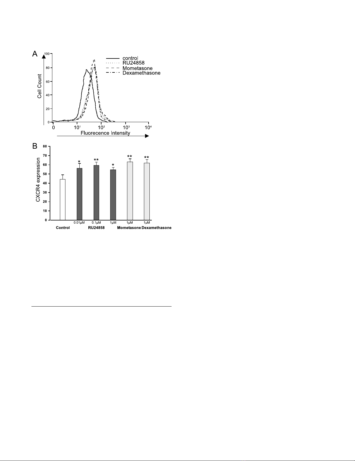
The effects of RU24858, mometasone and dexamethasone on eosinophil CXCR4 surface expressionFigure 1
The effects of RU24858, mometasone and dexameth-
asone on eosinophil CXCR4 surface expression. Cells
were stained and analyzed by using flow cytometry. A typical
experiment showing an increase in CXCR4 expression fol-
lowing treatment of cells with mometasone, dexamethasone
and RU24858 is indicated in (A). (B) Summary of results
expressed as mean fluorescence intensity. Values are the
mean ± S.E.M., n = 6. * Indicates P < 0.05, ** P < 0.01 as com-
pared with the respective control in the absence of glucocor-
ticoids.
Journal of Inflammation 2006, 3:10 http://www.journal-inflammation.com/content/3/1/10
Page 4 of 10
(page number not for citation purposes)
and propidium iodide were purchased from Sigma Chem-
ical Co. (St. Louis, MO). Other reagents were obtained as
follows: antibiotics, fetal calf serum, RPMI 1640 (Gibco
BRL, Paisley, Scotland, UK), anti-CD16 microbeads and
magnetic cell separation system (Miltenyi Biotec Ltd., Sur-
rey, UK), human recombinant IL-5, GM-CSF and DuoSet
ELISA Development System for IL-8 and MCP-1 (R&D sys-
tem Europe, Abingdon, UK), May-Grünwald (Merck,
Darmstadt, Germany), and Giemsa (J.T. Baker, Deventer,
Holland). PE-conjugated CXCR4 mAb (12G5), R-PE-con-
jugated IgG2a isotype control, Annexin I mAb, IgG1 isotype
standard and R-PE-conjugated anti-mouse IgG1 mono-
clonal antibody were all purchased from BD Pharmingen
(Temse, Belgium).
Statistics
Data are expressed as mean ± SEM. Differences were ana-
lyzed by analysis of variance supported by Student-New-
man-Keuls test and were considered significant if P < 0.05.
Results
The effect of RU24858 on CXCR4 expression on the
surface of eosinophils
Dexamethasone has previously been shown to induce
CXCR4 expression in human eosinophils.[27] As
expected, mometasone (1 µM) and dexamethasone (1
µM) induced CXCR4 expression in human eosinophils
(Figure 1a &1b). To our surprise RU24858 (0.01–1 µM)
also induced CXCR4 expression in a manner similar to
classical glucocorticoids (Figure 1a &1b). To exclude the
possibility that RU24858 affects the fluorescence proper-
ties of the PE-conjugated antibody, its effects were studied
on cells labelled with PE-conjugated isotype control anti-
body. RU24858 (0.01 µM–1 µM), mometasone (1 µM)
and dexamethasone (1 µM) had no effect on fluorescence
of PE-conjugated isotype control (n = 6, data not shown).
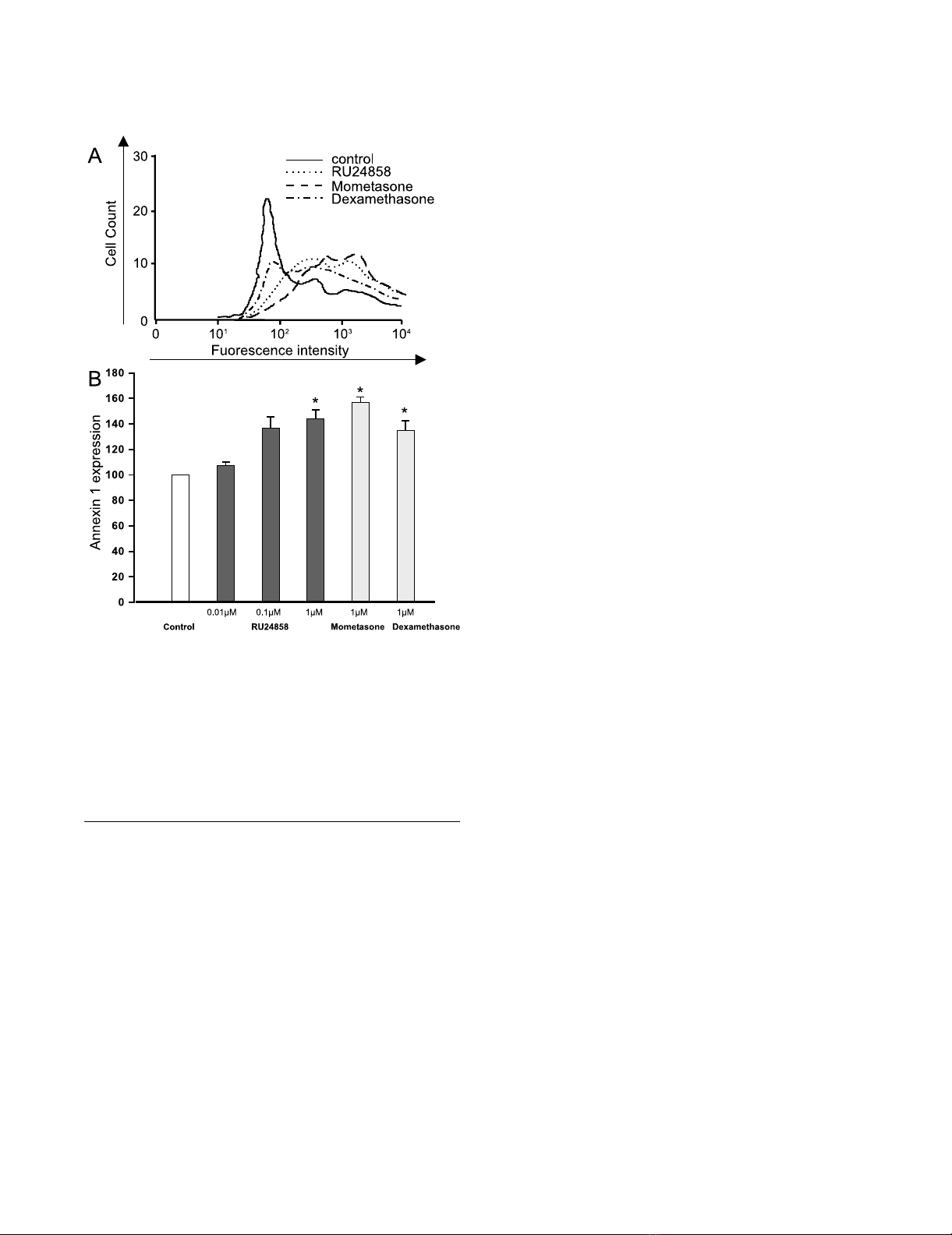
The effect of RU24858 on Annexin 1 expression on the
surface of eosinophils
It has been recently demonstrated that glucocorticoids
induce surface expression of Annexin 1 on human eosi-
nophils.[29] To further analyse the transactivation ability
of RU24858 we investigated whether it has a similar effect
on Annexin 1 expression as mometasone and dexametha-
sone. RU24858 significantly increased Annexin 1 expres-
sion to a similar extent to that seen with mometasone and
dexamethasone (both at 1 µM) (Figure 2a &2b). In addi-
tion, mometasone (1 µM), dexamethasone (1 µM) and
RU24858 (0.01 µM–1 µM) had no effect on the fluores-
cence of isotype and/or secondary antibody controls (n =
6, data not shown).
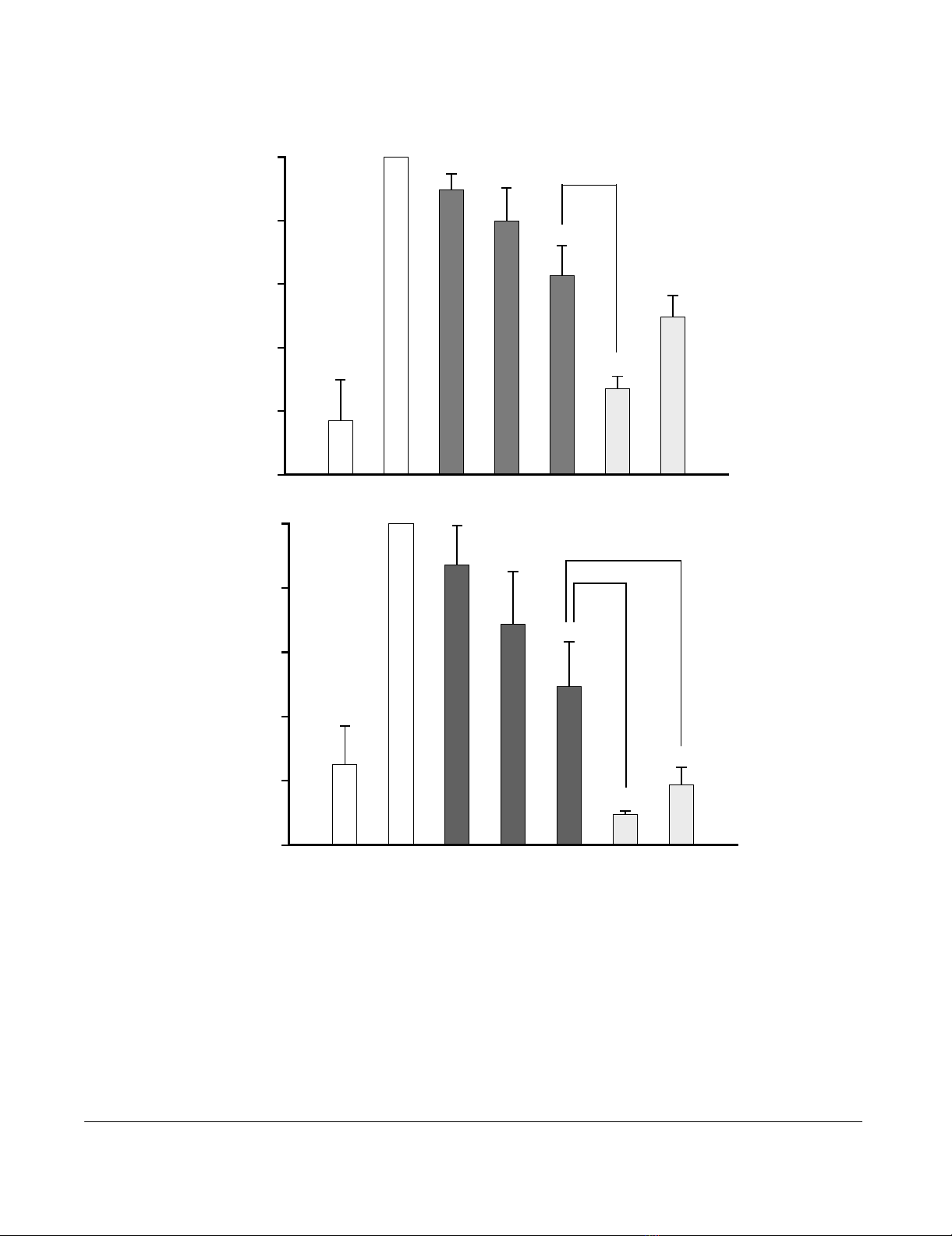
The Effect of RU24858 on cytokine production
Glucocorticoids have previously been reported to inhibit
IL-8 and MCP-1 production in eosinophils.[31] There-
fore, to study the transrepression capability of RU24858
we measured IL-8 and MCP-1 production from superna-
tants collected from ionomycin-treated eosinophils.
Spontaneous IL-8 and MCP-1 production was low and
ionomycin (1 µM) increased MCP-1 and IL-8 production
6–10-fold (figure 3a &3b). Mometasone (1 µM) and dex-
amethasone (1 µM) inhibited IL-8 and MCP-1 generation
as expected (figure 3a &3b) as also RU24858 (1 µM) did.
The effect of RU24858 on MCP-1 production was signifi-
cantly smaller than that of dexamethasone and mometa-
sone. Mometasone was also more potent than RU24858
in inhibiting IL-8 production, whereas the difference
between dexamethasone and RU24858 did not reach sta-
tistical significance (Figure 3a &3b). Taken together, the
present data suggest that RU24858 is a non selective com-
The effects of RU24858, mometasone and dexamethasone on eosinophil Annexin 1 surface expressionFigure 2
The effects of RU24858, mometasone and dexameth-
asone on eosinophil Annexin 1 surface expression.
Cells were stained and analyzed by using flow cytometry. A
typical experiment showing an increase in Annexin 1 expres-
sion following treatment of cells with mometasone, dexame-
thasone and RU24858 is indicated in (A). (B) Summary of
results expressed as mean fluorescence intensity. Values are
the mean ± S.E.M., n = 3. * Indicates P < 0.05 as compared
with the respective control in the absence of glucocorticoids.
Journal of Inflammation 2006, 3:10 http://www.journal-inflammation.com/content/3/1/10
Page 5 of 10
(page number not for citation purposes)
The effects of RU24858, mometasone and dexamethasone on eosinophil cytokine productionFigure 3
The effects of RU24858, mometasone and dexamethasone on eosinophil cytokine production. (A) IL-8 and (B)
MCP-1. Ionomycin was used to stimulate the IL-8 and MCP-1 production in cells. IL-8 and MCP-1 were analyzed by ELISA
based methods. Values are the mean ± S.E.M., n = 6. Results are expressed as % of stimulated eosinophils. * Indicates P < 0.05,
** P < 0.01 and *** P < 0.001 as compared with the respective control in the absence of glucocorticoids. # Indicates P < 0.05,
## P < 0.01 and ### P < 0.001 as compared with RU24858 (1 µM).
IL-8 production
% of stimulated eosinophils
% of stimulated eosinophils
0.01 0.1
1
1
1
MCP-1 production
***
**
**
*
***
***
***
###
##
#
Ionomycin
RU24858
Mom (µM)
Dex (µM
)
(µM)
-
-
--
--
--
---
-
--
--
-
++++++
0
20
40
60
80
100
0
20
40
60
80
100


