
Open Access
Available online http://arthritis-research.com/content/11/2/R39
Page 1 of 11
(page number not for citation purposes)
Vol 11 No 2
Research article
Serum levels of soluble receptor for advanced glycation end
products and of S100 proteins are associated with inflammatory,
autoantibody, and classical risk markers of joint and vascular
damage in rheumatoid arthritis
Yueh-Sheng Chen1, Weixing Yan2, Carolyn L Geczy2, Matthew A Brown1 and Ranjeny Thomas1
1Diamantina Institute, University of Queensland, Princess Alexandra Hospital, Ipswich Road, Woolloongabba, 4102, Australia
2Centre for Infection and Inflammation Research, School of Medical Sciences, University of New South Wales, Sydney, 2052, Australia
Corresponding author: Ranjeny Thomas, r.thomas1@uq.edu.au
Received: 16 Dec 2008 Revisions requested: 11 Feb 2009 Revisions received: 25 Feb 2009 Accepted: 11 Mar 2009 Published: 11 Mar 2009
Arthritis Research & Therapy 2009, 11:R39 (doi:10.1186/ar2645)
This article is online at: http://arthritis-research.com/content/11/2/R39
© 2009 Chen et al.; licensee BioMed Central Ltd.
This is an open access article distributed under the terms of the Creative Commons Attribution License (http://creativecommons.org/licenses/by/2.0),
which permits unrestricted use, distribution, and reproduction in any medium, provided the original work is properly cited.
Abstract
Introduction The receptor for advanced glycation end products
(RAGE) is a member of the immunoglobulin superfamily of cell
surface receptor molecules. High concentrations of three of its
putative proinflammatory ligands, S100A8/A9 complex
(calprotectin), S100A8, and S100A12, are found in rheumatoid
arthritis (RA) serum and synovial fluid. In contrast, soluble RAGE
(sRAGE) may prevent proinflammatory effects by acting as a
decoy. This study evaluated the serum levels of S100A9,
S100A8, S100A12 and sRAGE in RA patients, to determine
their relationship to inflammation and joint and vascular damage.
Methods Serum sRAGE, S100A9, S100A8 and S100A12
levels from 138 patients with established RA and 44 healthy
controls were measured by ELISA and compared by unpaired t
test. In RA patients, associations with disease activity and
severity variables were analyzed by simple and multiple linear
regressions.
Results Serum S100A9, S100A8 and S100A12 levels were
correlated in RA patients. S100A9 levels were associated with
body mass index (BMI), and with serum levels of S100A8 and
S100A12. S100A8 levels were associated with serum levels of
S100A9, presence of anti-citrullinated peptide antibodies
(ACPA), and rheumatoid factor (RF). S100A12 levels were
associated with presence of ACPA, history of diabetes, and
serum S100A9 levels. sRAGE levels were negatively associated
with serum levels of C-reactive protein (CRP) and high-density
lipoprotein (HDL), history of vasculitis, and the presence of the
RAGE 82Ser polymorphism.
Conclusions sRAGE and S100 proteins were associated not
just with RA inflammation and autoantibody production, but also
with classical vascular risk factors for end-organ damage.
Consistent with its role as a RAGE decoy molecule, sRAGE had
the opposite effects to S100 proteins in that S100 proteins
were associated with autoantibodies and vascular risk, whereas
sRAGE was associated with protection against joint and
vascular damage. These data suggest that RAGE activity
influences co-development of joint and vascular disease in
rheumatoid arthritis patients.
Introduction
Rheumatoid arthritis (RA) is a chronic inflammatory disease
that leads to bone and cartilage destruction and extra-articular
complications, including atherosclerotic vascular disease and
premature mortality [1]. The receptor for advanced glycation
end products (RAGE) has been implicated in the pathogene-
sis of RA through its ability to amplify inflammatory pathways
[2,3]. A member of the immunoglobulin superfamily of cell sur-
face receptors, RAGE binds advanced glycation end products
(AGEs), which are non-enzymatically glycated or oxidized
ACPA: anti-citrullinated peptide antibodies; ACR: American College of Rheumatology; AGE: advanced glycation end product; BMI: body-mass index;
CrCl: creatinine clearance; CRP: C-reactive protein; CT: computed tomography; CV: cardiovascular; ECG: electrocardiogram; ESR: erythrocyte sed-
imentation rate; HDL: high-density lipoprotein; HMGB1: high mobility group box chromosomal protein; HR: hazard ratio; LDL: low density lipoprotein;
MI: myocardial infarction; PCR: polymerase chain reaction; RA: rheumatoid arthritis; RAGE: receptor for advanced glycation end products; RF: rheu-
matoid factor; SCr: serum creatinine; TG: triglyceride; TIA: transient ischemic attack; TNF: tumor necrosis factor; VLDL: very-low-density lipoprotein.

Arthritis Research & Therapy Vol 11 No 2 Chen et al.
Page 2 of 11
(page number not for citation purposes)
proteins, lipids and nucleic acids formed under conditions of
oxidative stress and hyperglycemia (reviewed in [4]). In addi-
tion to these, RAGE binds some proinflammatory ligands,
including members of the S100/calgranulin family, and high
mobility group box chromosomal protein 1 (HMGB-1), which
is implicated in cell signaling by synergizing with DNA CpG
motifs [5,6]. Several RAGE ligands are characteristically over-
expressed in RA and psoriatic arthritis, compared to healthy
controls [7-9]. S100A8/A9 (calprotectin) and S100A12 (cal-
granulin C, EN-RAGE) levels are significantly elevated in
serum and synovial fluid from RA patients compared to healthy
normal donors [3,10]. S100A8/A9 levels are also higher in
supernatants of cultured RA synoviocytes than of osteoarthri-
tis synoviocytes [11].
Soluble C-truncated RAGE (sRAGE) lacks the transmem-
brane and cytosolic domains of the full-length receptor and
can prevent proinflammatory effects of RAGE signaling by act-
ing as a decoy [12-14]. For example, in a collagen-induced
arthritis (CIA) murine model, treatment with murine sRAGE
significantly reduced joint inflammation and destruction [15].
Serum or plasma levels of sRAGE from patients with RA,
hypertension or metabolic syndrome were lower than those in
healthy subjects [16-18], suggesting that sRAGE levels may
identify those RA patients exposed to high levels of RAGE lig-
ands. A gain-of-function Gly82Ser polymorphism in the RAGE
gene (RAGE 82Ser) occurs more frequently in RA patients
than in healthy controls [19]. Monocytes expressing the RAGE
82Ser allele activated a stronger inflammatory response to
S100A12 in vitro [15]. Although this might be predicted to
contribute to enhanced proinflammatory mechanisms in RA,
we found no evidence that patients with the RAGE 82S allele
had higher levels of inflammation, or a greater likelihood of
complicating cardiovascular (CV) events [19].
Most S100 proteins have a mass between 9 and 14 kDa, and
are characterized by two calcium binding sites of the EF-hand
type (helix-loop-helix) [20]. S100A8 and S100A9, generally
functioning as the S100A8/A9 heterocomplex, and S100A12
are implicated in non-infectious chronic inflammatory diseases
such as RA, psoriasis and inflammatory bowel disease [21-
25]. Longitudinal and cross-sectional studies suggest a rela-
tionship between S100A12 and RA disease activity [26-28].
The S100A12 gene is rapidly upregulated in human monocy-
toid cells and blood monocytes by tumor necrosis factor (TNF)
and lipopolysaccharide (LPS), suggesting its production in
response to proinflammatory signals in RA [10,25]. S100A12
is a potent monocyte chemoattractant and activates mast
cells, which are important effector cells in RA and atheroscle-
rosis [25,29,30]. S100A12 is also proposed to promote proin-
flammatory activities by binding and activating RAGE [31].
However, these studies were established using a murine
model, and since it was later shown that mice have no
S100A12 in their genome [20], alternate receptors are impli-
cated [25]. In addition, recombinant S100 ligands may contain
contaminating endotoxin, and their effects may not always be
fully RAGE dependent [32].
S100A8 and S100A9 regulate leukocyte migration and adhe-
sion [33]. The S100A8/A9 complex has antimicrobial effects,
transports arachidonic acid to endothelial cells, and activates
expression of endothelial cell adhesion molecules [11,34,35].
Although the receptor for S100A8/A9 complex is still
unknown, RAGE has been implicated in some circumstances
[36]. Murine S100A8 stimulates proatherogenic activity, such
as uptake of low-density lipoprotein (LDL), in macrophages.
S100A8 is a key target of oxidation by peroxide, hypochlorite
and nitric oxide [37,38]. Furthermore, S100A9 and S100A12
are implicated in vascular damage, whereas sRAGE is associ-
ated with vascular protection in atherosclerosis [30,39-41].
The relationship between S100 protein levels and vascular
disease or risk factors in RA patients has not been examined
to date. We measured serum levels of S100A8, S100A9 het-
erocomplexes, S100A12 and sRAGE in a previously charac-
terized cohort of established RA patients to identify their
possible relationship to joint and vascular damage and risk fac-
tors in RA patients [19]. We report associations of each pro-
tein with both joint and vascular disease and their risk factors.
Materials and methods
Subjects
The cohort of RA patients met the American College of Rheu-
matology (ACR) 1987 revised criteria for the classification of
RA, and has been previously described [42]. These patients
presented for a scheduled appointment over a 5-month period
(July to November 2003) at our tertiary hospital rheumatology
clinic, as described previously [19]. Patients completed a
questionnaire detailing CV history, risk factors, treatment, and
details of RA. Each patient was clinically evaluated, with chart
review to confirm history, at least once. The study protocol was
approved by the Princess Alexandra Hospital Research Ethics
Committee. Healthy controls (n = 44) without RA or CV dis-
ease were recruited by advertisement. All patients and con-
trols signed informed consent to participate. No prospective
follow-up was carried out in this study.
Measurement of S100 proteins
The serum levels of S100A8, S100A9 and S100A12 levels
were measured using in-house affinity-purified rabbit polyclo-
nal sandwich ELISAs exactly as described for S100A12 [25].
Antibodies to S100A8 did not cross-react with S100A9 (and
did not recognize S100A8/A9 complexes) or S100A12, anti-
S100A9 detected free S100A9 and S100A9 as an S100A8/
A9 complex; anti-S100A12 was immunoadsorbed with
S100A8 and S100A9 [25] and did not cross-react with these
when tested by ELISA or immunoblotting. Standard curves
were constructed with the relevant recombinant S100
proteins.

Available online http://arthritis-research.com/content/11/2/R39
Page 3 of 11
(page number not for citation purposes)
Measurement of sRAGE
sRAGE levels in sera were determined by RAGE Immu-
noassay (R&D Systems, Minneapolis, MN, USA) in an ELISA
format, with wells coated with murine anti-human RAGE mAb
in which serum samples (50 l/well, normally 1:2 v/v dilution)
were incubated. A polyclonal capture antibody against the
extracellular domain of RAGE was used for detection. The min-
imum detectable sRAGE concentration is 4.12 pg/ml accord-
ing to the manufacturer, and the interassay coefficient of
variation is < 8% [41].
Ascertainment of CV events and risk factors, and
features of RA
To ascertain CV events, patients were asked for a history,
dates and treatments of myocardial infarction, angina, stroke,
transient ischemic attack or peripheral vascular disease, and
these events were verified by medical record review. Although
a number of patients had events prior to the diagnosis of RA,
only those CV events that occurred after RA diagnosis were
included in the current analysis. Patients with multiple events
had only one event counted per person. Myocardial infarction
was identified if subjects developed either of; (1) typical rise
and fall of biochemical markers (troponin or creatine kinase-
MB (CK-MB)) consistent with myocardial necrosis with at
least one of the following (a) ischemic symptoms, (b) develop-
ment of pathological Q waves on the electrocardiogram
(ECG), (c) ECG changes indicative of ischemia (ST segment
elevation or depression); (2) either new pathological Q waves
on serial ECGs or pathological changes of healed or healing
infarction [43]. Stroke or transient ischemic attack were iden-
tified if subjects had been admitted to the hospital with CT evi-
dence of ischemic occlusion or with carotid endarterectomy,
or the subject presented with stroke/transient ischemic attack
(TIA) symptoms with significant plaque on the carotid ultra-
sound and neurological sequelae, with exclusion of subarach-
noid hemorrhage and space occupying lesions. Peripheral
vascular disease was confirmed if Doppler ultrasonography
showed significant large vessel disease.
Cigarette smoking was assessed by questionnaire, which
included details about past and present smoking habits,
number of cigarettes smoked per day and smoking duration.
History of diabetes mellitus was identified if subjects had been
diagnosed by a physician, were taking anti-diabetic medica-
tions, or had an elevated fasting glucose at the time of the
assessment. Family history of CV disease or cerebrovascular
attack before age of 65 in first-degree relatives was deter-
mined by questionnaire. History was not included if a stroke
was deemed hemorrhagic. Body mass index (BMI) was calcu-
lated as weight in kilograms divided by the square of the height
in meters. Blood pressure was measured at the time of evalu-
ation. History of hypercholesterolemia and hypertension were
identified if the diagnoses were recorded in medical records
by a physician, if patients were taking lipid-lowering or antihy-
pertensive drugs, or if elevated blood pressure or fasting cho-
lesterol levels were found at the time of the evaluation. The
percentage risk of coronary heart disease over the next 10
years was estimated using the 'CVD Risk Calculator' based on
the Framingham Study [44] for patients between 30 and 74
years of age and without a history of coronary heart disease.
Metabolic syndrome (modified American Heart Association
(AHA) standard [45]) was identified by the presence of three
or more of these components: (1) BMI > 30; (2) fasting blood
triglycerides 150 mg/dl; (3) blood high-density lipoprotein
(HDL) cholesterol (men: < 40 mg/dl (1.03 mmol/l), women: <
50 mg/dl (1.3mmol/l)); (4) blood pressure 130/85 mmHg;
and (5) fasting glucose 100 mg/dl.
Laboratory data collected at the time of clinical evaluation
included fasting total cholesterol, LDL, HDL, very low-density
lipoprotein (VLDL), triglycerides, LDL/HDL cholesterol ratio,
glucose, creatinine, C-reactive protein (CRP), erythrocyte sed-
imentation rate (ESR), anti-citrullinated peptide antibodies
(ACPA) and rheumatoid factor (RF). A 12-lead ECG carried
out within the previous 12 months was scored for evidence of
Q waves to ascertain possible silent coronary disease. Creat-
inine clearance (CrCl) was estimated for each patient on the
basis of serum creatinine (SCr), age (years), and ideal body
weight (kg) using the Cockcroft and Gault method as follows:
CrCl (ml/min) = [(140 - age)(ideal wt)]/833 × SCr (mmol/l) ×
0.85 for females [46]. Hand radiographs carried out at the
time of evaluation were scored for erosions and joint space
narrowing using the modified Sharp score [47].
Genotyping
High resolution human leukocyte antigen (HLA)-DRB1 geno-
typing was carried out on buffy coat DNA using PCR and
sequence-specific oligonucleotide probes. PCR-based
restriction fragment length polymorphism (RFLP) analysis was
used to delineate the RAGE Gly82Ser and protein tyrosine
phosphatase, non-receptor type 22 (PTPN22) Cys1858Thr
polymorphisms as described [15,48]. Shared epitope was
considered positive when at least one DRB1 allele was one of
the RA susceptibility alleles, as previously described [49].
Statistical analysis
Data were analyzed using STATA 9.1 (StataCorp, College
Station, TX, USA). The variables included age, sex, BMI, cur-
rent and previous smoking status, RF, ACPA, history of CV
events, fasting glucose, homocysteine, cholesterol and triglyc-
eride, ESR, CRP, HDL, LDL, creatinine, CrCl, systolic and
diastolic blood pressure, history of diabetes or elevated blood
sugar level, history of hyperlipidemia or elevated cholesterol,
HLA-DRB1 genotype, Sharp erosion score, Sharp joint space
narrowing score, RAGE Gly82Ser polymorphism, history of
hypertension or elevated blood pressure, metabolic syndrome
(modified AHA standard), serum S100A9, S100A8,
S100A12 and sRAGE. Before further analysis, each variable
was examined for normal distribution by histogram and box
plot. If a variable was not normally distributed, it was

Arthritis Research & Therapy Vol 11 No 2 Chen et al.
Page 4 of 11
(page number not for citation purposes)
transformed (either logarithmic base e or square root transfor-
mation) before further analysis. Results are reported as mean
± standard deviation (SD).
Unpaired t tests compared the serum levels of S100A9,
S100A8, S100A12 and sRAGE between RA patients and
healthy controls. Simple linear regression analysis was used to
evaluate the relationship between a variable and the serum
concentration of sRAGE or S100 proteins. Variables with P <
0.1 using this method were then subjected to multiple linear
regression (MLR) analysis. An interaction and residual analysis
was also performed on the MLR data. P values < 0.05 (two-
tailed) were considered statistically significant.
Results
Clinical features of the RA cohort
We studied 138 patients with RA (mean age 64.0 years, range
17 to 87 years) and 44 healthy controls (mean age 62 years,
range 44 to 80 years) with neither RA nor CV disease. The RA
patients were characterized for RA clinical variables, CV risk
factors, and RA complications such as vasculitis, radiographic
changes, and CV events (Table 1).
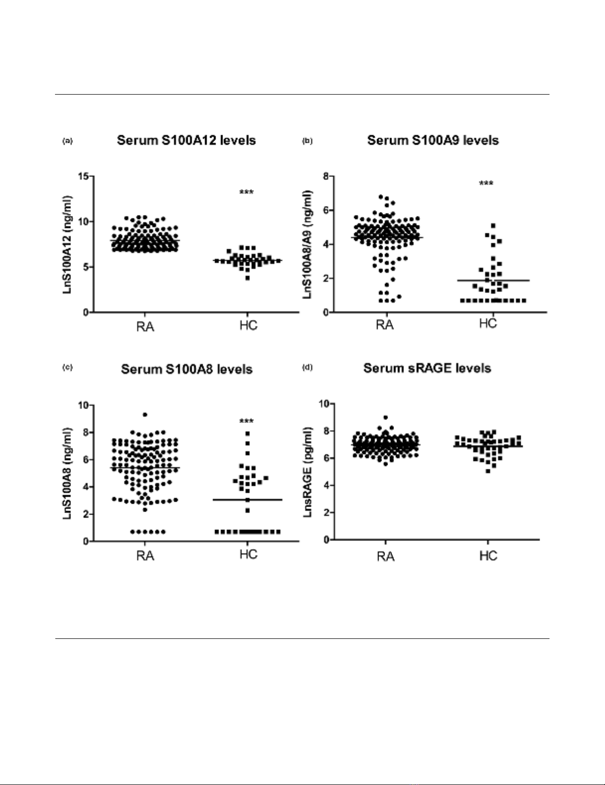
Increased serum concentrations of the S100 proteins,
but not sRAGE, in patients with established RA
Serum levels of S100A9, S100A8 and S100A12 in patients
with RA (n = 138) were increased relative to serum levels in
healthy controls (n = 44, P < 0.001). The S100A9 levels
detected in patient sera with an anti-S100A9 antibody that
detected S100A9, and S100A9 complexed with S100A8,
were some 100-fold lower than those reported in other studies
[26,27]. This could reflect differences in the specificity of the
anti-calprotectin (an antibody generated against the S100A8/
A9 complex) used by others; the anti-S100A9 used by us was
generated against pure S100A9. In contrast to the S100 pro-
teins, serum levels of sRAGE were not different (Figure 1a–d).
Table 1
Demographic details, cardiovascular risk factors, features of
rheumatoid arthritis (RA) and its control in the study
population (n = 138)
Parameter Value
Demographics:
Age (years) 64.0 (10.9)
Females, n (%) 92 (66.7)
Duration of RA (years) 17.6 (13.6)
RF positive, n (%) 83 (61.0)
CV disease:
History of MI, n (%) 14 (10.1)
History of angina, n (%) 11 (8.0)
History of stroke/TIA, n (%) 9 (6.5)
History of PVD, n (%) 6 (4.4)
Any vascular event, n (%) 26 (18.8)
Risk factors for CV diseases:
Smoking pack-year history 18 (24)
Current smoker, n (%) 25 (18.1)
History of hypertension, n (%) 47 (34.1)
History of hyperlipidemia, n (%) 33 (23.9)
History of diabetes, n (%) 19 (13.8)
Family history CV disease, n (%) 38 (27.5)
Clinical findings:
BMI (kg/m2) 27.5 (6.9)
Systolic BP (mmHg) 132 (19)
Diastolic BP (mmHg) 78 (10)
Laboratory tests:
ESR (mm/h) 25 (18)
CRP (mg/l) 13.6 (18.6)
Total cholesterol (mmol/l) 5.3 (1.0)
HDL cholesterol (mmol/l) 1.5 (0.4)
LDL cholesterol (mmol/l) 3.0 (0.9)
TG (mmol/l) 1.6 (1.0)
Homocysteine (mol/l) 12 (5)
Fasting glucose (mmol/l) 5.6 (1.6)
Serum creatinine (mmol/l) 0.08 (0.05)
CrCl (ml/min) 79.1 (29.9)
Framingham score (%) 11.1 (9.5)
ECG evidence of ischemia, n (%) 2 (1.5)
Severity and feature of RA:
Radiographic erosion score 24 (35)
Joint space narrowing score 21 (28)
Presence of erosive disease, n (%) 97 (71.3)
History of vasculitis, n (%) 15 (10.9)
Shared epitope, n (%) 103 (75.2)
> 10 mg/day of prednisone, n (%) 9 (6.5)
RAGE polymorphism, n (%) 29 (21.0)
BP, blood pressure; CrCl, creatinine clearance; CRP < C-reactive
protein; CV, cardiovascular; ECG, electrocardiogram; ESR,
erythrocyte sedimantation rate; HDL, high-density lipoprotein; LDL,
low-density lipoprotein; MI, myocardial infarction; PVD, peripheral
vascular disease; RAGE, receptor for advanced glycation end
products; RF, rheumatoid factor; TG, triglyceride; TIA, transient
ischemic attack.
Table 1 (Continued)
Demographic details, cardiovascular risk factors, features of
rheumatoid arthritis (RA) and its control in the study
population (n = 138)
Available online http://arthritis-research.com/content/11/2/R39
Page 5 of 11
(page number not for citation purposes)
Factors associated with serum levels of S100A9, S100A8
and S100A12 in patients with RA
We analyzed the cohort of 138 RA patients for associations
between serum levels of S100A9, S100A8, S100A12 and
sRAGE with RA clinical variables, CV risk factors, and with
complications such as vasculitis, radiographic changes, and
CV events. In simple linear regression analysis, we found that
serum levels of S100A9 in RA patients were positively associ-
ated with the presence of the PTPN22 Cys1858Thr genetic
polymorphism, serum levels of S100A12, and serum levels of
S100A8 (Table 2, P < 0.05). Serum levels of S100A9 in MLR
model analysis were positively associated with body mass
index, and with serum levels of S100A8 and S100A12 (Table
2, P < 0.05).
Figure 1
Serum sRAGE, S100A9, S100A8 and S100A12 levels in rheumatoid arthritis (RA) patients and healthy controlsSerum sRAGE, S100A9, S100A8 and S100A12 levels in rheumatoid arthritis (RA) patients and healthy controls. Levels of S100A12 (a), S100A9
(b), S100A8 (c), and soluble receptor for advanced glycation end products (sRAGE) (d) were measured in serum of 138 patients with established
RA and 44 healthy controls by ELISA. The horizontal line represents the mean value. *** P < 0.001, * P < 0.05.


