
Open Access
Available online http://arthritis-research.com/content/9/5/R104
Page 1 of 9
(page number not for citation purposes)
Vol 9 No 5
Research article
Glucosamine affects intracellular signalling through inhibition of
mitogen-activated protein kinase phosphorylation in human
chondrocytes
Anna Scotto d'Abusco1, Valentina Calamia1, Claudia Cicione1, Brunella Grigolo2, Laura Politi1 and
Roberto Scandurra1
1Department of Biochemical Sciences, Sapienza University of Roma, P.le Aldo Moro 5, 00185 Roma, Italy
2Laboratory of Immunology and Genetics, Istituto di Ricerca Codivilla Putti, Istituti Ortopedici Rizzoli, Via di Barbiano 1/10, 40136, Bologna Italy
Corresponding author: Anna Scotto d'Abusco, anna.scottodabusco@uniroma1.it
Received: 9 May 2007 Revisions requested: 22 Jun 2007 Revisions received: 17 Sep 2007 Accepted: 9 Oct 2007 Published: 9 Oct 2007
Arthritis Research & Therapy 2007, 9:R104 (doi:10.1186/ar2307)
This article is online at: http://arthritis-research.com/content/9/5/R104
© 2007 Scotto d'Abusco et al.; licensee BioMed Central Ltd.
This is an open access article distributed under the terms of the Creative Commons Attribution License (http://creativecommons.org/licenses/by/2.0),
which permits unrestricted use, distribution, and reproduction in any medium, provided the original work is properly cited.
Abstract
The aim of this study was to determine the effects of
glucosamine on matrix metalloprotease (MMP) production, on
mitogen-activated protein kinase (MAPK) phosphorylation, and
on activator protein (AP)-1 transcription factor activation in
human chondrocytes. The human immortalized cell line lbpva55
and healthy human chondrocytes (obtained from healthy
donors) were subjected to challenge with 10 ng/ml IL-1β after
pretreatment with 2.5 or 10 mmol/l glucosamine. MMP mRNA
expression levels were evaluated using quantitative real-time
PCR, and MMP protein production levels were evaluated in the
culture supernatant using ELISA. MAPK phosphorylation was
evaluated using Western blotting. AP-1 transcription factor
activation was evaluated by measuring AP-1 DNA-binding
activity. After IL-1β stimulation, levels of MMP-1, MMP-3 and
MMP-13 production were markedly increased. Treatment with
2.5 and 10 mmol/l glucosamine reduced expression of these
metalloproteases. MMP expression is regulated by transcription
factors such as the AP-1 complex, which is activated by
phosphorylated MAPKs. IL-1β stimulated phosphorylation of c-
jun amino-terminal kinase, p38 MAPK and extracellular signal-
regulated kinase-1/2. Glucosamine inhibited c-jun amino-
terminal kinase and p38 phosphorylation, and consequently c-
jun binding activity. These findings demonstrate, for the first
time, that glucosamine inhibits IL-1β-stimulated MMP
production in human chondrocytes by affecting MAPK
phosphorylation.
Introduction
The pharmacological treatment of osteoarthritis (OA), a joint
disorder characterized by slow, progressive degradation of the
cartilage, includes analgesic agents and nonsteroidal antin-
flammatory drugs. During recent years there has been growing
interest in alternative treatments for OA, such as glucosamine.
In particular, glucosamine was found to be effective in reduc-
ing joint space narrowing compared with placebo in clinical tri-
als conducted over a period of 3 years [1-4]. It was also found
to be effective in decreasing pain compared with analgesic
agents in OA of the knee [5,6]. A recent trial showed that glu-
cosamine was ineffective in reducing pain in patients with
severe knee OA, but it was more effective when it was used in
combination with chondroitin sulphate in patients with moder-
ate-to-severe pain [7].
Cartilage degradation in OA is due to an imbalance between
synthesis and degradation of extracellular matrix components.
Proinflammatory cytokines, such as IL-1β, which are produced
in OA, trigger several biological effects by stimulating mitogen-
activated protein kinase (MAPK) phosphorylation. The latter
results in activation of transcription factors [8-10], which in
turn upregulate the production of several molecules such as
matrix metalloproteases (MMPs) and aggrecanases.
Increased enzymatic activity of MMPs and aggrecanases is the
major factor responsible for matrix degradation [11,12].
AP = activator protein; Ct = threshold cycle; DMEM = Dulbecco's modified Eagle's medium; ERK = extracellular signal-regulated kinase; FBS = foetal
bovine serum; HPC = human primary chondrocyte; IL = interleukin; JNK = c-jun amino-terminal kinase; MAPK = mitogen-activated protein kinase;
MMP = matrix metalloprotease; OA = osteoarthritis; PBS = phosphate-buffered saline; PCR = polymerase chain reaction.

Arthritis Research & Therapy Vol 9 No 5 Scotto d'Abusco et al.
Page 2 of 9
(page number not for citation purposes)
Several studies have examined the effects of glucosamine on
MMP expression and activity in stimulated chondrocytes,
obtained from various sources. The addition of glucosamine to
cells appears to decrease the activity of MMPs [13-19]. More-
over, most in vitro studies conducted to elucidate the molecu-
lar basis of the effect of glucosamine on cartilage cells [20-24]
demonstrated an anti-inflammatory and chondroprotective role
for this molecule. However, the mechanisms responsible for
these activities are not entirely understood.
To address whether glucosamine can inhibit production of
MMPs by affecting IL-1β-induced MAPK activation, we inves-
tigated the phosphorylation of c-jun amino-terminal kinase
(JNK), p38 and extracellular signal-regulated kinase (ERK)1/2
after pretreatment with glucosamine and stimulation with IL-
1β. Moreover, we analyzed the activation of some activator
protein (AP)-1 transcription factor components. We con-
ducted the study both in the human immortalized chondrocyte
cell line lbpva55 (derived from adult articular healthy cartilage),
which has been demonstrated to be a useful tool for studying
the biology of chondrocytes [25-27], and in human primary
chondrocytes (HPCs) from healthy donors as a further control.
Materials and methods
Cell culture
lbpva55 cell culture was conducted as described previously
[25]. Briefly, human immortalized chondrocytes, from the
lbpva55 cell line, were grown to 80% confluence in Dul-
becco's modified Eagle's medium (DMEM; Sigma, St. Louis,
MO, USA) supplemented with L-glutamine, penicillin/strepto-
mycin (HyClone, Logan, UT, USA) and gentamycin (Roche
Diagnostic, Mannheim, Germany), along with 20% foetal
bovine serum (FBS). The cells were then transferred in DMEM
plus 10% FBS. After overnight incubation, the monolayer was
rinsed with phosphate-buffered saline (PBS; Sigma) and incu-
bated with culture medium containing 1% Nutridoma-SP
(Roche). Medium was changed twice a week and the cells
were split once. In these culture conditions, after 14 days the
cells re-expressed the differentiated chondrocyte phenotype
(namely collagen type IIA1 mRNA) [25].
HPCs were isolated from cartilage obtained from six healthy
donors. Full informed consent was obtained from all donors
and families.
Articular cartilages were aseptically dissected. Chondrocytes
were obtained after sequential digestion with protease type IV
(Sigma; 1 mg/ml) for 30 minutes and collagenase type II
(Sigma; 1 mg/ml) for 90 minutes, both in Hank's medium
(Hyclone). Chondrocytes were grown to 80% confluence in
DMEM, supplemented as described above, along with 10%
FBS. Experiments were performed with first passage cells in
DMEM containing 1% FBS and were repeated in HPCs
derived from the six donors, analyzing each sample separately.
Cell treatment
lbpva55 cell line and HPCs were seeded in 60 mm plates at
density of about 3 × 10⁶ per plate. Cells were left untreated or
treated with 10 ng/ml recombinant IL-1β (PeproTech House,
London, UK) or pretreated for 2 hours with 2.5 or 10 mmol/l
(0.54 and 2.16 mg/ml, respectively) glucosamine (Sigma) and
then stimulated with 10 ng/ml IL-1β for 22 hours. Culture
supernatants were collected and analyzed by ELISA, and cells
were harvested and processed for quantitative real-time PCR.
To analyze early responsive proteins, JNK, p38, ERK1/2 and
AP-1 components, lbpva55 cells and HPCs were pre-incu-
bated for 2 hours in 2.5 or 10 mmol/l glucosamine containing
medium and then stimulated with 10 ng/ml IL-1β for 15 min-
utes. Cells were harvested and conveniently processed for
Western blot analysis or for DNA-binding activity.
RNA extraction and reverse-transcription
Total RNA was extracted using TRIZOL reagent (Invitrogen,
Carlsbad, CA, USA), in accordance with the manufacturer's
instructions. Briefly, a confluent 60 mm plate, either of lbpva55
or HPCs, was washed with PBS and homogenized in 1 ml TRI-
ZOL reagent. RNA was stored at -80°C until use.
cDNA was synthesized from 1 μg total RNA, using reverse
transcriptase Improm II enzyme (Promega Corporation, Madi-
son, WI, USA) in accordance with the manufacturer's instruc-
tions, and analyzed by quantitative real-time PCR.
Real-time PCR
Quantitative real-time PCR analysis was performed using an
ABI Prism 7300 (Applied Biosystems, Foster City, CA, USA).
Amplification was carried out with 50 ng cDNA, in 96-well
plates, using SYBR Green PCR Master mix (Applied Biosys-
tems) in a 25 μl volume. Each sample was analyzed in tripli-
cate. PCR conditions were as follows: 94°C for 10 minutes
followed by 40 cycles of 94°C for 15 seconds and 60°C for 1
minute. Primers were designed using Primer Express software
(Applied Biosystems) and were synthesized by Primm (Milan,
Italy). The primer sequences are summarized in Table 1. The
results were analyzed using Sequence Detection Systems
software (Applied Biosystems), which automatically records
the threshold cycle (Ct). The untreated cell sample (control)
was used as a calibrator; the fold change for control was 1.0.
Target gene Ct values were normalized against GAPDH. Data
were analyzed using the 2-ΔΔCt method and expressed as fold
change compared to control.
ELISA
For quantification of MMP levels in the culture medium, cells
were treated as described above. Twenty-four hours after
treatment, supernatants were collected and stored at -80°C
until analysis using ELISA. Human MMP-1 ELISA kits were
purchased from Chemicon International, Inc. (Temecula, CA,
USA), and human MMP-3 and MMP-13 ELISA kits were

Available online http://arthritis-research.com/content/9/5/R104
Page 3 of 9
(page number not for citation purposes)
purchased from Amersham Biosciences (GE Healthcare
Europe, Milan, Italy). The experiments were performed in
accordance with the manufacturers' instructions.
Western blotting
To analyze MAPK phosphorylation, we performed Western
blotting experiments. Cells, treated as described above, were
washed with PBS and then scraped in 2× denaturing SDS
buffer (Sigma). Extracts were heated to 100°C for 5 minutes
and resolved on 10% SDS-PAGE. Gels were transferred to
Hybond C membranes (GE Healthcare) by electroblotting
(Bio-Rad Laboratories, Hercules, CA, USA) and probed with
specific antibodies, in accordance with the manufacturers'
instructions. Antibodies to JNK, phosphorylated-JNK and p38
were purchased from Santa Cruz Biotechnology, Inc. (Santa
Cruz, CA, USA), antibodies to ERK1/2 and phosphorylated-
ERK1/2 were from Biosource International (Camarillo, CA,
USA), and antibodies to phosphorylated-p38 were from
Chemicon International, Inc.
AP-1 binding assay
Nuclear proteins were obtained from HPCs, treated as
described above, using the Nuclear Extracts Kit (Active Motif,
Carlsbad, CA, USA), in accordance with the manufacturer's
instructions. Pellets were resuspended in 22 μl of Active Motif
lysis buffer and proteins were measured (Bio-Rad Protein
Assay). AP-1 consensus nucleotide binding activity from
nuclear extracts (8 μg) was assessed using the TransAM AP-
1 family kit (Active Motif), as recommended by the manufac-
turer. Nuclear extract was added to the immobilized oligonu-
cleotides, followed by primary transcription factor antibody,
secondary horse radish peroxidase (HRP)-conjugated anti-
body and HRP substrate, and colorimetric values (measured
at 450 nm) were plotted.
Statistical analysis
Each experiment was repeated at least three times. The statis-
tical significance of the differences between mean values was
determined using a two-tailed t-test. P ≤ 0.05 was considered
statistically significant. Where appropriate, results are
expressed as the mean ± standard error.
Results
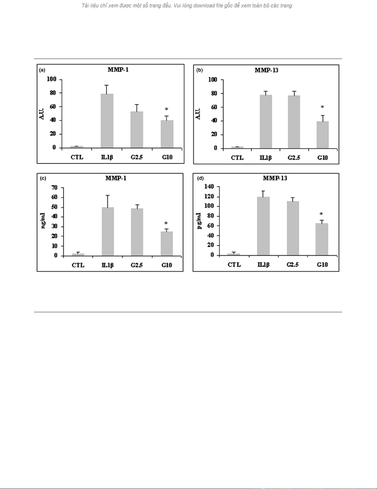
Effect of glucosamine on expression of MMPs in the
lbpva55 cell line
After stimulation with IL-1β, MMP-1 and MMP-13 mRNA levels
were markedly upregulated (both MMPs almost 80-fold). Pre-
treatment with 2.5 mmol/l and 10 mmol/l glucosamine inhib-
ited MMP-1 and MMP-13 mRNA expression (Figure 1a,b), but
only the treatment with 10 mmol/l glucosamine yielded a sta-
tistically significant effect (P < 0.05 for MMP1 and P < 0.03
for MMP-13). MMP-8 mRNA expression was not upregulated
by IL-1β (data not shown). Consistent with quantitative real-
time PCR findings, the ELISA assay demonstrated that levels
of MMP-1 and MMP-13 protein secreted into the media were
significantly decreased by 10 mmol/l glucosamine (P < 0.05;
Figure 1c,d).
Table 1
Sequences of primers used to quantify gene expression by real-time PCR
Gene Primers GenBank
GAPDH Forward: GGAGTCAACGGATTTGGTCGTA NM_002046
Reverse: GGCAACAATATCCACTTTACCAGAGT
MMP-1 Forward: GATGGACCTGGAGGAAATCTTG NM_002421
Reverse: TGAGCATCCCCTCCAATACC
MMP-2 Forward: GCACCCATTTACACCTACACCAA NM_004530
Reverse: AGAGCTCCTGAATGCCCTTGA
MMP-3 Forward: CCTGGTACCCACGGAACCT NM_002422
Reverse: AGGACAAAGCAGGATCACAGTTG
MMP-8 Forward: GACCAACACCTCCGCAAATT NM_002424
Reverse: CCCCAAAGAATGGCCAAAT
MMP-9 Forward: GGACGATGCCTGCAACGT NM_004994
Reverse: ACAAATACAGCTGGTTCCCAATC
MMP-13 Forward: TTCTTGTTGCTGCGCATGA NM_002427
Reverse: TGCTCCAGGGTCCTTGGA
GAPDH, glyceraldehyde-3-phosphate dehydrogenase; MMP, matrix metalloprotease.
Arthritis Research & Therapy Vol 9 No 5 Scotto d'Abusco et al.
Page 4 of 9
(page number not for citation purposes)
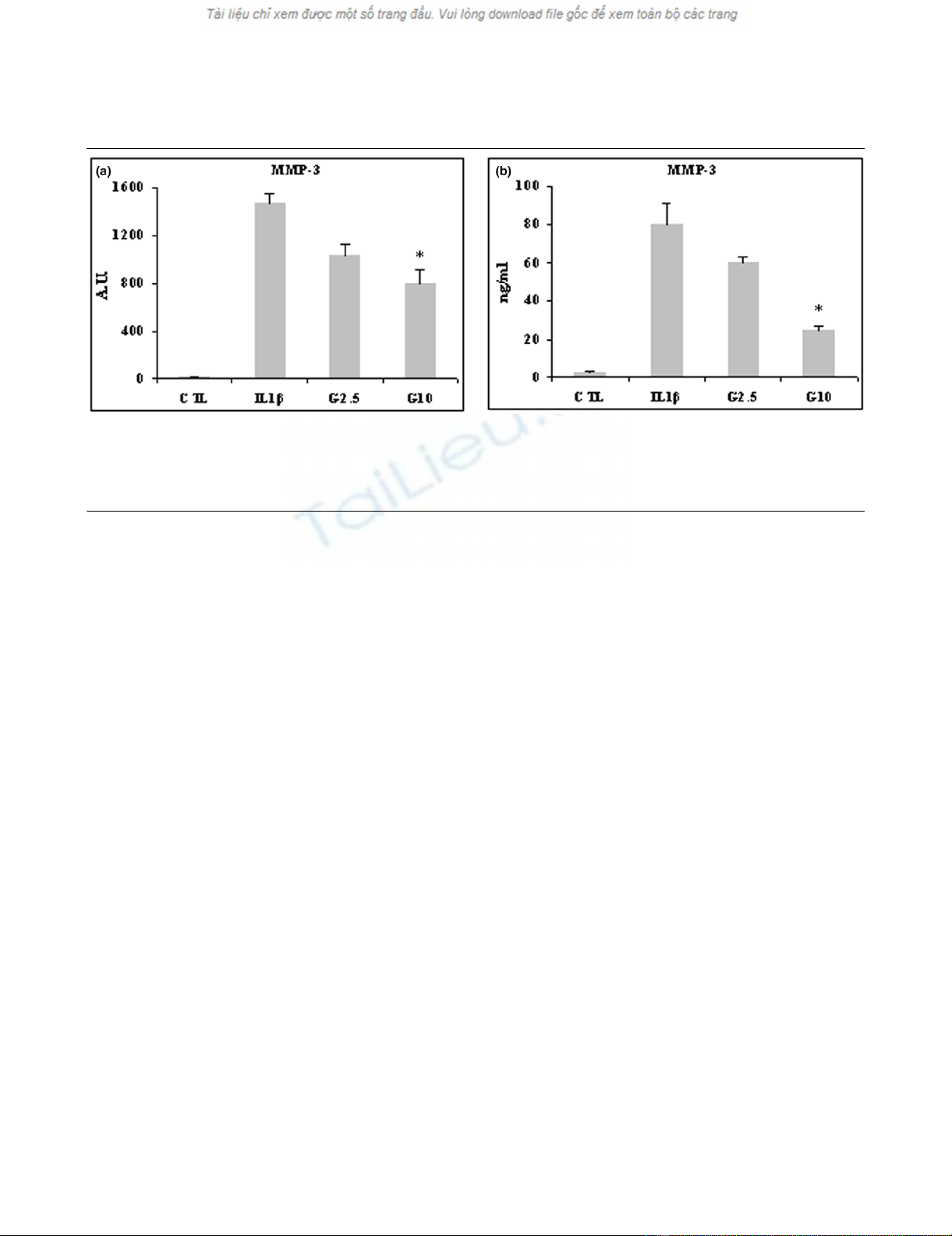
IL-1β stimulated by 1,500-fold the expression of MMP-3
mRNA. This stimulation was significantly counteracted by 10
mmol/l glucosamine (P < 0.05; Figure 2a). IL-1β also stimu-
lated secretion of MMP-3, which was counteracted by 2.5
mmol/l glucosamine and significantly so by 10 mmol/l glu-
cosamine (P < 0.03; Figure 2b).
Levels of MMP-2 and MMP-9 mRNA expression were not
upregulated by cytokine stimulation (data not shown) and their
protein levels were not analyzed.
Effect of glucosamine on expression of MMPs in HPCs
MMP-1, MMP-3 and MMP-13 were also stimulated by IL-1β in
HPCs at both mRNA and protein levels, and the stimulation
was counteracted by glucosamine treatment. MMP-1 mRNA
expression level was stimulated 140-fold, MMP-3 180-fold
and MMP-13 170-fold. All three MMPs were downregulated
by 2.5 mmol/l and significantly so by 10 mmol/l glucosamine
(P < 0.01 for MMP-1 and P < 0.05 for MMP-13 [Figure 3a,b]
and P < 0.03 for MMP-3 [Figure 4a]). Levels of MMP-1 and
MMP-3 secretion induced by IL-1β were higher compared
than those of MMP-13. At any rate, 10 mmol/l glucosamine
was effective in significantly downregulating all three MMPs (P
< 0.05 for MMP-1 and P < 0.01 for MMP-13 [Figure 3c,d] and
P < 0.03 for MMP-3 [Figure 4b]).
Effect of glucosamine on IL-1β-induced phosphorylation
of JNK, p38 and ERK1/2 MAP kinases in lbpva55 cell line
and HPCs
We analyzed the phosphorylation levels of three MAPKs,
namely JNK, p38 and ERK1/2, by Western blotting. Time
course experiments showed that 15 minutes of stimulation
with IL-1β was able to induce phosphorylation of all three
kinases analyzed in lbpva55 cells and HPCs (data not shown).
Two hours of pretreatment with 2.5 or 10 mmol/l glucosamine
prevented the phosphorylations of JNK (Figure 5a) and p38
(Figure 5b) in lbpva55 cells. Glucosamine was ineffective in
counteracting the phosphorylation of ERK1/2 (Figure 5c).
Figure 1
Effect of glucosamine on MMP-1 and MMP-13 expression in lbpva55 cells stimulated with 10 ng/ml IL-1βEffect of glucosamine on MMP-1 and MMP-13 expression in lbpva55 cells stimulated with 10 ng/ml IL-1β. Cells were pretreated for 2 hours with 2.5
or 10 mmol/l glucosamine (G2.5 and G10, respectively), and then stimulated with IL-1β for 22 hours. mRNA was extracted and analyzed by quanti-
tative real-time PCR, and cell supernatant was analyzed by ELISA. Shown are (a) matrix metalloprotease (MMP)-1 and (b) MMP-13 mRNA levels,
and (c) MMP-1 and (d) MMP-13 protein amounts. Quantitative real-time PCR results are expressed in relative arbitrary units (AU), and ELISA results
are expressed in ng/ml or pg/ml. Results are expressed as mean ± standard error, obtained in three different experiments. *P ≤ 0.05. CTL, control.
Available online http://arthritis-research.com/content/9/5/R104
Page 5 of 9
(page number not for citation purposes)
Similar findings were obtained in HPCs; IL-1β-induced phos-
phorylation of JNK and p38 was prevented by both 2.5 and 10
mmol/l glucosamine, which had no effect on ERK1/2 phos-
phorylation (Figure 6a,b,c).
Effect of glucosamine on AP-1 transcription factor
activation
We next examined the effects of glucosamine on AP-1 compo-
nents in HPCs. In our experimental conditions, 15 minutes of
stimulation with IL-1β induced c-jun and junD DNA binding
activity. Two hours of pretreatment with 2.5 and 10 mmol/l glu-
cosamine significantly reduced c-jun DNA binding activity (P
< 0.03; Figure 7), whereas junD binding activity was reduced
to a lower degree (data not shown).
Discussion
Imbalance between catabolic and anabolic factors in OA leads
to degradation of articular cartilage. Catabolic factors include
proinflammatory cytokines IL-1β and tumour necrosis factor-α,
which stimulate intracellular signalling such as MAPK activa-
tion, which results in overproduction of several molecules,
including MMPs [8-10]. In our experimental model, stimulation
of the lbpva55 cell line with 10 ng/ml IL-1β resulted in upreg-
ulation of mRNA and protein expression of collagenases
MMP-1 and MMP-13 and the stromelysin MMP-3. Pretreat-
ment with 2.5 and 10 mmol/l glucosamine was able, to differ-
ing degrees, to downregulate mRNA and protein levels of
MMP-1, MMP-3 and MMP-13.
These findings were confirmed in HPCs. In these cells as well,
over-expression of MMP-1, MMP-3 and MMP-13 induced by
IL-1β stimulation was downregulated by 2.5 and 10 mmol/l
glucosamine pretreatment at both mRNA and protein levels.
These findings are in agreement with those obtained by Naka-
mura and coworkers [18] in healthy and OA human chondro-
cytes. The lbpva55 cell line produces higher basal amounts of
MMPs [27] as compared with those produced by HPCs, but
MMP production is similarly stimulated by IL-1β and inhibited
by glucosamine both in lbpva55 and HPCs.
To address the hypothesis that glucosamine can downregu-
late MMP production by affecting MAPK activation and there-
fore activation of transcription factors, we analyzed
phosphorylation of JNK, p38 and ERK1/2 MAPKs. Phosphor-
ylation of ERK1/2 was increased by IL-1β but was not
decreased by glucosamine pretreatment. During the early
phases of OA, proinflammatory cytokines promote chondro-
cyte proliferation. The ERK1/2 pathway is essential in proc-
esses that involve cellular proliferation and differentiation
[28,29]. Chondrocytes stimulated in vitro with IL-1β mimic
early phase OA, thus explaining the increased level of phos-
phorylation of ERK1/2. Nevertheless, glucosamine was inef-
fective in preventing phosphorylation of this kinase. JNK and
p38 are involved in cellular stress; they are phosphorylated in
response to proinflammatory cytokines, resulting in activation
of transcription factors such as AP-1 complex [30], which are
involved in expression of MMPs [8-10]. We found increased
phosphorylation of JNK and p38 in samples stimulated with IL-
1β, and decreased phosphorylation level in samples pre-
treated with 2.5 or 10 mmol/l glucosamine and then stimulated
with IL-1β. Moreover, c-jun and junD activity were affected by
glucosamine pretreatment in HPCs. Both c-jun [8-10] and
junD [31] are involved in transcription of MMPs; inhibition of
their activity is in accordance with decreased MMP produc-
Figure 2
Effect of glucosamine on MMP-3 expression in lbpva55 cell line stimulated with 10 ng/ml IL-1βEffect of glucosamine on MMP-3 expression in lbpva55 cell line stimulated with 10 ng/ml IL-1β. Cells were pretreated for 2 hours with 2.5 or 10
mmol/l glucosamine (G2.5 and G10, respectively), and then stimulated with IL-1β for 22 hours. mRNA was extracted and analyzed by quantitative
real-time PCR, and cell supernatant was analyzed by ELISA. Shown are (a) matrix metalloprotease (MMP)-3 mRNA and (b) MMP-3 protein levels.
Quantitative real-time PCR results are expressed in relative arbitrary units (AU) and ELISA results are expressed in ng/ml. Results are expressed as
mean ± standard error, obtained in three different experiments. *P ≤ 0.05. CTL, control.


