
BioMed Central
Page 1 of 12
(page number not for citation purposes)
Retrovirology
Open Access
Review
HIV interactions with monocytes and dendritic cells: viral latency
and reservoirs
Christopher M Coleman and Li Wu*
Address: Department of Microbiology and Molecular Genetics, Medical College of Wisconsin, 8701 Watertown Plank Road, Milwaukee, WI 53226,
USA
Email: Christopher M Coleman - ccoleman@mcw.edu; Li Wu* - liwu@mcw.edu
* Corresponding author
Abstract
HIV is a devastating human pathogen that causes serious immunological diseases in humans around
the world. The virus is able to remain latent in an infected host for many years, allowing for the
long-term survival of the virus and inevitably prolonging the infection process. The location and
mechanisms of HIV latency are under investigation and remain important topics in the study of viral
pathogenesis. Given that HIV is a blood-borne pathogen, a number of cell types have been
proposed to be the sites of latency, including resting memory CD4+ T cells, peripheral blood
monocytes, dendritic cells and macrophages in the lymph nodes, and haematopoietic stem cells in
the bone marrow. This review updates the latest advances in the study of HIV interactions with
monocytes and dendritic cells, and highlights the potential role of these cells as viral reservoirs and
the effects of the HIV-host-cell interactions on viral pathogenesis.
Background
Human immunodeficiency virus (HIV) remains a devas-
tating human pathogen responsible for a world-wide pan-
demic of acquired immunodeficiency syndrome (AIDS).
Despite extensive research of HIV since the virus was iden-
tified over 25 years ago, eradication of HIV-1 infection
and treatment of AIDS remain a long-term challenge
[1,2]. The AIDS pandemic has stabilised on a global scale.
In 2007, it was estimated that 30 to 36 million people
world-wide were living with HIV, and 2.7 million people
were newly infected with HIV. Moreover, AIDS-related
deaths were increased from an estimated 1.7 million peo-
ple in 2001 to 2.0 million in 2007. Africa continues to be
over-represented in the statistics, with 68% of all HIV-pos-
itive people living in sub-Saharan countries. The young
generation represents a large proportion of newly infected
population who may contribute to the overall spread of
HIV in the future [3].
There are two types of HIV, HIV-1 and HIV-2; both are
capable of causing AIDS, but HIV-2 is slightly attenuated
with regards to disease progression [4]. Given the relative
severity of HIV-1 infection, the majority of studies have
been done using HIV-1. The infection dynamics of HIV-1
are very interesting. Upon initial HIV-1 infection, there is
a period of continuous viral replication and strong
immune pressure against the virus, resulting in a relatively
low steady state of viral load. The virus then enters a
chronic stage, wherein there is limited virus replication
and no outward signs of disease. This clinical phase can
last many years, ultimately leading to destruction of the
host immune system due to chronic activation or viral
Published: 1 June 2009
Retrovirology 2009, 6:51 doi:10.1186/1742-4690-6-51
Received: 27 March 2009
Accepted: 1 June 2009
This article is available from: http://www.retrovirology.com/content/6/1/51
© 2009 Coleman and Wu; licensee BioMed Central Ltd.
This is an Open Access article distributed under the terms of the Creative Commons Attribution License (http://creativecommons.org/licenses/by/2.0),
which permits unrestricted use, distribution, and reproduction in any medium, provided the original work is properly cited.

Retrovirology 2009, 6:51 http://www.retrovirology.com/content/6/1/51
Page 2 of 12
(page number not for citation purposes)
replication. This results in the onset of the AIDS stage with
opportunistic infections and inevitable death in the vast
majority of untreated patients [4].
Unfortunately, there is no effective AIDS vaccine currently
available, and antiretroviral therapy is limited in its ability
to fully control viral replication in infected individuals.
Recent progress suggests that understanding how HIV
interacts with the host immune cells is vitally important
for the development of new treatments and effective vac-
cination regimens [1,2]. Monocytes, monocyte-differenti-
ated dendritic cells (DCs) and macrophages are critical
immune cells responsible for a wide range of both innate
and adaptive immune functions [5]. These cell types also
play multifaceted roles in HIV pathogenesis (Table 1). In
this review, the potential roles of monocytes and DCs as
HIV reservoirs and in latency will be discussed in detail.
Monocytes interact with HIV-1
Monocyte distribution and function
Monocytes are vitally important cells in the immune sys-
tem, as they are the precursor cells to professional antigen-
presenting cells (APCs), such as macrophages and DCs.
These types of immune cells patrol the bloodstream and
tissues, replenishing dying APCs or, in an infection, pro-
viding enough of these cells for the body to effectively
combat an invading pathogen [5]. Undifferentiated
monocytes live for only a few days in the bloodstream.
Upon differentiation or activation, the life-span of mono-
cytes is significantly prolonged for up to several months
[6].
There are two major subtypes of monocytes, those that are
highly CD14-positive (CD14++CD16-) and those that are
CD16-positive (CD14+CD16+). CD16+ cells make up only
a small percentage (around 5%) of the total monocyte
population, but they are characterised as more pro-
inflammatory and having a greater role in infections than
the CD14++CD16- cells [7].
HIV infection of monocyte
Although monocytes express the required HIV-1 receptors
and co-receptors for productive infection [8,9], they are
not productively infected by HIV-1 in vitro. This is possibly
due to an overall inefficiency in each of the steps required
for virus infection, ranging from viral entry to proviral
DNA integration [10-12], but not due to a viral nucleocap-
sid uncoating defect [13]. Recent studies have suggested a
role for naturally occurring anti-HIV micro-RNA (miRNA)
in suppressing HIV-1 replication in peripheral blood
mononuclear cells or purified monocytes [14-17]. This
mechanism could allow for further studies utilising miR-
Table 1: Myeloid lineage cell types and their potential roles and proposed mechanisms in HIV-1 latency
Cell types Primary Locations Cellular markers Potential role in HIV latency and
proposed mechanisms
References
Monocytes Peripheral blood CD14++
or CD16+CD14+
YES, but possibly mainly in CD16+ cells
• Restricted HIV-1 replication at different
steps of viral life-cycle
• Low molecular weight APOBEC3G (CD16+
only)
• Low level or undetectable Cyclin T1
• Impaired phosphorylation of CDK9
[10-12,87-92,94]
Macrophages Mucosal surface/tissues CD14-
EMR1+
CD68+
NO
• High level Cyclin T1
• Phosphorylation of CDK9 and active P-TEFb
[14,18,94,97]
Myeloid DCs Peripheral blood (immature)
Lymph node (mature)
CD11c+
CD123-
BDCA1+
YES
• Low level virus replication
• Lymph node biopsies reveal presence
• Unknown mechanism
[101,107,112]
Plasmacytoid DCs Peripheral blood (immature)
Lymph node (mature)
CD11c-
CD123+
BDCA2+
BDCA4+
Unlikely
• Inhibiting HIV-1 replication through the
secretion of IFNα and an unidentified small
molecule
• Unknown mechanism
[49,50,101]
Langerhans cells Mucosal surface and epidermal tissue CD1a+
Langerin+
Unlikely
• Langerin inhibits virus transmission and
enhances virus take-up and degradation
• May act differently in co-infections
[40,41,113]
EMR1, epidermal growth factor module-containing mucin-like receptor 1 (a G-protein coupled receptor); BDCA, blood DC antigen.

Retrovirology 2009, 6:51 http://www.retrovirology.com/content/6/1/51
Page 3 of 12
(page number not for citation purposes)
NAs as inhibitors of HIV-1 [15]. However, it has also been
shown that HIV-1 is capable of suppressing some inhibi-
tory miRNAs [16], which may reflect an evolutional inter-
action between HIV-1 and host factors. Further studies are
required to understand this interaction and develop a
therapeutic approach against HIV-1 infection using miR-
NAs.
Differentiation of monocytes into macrophages or DCs in
vitro enables productive HIV-1 replication in the differen-
tiated cells [14,18,19]. Based on current understanding,
vaginal macrophages are more monocyte-like than intes-
tinal macrophages and show increased HIV-1 susceptibil-
ity [20]. Hence, some monocyte characteristics might be
required for efficient infection, and these traits may be lost
in fully differentiated tissue macrophages.
Monocyte-HIV interactions that impact immune function
Given the role of monocytes in the immune system and in
HIV-1 replication, a number of HIV-1 proteins have been
shown to affect the biology of monocytes.
HIV-1 Tat-mediated transactivation of the viral promoter
is essential for HIV-1 transcription [21]. Exogenous
recombinant HIV-1 Tat protein has been shown to
increase monocyte survival through increased expression
of the anti-apoptotic protein Bcl-2 [22]. Using an in vitro
model of monocyte death mediated by TRAIL (tumour
necrosis factor-alpha-related apoptosis inducing ligand),
it has been shown that HIV-1 Tat encourages the survival
of monocytes in situations where they would normally be
cleared [22]. Exogenous HIV-1 Tat has been shown to
cause production of the cytokine interleukin (IL)-10 from
monocytes in vitro [23,24]. Significantly increased IL-10
levels were also observed in HIV/AIDS patients compared
with healthy controls [25]. Furthermore, up-regulation of
IL-10 production in HIV/AIDS patients has been corre-
lated with increased levels of monocyte-secreted myeloid
differentiation-2 and soluble CD14 [25]; both proteins
are key molecules in the immune recognition of gram-
negative bacterial lipopolysaccharide (LPS). Given that
high levels of secreted CD14 have been associated with
impaired responses to LPS [26], it has been proposed that
the release of general immunosuppressant IL-10 by
monocytes [27] facilitates the progression to AIDS [25].
HIV-1 Nef is a multifunctional accessory protein that
plays an important role in viral pathogenesis [28]. Retro-
viral-mediated HIV-1 Nef expression in primary mono-
cytes and a promonocytic cell line inhibits LPS-induced
IL-12p40 transcription by inhibiting the JNK mitogen-
activated protein kinases [29]. As an inducible subunit of
biologically active IL-12, IL-12p40 plays a critical role in
the development of cellular immunity, and its production
is significantly decreased during HIV-1 infection [29]. This
study implicates the importance of HIV-1 Nef in the loss
of immune function and progression to AIDS.
HIV-1 matrix protein (p17) regulates a number of cellular
responses and interacts with the p17 receptor (p17R)
expressed on the surface of target cells [30]. Upon binding
to the cell surface receptor p17R, exogenous HIV-1 matrix
protein causes secretion of the chemokine monocyte
chemotactic protein-1 (MCP-1, also known as CCL2)
from monocytes [30]. MCP-1 potentially increases mono-
cyte recruitment to the sites of HIV-1 infection, increasing
the available monocyte pool for infection by HIV-1; this
recruitment may be of critical importance given the rela-
tively low rate of infection of this cell type [10-12].
HIV-1 and HIV-1-derived factors have been shown also to
induce up-regulation of programmed death ligand-1 on
monocytes in vitro [31,32]. This ligand, in complex with
its receptor, programmed death-1, causes apoptosis of all
T cell types [33] and a loss of anti-viral function in a man-
ner similar to known immunosuppressive cytokines [34].
Together, these studies suggest that HIV-1 can impair
virus-specific immunity by modulating immuno-regula-
tory molecules of monocytes and T cells.
Of the studies discussed above, those involving Tat,
matrix protein and HIV-1-derived factors, were performed
using recombinant or purified proteins, whereas the Nef
study and the reports on the programmed death ligand-1
were performed using infectious viruses and nef-deleted
HIV-1 mutants. Although these results shed light on the
influence of individual viral proteins on monocytes in
vitro, synergistic or antagonistic effects of HIV-1 proteins
on cellular responses cannot be ruled out, nor can the
roles played by other host factors in vivo be excluded.
Overall, HIV-1 appears to promote the survival of mono-
cytes as a key step for viral persistence. The interactions
between the virus and monocytes may contribute key
functions in establishing chronic HIV-1 infection and
facilitating the progression to AIDS. These outcomes are
likely influenced by the altered immunological function
of monocytes and their interactions with other types of
HIV-1 target cells (Figure 1).
DCs interact with HIV
Immune function of DCs
DCs are professional APCs that are differentiated from
monocytes in specific cytokine environments. DCs bridge
the innate and adaptive immune responses, as they endo-
cytose and break down invading pathogens in the
endolysosome or proteasome and present antigen frag-
ments to T cells, usually in the context of major histocom-
patability complexes [5]. There are three major DC
subtypes: myeloid DCs, plasmacytoid DCs (pDC), and
Retrovirology 2009, 6:51 http://www.retrovirology.com/content/6/1/51
Page 4 of 12
(page number not for citation purposes)
Langerhans cells. These DC subtypes are characterised
based on their locations, surface markers and cytokine
secretion profiles [5].
DC life-span and survival are highly dependent on their
anatomical locations and the DC subtypes [35]. In gen-
eral, DC half-lives measure up to a few weeks, and they
can be replaced through proliferating hematopoietic pro-
genitors, monocytes, or tissue resident cells [35]. It has
been shown that productive HIV-1 replication occurs in
human monocyte-derived DCs for up to 45 days [36].
DCs may survive longer within the lymph nodes due to
cytokine stimulation in the microenvironment, which
may help spread HIV-1 infection and maintain viral reser-
voirs.
HIV infection of DCs
HIV-1 is capable of directly infecting different DC sub-
types (known as cis infection), but at a lower efficiency
than HIV-1's ability to infect activated CD4+ T cells; there-
fore, only a small percentage of circulating DCs are posi-
tive for HIV in infected individuals [19]. Productive HIV-
1 replication is dependent on fusion-mediated viral entry
in monocyte-derived DCs [37], and mature HIV-1 parti-
cles are localised to a specialised tetraspanin-enriched
sub-compartment within the DC cytoplasm [38].
Langerhans cells are present in the epidermis or mucosal
epithelia as immune sentinels [39]. It is interesting that
Langerhans cells have been shown to be resistant to HIV-
1 infection [40]. This resistance appears to be due to the
expression of Langerin, which causes internalisation and
break-down of HIV-1 particles and blocks viral transmis-
sion [40]. However, in the context of co-infection with
other sexually transmitted organisms, such as the bacte-
rium Neisseria gonorrhoeae and/or the fungus Candida alba-
cans [41] or when stressed by skin abrasion [42],
Langerhans cells can become more susceptible to HIV-1
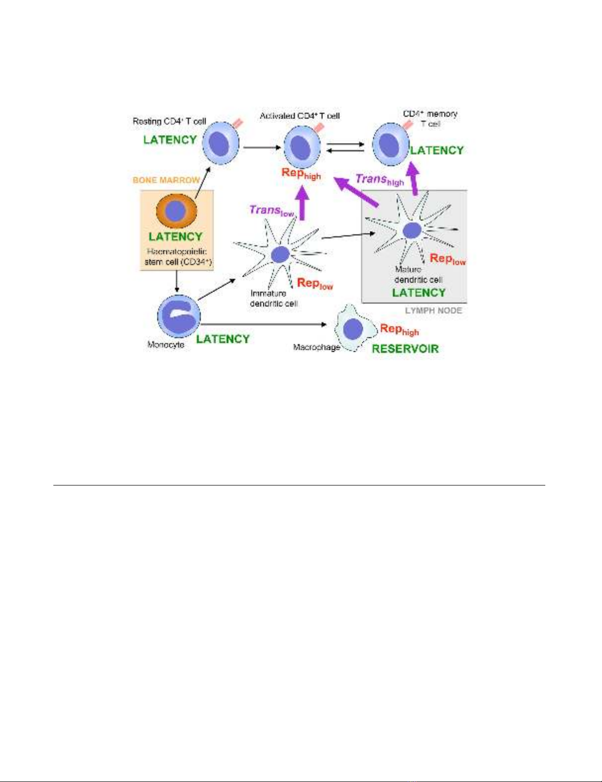
Locations of HIV-1 replication and latency and routes of transmission between haematopoietic cell populationsFigure 1
Locations of HIV-1 replication and latency and routes of transmission between haematopoietic cell popula-
tions. All cell types shown are susceptible to HIV-1 entry and integration of the proviral DNA. Some anatomical locations are
shown; those outside of marked areas are in the bloodstream, lymphatic system and/or tissues. Black arrows represent differ-
entiation and/or maturation and may represent more than one step and could involve multiple intermediate cell types. Purple
arrows represent routes of trans infection, and relative rates are shown as high or low. "Rep" indicates productive HIV-1 repli-
cation with relative rates shown as high or low. HIV-1 cis infection routes are not shown, as any susceptible cell may be
infected by productive replication from another cell. Those cells in which HIV-1 latency is thought to occur should be consid-
ered as putative viral reservoirs and therapeutic targets.

Retrovirology 2009, 6:51 http://www.retrovirology.com/content/6/1/51
Page 5 of 12
(page number not for citation purposes)
infection and are able to transmit HIV-1 to CD4+ T cells
effectively [42].
Drug abuse can significantly facilitate HIV infection,
transmission and AIDS progression through drug-medi-
ated immunomodulation. Recent studies have suggested
that the recreational drug, methamphetamine, increases
susceptibility of monocyte-derived DCs to HIV-1 infec-
tion in vitro [43] and blocks the antigen presentation func-
tion of DCs [44]. Although its relevance to the in vivo
situation is unclear, this finding is potentially a further
risk factor (aside from the use of contaminated needles,
etc.) associated with drug use and may explain the high
levels of HIV-prevalence among drug abusers.
HIV-1 infection of DCs likely contributes to viral patho-
genesis. Notably, HIV-2 is much less efficient than HIV-1
at infecting both myeloid DCs and pDCs, whilst retaining
its infectivity of CD4+ T cells [45]. This observation offers
an explanation for the decreased pathogenicity of HIV-2,
since HIV-2 will need to infect CD4+ T cells directly and,
perhaps more importantly, resting or memory CD4+ T
cells to ensure long-term survival of the virus.
DC-HIV interactions that impact the immune function
Given the important roles DCs play in the immune
response, it is reasonable that HIV-1 proteins or the virus
itself have been shown to affect the function of DCs in
vitro. Both HIV-1 matrix and Nef proteins have been
shown to cause only partial maturation of pDCs in vitro
[46,47]. In the presence of these viral proteins, DCs
acquire a migratory phenotype, facilitating travel to the
lymph nodes. However, these DCs do not express
increased levels of activation markers, such as the T cell
co-stimulatory molecules CD80 and CD86, or MHC class
II, that would lead to a protective immune response
[46,47]. It is possible, therefore, that the DCs are trapped
in the lymph nodes and unable to initiate a protective
immune response against the virus. The study of Nef pro-
tein's effects on DCs [47] was performed using a mouse
DC model in vitro and an immortalised cell line; hence the
full relevance of this finding to the in vivo situation is
unclear.
Conversely, recombinant Nef protein appears to cause DC
activation and differentiation by up-regulating the expres-
sion of CD80, CD86, MHC class II and other markers, as
well as various cytokines and chemokines associated with
T cell activation [48]. These effects have led to the propo-
sition that Nef protein is capable of causing bystander
activation of T cells via DCs [48], although this activity has
not been demonstrated experimentally. Of note, the
above study was performed using recombinant Nef alone.
DCs could contribute largely to an anti-HIV innate immu-
nity. It has been demonstrated that pDCs are capable of
inhibiting HIV-1 replication in T cells when cultured
together in vitro [49,50], implicating the importance of
pDCs for viral clearance. HIV-1 infected individuals are
known to have lower levels of circulating pDCs compared
with those of uninfected individuals [51]. It has been con-
firmed that HIV-1 is capable of directly killing pDCs [49],
illustrating that the virus can remove a potential block to
its replication and dissemination in pDCs.
HIV-1 can block CD4+ T cell proliferation or induce the
differentiation of naive CD4+ T cells into T regulatory cells
through pDCs [52,53]. These mechanisms involve HIV-1-
induced expression of indoleamine 2,3-deoxygenase in
pDCs. Indoleamine 2,3-deoxygenase is a CD4+ T cell sup-
pressor and regulatory T cell activator [52,53]. HIV-1
envelope protein gp120 has also been shown to inhibit
activation of T cells by monocyte-derived DCs [54], sug-
gesting that gp120 may also have a role in the suppression
of T cell function and progression to AIDS.
In addition, HIV-1 has been shown to suppress the
immune function of pDCs in general by suppressing acti-
vation of the anti-viral toll-like receptor 7 (TLR7) and
TLR8 [55], and by blocking the release of the anti-viral
interferon alpha [56]. A recent study indicated that diver-
gent TLR7 and TLR9 signalling and type I interferon pro-
duction in pDCs contribute to the pathogenicity of simian
immunodeficiency virus (SIV) infection in different spe-
cies of macaques [57]. These results suggest that chronic
stimulation of pDCs by SIV or HIV in non-natural hosts
may induce immune activation and dysfunction in AIDS
progression [57]. Overall, HIV-1 inhibits the function of
pDCs to allow maintenance of the virus within the host.
DC-mediated HIV-1 trans infection
The most interesting aspect of HIV-1 infection in DCs is
the ability of the cells to act as mediators of trans infection
of activated CD4+ T cells, which is the most productive cell
type for viral replication. DC-mediated HIV-1trans infec-
tion of CD4+ T cells is functionally distinct from cis infec-
tion [58,59] and involves the trafficking of whole virus
particles from the DCs to the T cells via a 'virological syn-
apse' [59,60]. Previous reviews have summarised the
understanding of HIV-DC interactions [19,61]; so here we
focus on discussing the latest progress in this field.
DC-mediated HIV-1 trans infection of CD4+ T cells is
dependent on, or enhanced by, a number of other cellular
and viral factors. CD4 co-expression with DC-SIGN (DC-
specific intercellular adhesion molecule 3-grabbing non-
integrin), a C-type lectin expressed on DCs, inhibits DC-
mediated trans infection by causing retention of viral par-
ticles within the cytoplasm [62]. HIV-1 Nef appears to
enhance DC-mediated HIV-1 trans-infection. Nef-
enhanced HIV-1 transmission efficiency correlates with
significant CD4 down-regulation in HIV-1-infected DCs


![PET/CT trong ung thư phổi: Báo cáo [Năm]](https://cdn.tailieu.vn/images/document/thumbnail/2024/20240705/sanhobien01/135x160/8121720150427.jpg)


%20--%3e%3cdefs%3e%3cstyle%3e%20.st0%20{%20fill:%20%23fff;%20}%20.st1%20{%20fill:%20%237800fa;%20}%20%3c/style%3e%3c/defs%3e%3cpath%20class='st1'%20d='M117.78,12.18H43.11c2.9,3.47,4.65,7.94,4.65,12.82,0,5.6-2.3,10.66-6.01,14.29h76.02l7.22-13.56-7.22-13.56Z'/%3e%3cg%3e%3cpath%20class='st0'%20d='M53.58,26.17h-.59v-1.46h.59v-4.96h2.83c1.78,0,2.67.94,2.67,2.82v5.76c0,1.87-.89,2.81-2.67,2.81h-2.83v-4.96ZM55.36,21.37v3.34h1.1v1.46h-1.1v3.34h1.01c.61,0,.91-.37.91-1.1v-5.93c0-.74-.3-1.1-.91-1.1h-1.01Z'/%3e%3cpath%20class='st0'%20d='M65.99,31.14h-1.8l-.31-2.07h-2.19l-.31,2.07h-1.64l1.82-11.39h2.62l1.82,11.39ZM65.28,18.04c-.25.46-.51.77-.75.94-.21.15-.47.22-.79.22-.26,0-.57-.07-.92-.22l-.38-.15c-.14-.05-.26-.07-.37-.07-.3,0-.53.18-.71.54l-.91-.68c.25-.46.51-.77.75-.94.21-.14.48-.21.79-.21.26,0,.57.07.92.21l.38.15c.14.05.26.07.37.07.3,0,.53-.18.71-.54l.91.68ZM61.91,27.52h1.73l-.87-5.76-.87,5.76Z'/%3e%3cpath%20class='st0'%20d='M74.53,26.89v1.52c0,1.91-.89,2.86-2.67,2.86s-2.67-.95-2.67-2.86v-5.93c0-1.91.89-2.86,2.67-2.86s2.67.95,2.67,2.86v1.11h-1.69v-1.22c0-.75-.31-1.12-.93-1.12s-.93.37-.93,1.12v6.15c0,.74.31,1.11.93,1.11s.93-.37.93-1.11v-1.63h1.69Z'/%3e%3cpath%20class='st0'%20d='M81.4,31.14h-1.8l-.31-2.07h-2.19l-.31,2.07h-1.64l1.82-11.39h2.62l1.82,11.39ZM75.9,19.2l1.52-1.91h1.71l1.51,1.91h-1.61l-.76-.95-.75.95h-1.61ZM77.32,27.52h1.73l-.87-5.76-.87,5.76ZM83.1,15.99l-1.76,1.91h-1.26l1.17-1.91h1.86Z'/%3e%3cpath%20class='st0'%20d='M84.86,19.75c1.78,0,2.67.94,2.67,2.82v1.48c0,1.87-.89,2.81-2.67,2.81h-.85v4.28h-1.79v-11.39h2.64ZM84.01,21.37v3.86h.85c.58,0,.87-.36.87-1.08v-1.71c0-.71-.29-1.07-.87-1.07h-.85Z'/%3e%3cpath%20class='st0'%20d='M93.51,19.75c1.78,0,2.67.94,2.67,2.82v1.48c0,1.87-.89,2.81-2.67,2.81h-.85v4.28h-1.79v-11.39h2.64ZM92.66,21.37v3.86h.85c.58,0,.87-.36.87-1.08v-1.71c0-.71-.29-1.07-.87-1.07h-.85Z'/%3e%3cpath%20class='st0'%20d='M98.8,31.14h-1.79v-11.39h1.79v4.88h2.03v-4.88h1.83v11.39h-1.83v-4.88h-2.03v4.88Z'/%3e%3cpath%20class='st0'%20d='M105.36,24.55h2.46v1.62h-2.46v3.34h3.09v1.63h-4.88v-11.39h4.88v1.63h-3.09v3.18ZM108.17,17.29l-1.76,1.91h-1.26l1.17-1.91h1.86Z'/%3e%3cpath%20class='st0'%20d='M112.2,19.75c1.78,0,2.67.94,2.67,2.82v1.48c0,1.87-.89,2.81-2.67,2.81h-.85v4.28h-1.79v-11.39h2.64ZM111.35,21.37v3.86h.85c.58,0,.87-.36.87-1.08v-1.71c0-.71-.29-1.07-.87-1.07h-.85Z'/%3e%3c/g%3e%3ccircle%20class='st1'%20cx='25'%20cy='25'%20r='20'/%3e%3cpath%20class='st0'%20d='M32.78,19.27c2.92,0,4.43,2.55,5.28,5.33l.71,2.17c.14.38-.33.75-.71.75h-5.61c.19-.33.24-.71.09-1.08l-.75-2.45c-.43-1.32-.99-2.64-1.79-3.77.75-.57,1.65-.94,2.78-.94h0ZM25,18.38c3.25,0,4.9,2.78,5.89,5.89l.76,2.45c.14.42-.33.8-.8.8h-11.69c-.42,0-.94-.38-.8-.8l.75-2.45c.99-3.11,2.64-5.89,5.89-5.89h0ZM25,11.35c1.74,0,3.11,1.37,3.11,3.11s-1.37,3.11-3.11,3.11-3.11-1.41-3.11-3.11,1.41-3.11,3.11-3.11h0ZM17.27,19.27c1.08,0,1.98.38,2.73.94-.8,1.13-1.37,2.45-1.74,3.77l-.8,2.45c-.14.38-.05.75.09,1.08h-5.56c-.42,0-.9-.38-.75-.75l.71-2.17c.9-2.78,2.41-5.33,5.33-5.33h0ZM17.27,12.91c1.51,0,2.78,1.27,2.78,2.83s-1.27,2.83-2.78,2.83-2.83-1.27-2.83-2.83,1.27-2.83,2.83-2.83h0ZM32.78,12.91c1.56,0,2.78,1.27,2.78,2.83s-1.23,2.83-2.78,2.83-2.83-1.27-2.83-2.83,1.27-2.83,2.83-2.83h0ZM27.07,28.56v.09c0,.57-.24,1.08-.61,1.46h0v.05c-.38.33-.9.57-1.46.57s-1.08-.24-1.46-.61h0c-.38-.38-.61-.9-.61-1.46v-.09h1.41v.09c0,.19.05.38.19.47v.05c.09.09.28.19.47.19s.38-.09.47-.19v-.05c.14-.09.24-.28.24-.47t-.05-.09h1.41ZM30.99,28.56v.09c0,1.65-.66,3.16-1.74,4.24-1.08,1.08-2.59,1.79-4.24,1.79s-3.16-.71-4.24-1.79l-.05-.05c-1.04-1.08-1.7-2.55-1.7-4.2v-.09h1.41v.09c0,1.27.47,2.4,1.27,3.25h.05c.85.85,1.98,1.37,3.25,1.37s2.4-.52,3.25-1.37c.85-.8,1.37-1.98,1.37-3.25v-.09h1.37ZM34.99,28.56v.09c0,2.78-1.13,5.28-2.92,7.07-1.79,1.79-4.29,2.92-7.07,2.92s-5.23-1.13-7.07-2.92c-1.79-1.79-2.92-4.29-2.92-7.07v-.09h1.41v.09c0,2.4.94,4.53,2.5,6.08,1.56,1.56,3.72,2.5,6.08,2.5s4.52-.94,6.08-2.5c1.56-1.56,2.5-3.68,2.5-6.08v-.09h1.41Z'/%3e%3c/svg%3e)