
RESEARCH Open Access
Impaired resolution of inflammatory response in
the lungs of JF1/Msf mice following carbon
nanoparticle instillation
Koustav Ganguly
1,6
, Swapna Upadhyay
1,6
, Martin Irmler
2
, Shinji Takenaka
1
, Katrin Pukelsheim
1
, Johannes Beckers
2,3
,
Martin Hrabé De Angelis
2,3
, Eckard Hamelmann
4,5
, Tobias Stoeger
1*†
and Holger Schulz
1,7†
Abstract
Background: Declined lung function is a risk factor for particulate matter associated respiratory diseases like
asthma and chronic obstructive pulmonary disease (COPD). Carbon nanoparticles (CNP) are a prominent
component of outdoor air pollution that causes pulmonary toxicity mainly through inflammation. Recently we
demonstrated that mice (C3H/HeJ) with higher than normal pulmonary function resolved the elicited pulmonary
inflammation following CNP exposure through activation of defense and homeostasis maintenance pathways. To
test whether CNP-induced inflammation is affected by declined lung function, we exposed JF1/Msf (JF1) mice with
lower than normal pulmonary function to CNP and studied the pulmonary inflammation and its resolution.
Methods: 5μg, 20 μg and 50 μg CNP (Printex 90) were intratracheally instilled in JF1 mice to determine the dose
response and the time course of inflammation over 7 days (20 μg dosage). Inflammation was assessed using
histology, bronchoalveolar lavage (BAL) analysis and by a panel of 62 protein markers.
Results: 24 h after instillation, 20 μg and 50 μg CNP caused a 25 fold and 19 fold increased polymorphonuclear
leucocytes (PMN) respectively while the 5 μg represented the ‘no observable adverse effect level’as reflected by
PMN influx (9.7 × 10E3 vs 8.9 × 10E3), and BAL/lung concentrations of pro-inflammatory cytokines. Time course
assessment of the inflammatory response revealed that compared to day1 the elevated BAL PMN counts (246.4 ×
10E3) were significantly decreased at day 3 (72.9 × 10E3) and day 7 (48.5 × 10E3) but did not reach baseline levels
indicating slow PMN resolution kinetics. Strikingly on day 7 the number of macrophages doubled (455.0 × 10E3 vs
204.7 × 10E3) and lymphocytes were 7-fold induced (80.6 × 10E3 vs 11.2 × 10E3) compared to day1. At day 7
elevated levels of IL1B, TNF, IL4, MDC/CCL22, FVII, and vWF were detected in JF1 lungs which can be associated to
macrophage and lymphocyte activation.
Conclusion: This explorative study indicates that JF1 mice with impaired pulmonary function also exhibits delayed
resolution of particle mediated lung inflammation as evident from elevated PMN and accumulation of
macrophages and lymphocytes on day7. It is plausible that elevated levels of IL1B, IL4, TNF, CCL22/MDC, FVII and
vWF counteract defense and homeostatic pathways thereby driving this phenomenon.
* Correspondence: tobias.stoeger@helmholtz-muenchen.de
†Contributed equally
1
Comprehensive Pneumology Center, Institute of Lung Biology and Disease,
Helmholtz Zentrum München, German Research Center for Environmental
Health, Neuherberg/Munich, Germany
Full list of author information is available at the end of the article
Ganguly et al.Respiratory Research 2011, 12:94
http://respiratory-research.com/content/12/1/94
© 2011 Ganguly et al; licensee BioMed Central Ltd. This is an Open Access article distributed under the terms of the Creative Commons
Attribution License (http://creativecommons.org/licenses/by/2.0), which permits unrestricted use, distribution, and reproduction in
any medium, provided the original work is properly cited.

Introduction
A major component of urban air pollution is particulate
matter (PM). Various epidemiological and clinical studies
have shown the correlation between ambient PM con-
centration and adverse respiratory health effects through-
out the developing countries and the industrialized
world. Exposure to PM has been associated with an
increased risk of various respiratory and cardiopulmonary
diseases, increased mortality, and emergency room visits
due to respiratory problems and restricted lung function
[1-3]. Interestingly, several studies show that individuals
with poor pulmonary function are expected to be at
higher risk to respiratory diseases [4-6] like asthma or
chronic obstructive pulmonary disease (COPD). Carbon
black is an ingredient in rubber, plastics, inks and paints
with an annual production of 10 million tons [7] indicat-
ing its wide usage and potentially massive exposure in
day to day life among people of various working class.
Carbon nanoparticles (CNP) also constitutes the core of
combustion derived particles [8] and represents relevant
surrogates for exhaust particles from modern diesel
engines [9,10].
Inflammatory responses, triggered by pro-oxidative par-
ticle properties, are considered to attribute significantly to
chronic pulmonary disease processes, such as COPD.
Beside exposure to cigarette smoke, traffic and domestic
heating as well as indoor house cooking are major sources
of local combustion related particle exposures [11,12]. In
this context ultrafine carbon particles are an important
component of air pollution with respect to particle num-
ber and surface area. An increasing use of engineered
nanoparticles in all spheres of life makes CNP also an
evolving source of human exposure [13]. CNPs regardless
of their different sources exhibit properties not displayed
by their macroscopic counterparts. The high pulmonary
deposition efficiency along with their large specific surface
area, ready to interact with biological material and their
potential to evade lung clearance by entering pulmonary
cells is considered to be important in driving the emerging
health effects of CNP linked to respiratory toxicity [14,15].
Previously C3H/HeJ (C3) and JF1/Msf (JF1) were
detected to be the most divergent inbred mouse strains
based on a pulmonary function screen [16-18]. For
example, JF1 mice have smaller and less compliant
lungs with larger conducting airway volumes compared
to C3 mice. Therefore to approach experimentally the
epidemiological finding of higher susceptibility for
respiratory diseases among individuals with lower basal
pulmonary function we have selected JF1, a strain pre-
viously characterized for limited pulmonary function as
the physiological base [16-18] and physically-chemically
well characterized, endotoxin free moderately toxic car-
bon nanoparticles (Printex 90) as the toxicological base
in this study [19,20]. Through this broad explorative
study our prime aim was to identify the potentially
important molecular events taking place during the
acute inflammatory response and its resolution following
CNP exposure in JF1 mice and to compare the response
to that of recently assessed in C3 mice, a strain with
higher basal pulmonary function [21].
Methods
The experiments were carried out using identical meth-
odology as previously described by Ganguly et al [21].
Particles
For CNP instillation endotoxin free Printex 90 particles
obtained from Degussa (Frankfurt, Germany) were used as
described earlier [21]. The primary particle size of Printex
90 is 14 nm, the specific surface area about 300 m
2
/g, and
the organic content low, 1-2% [20]. Vials of 5 μg, 20 μg
and 50 μg CNP particles in 50 μl were prepared just before
use by suspending in pyrogen-free distilled water (Braun,
Germany). The suspension of particles was sonicated on
ice for 1 min prior to instillation, using a SonoPlus HD70
(Bachofer, Berlin, Germany) at a moderate energy of 20
Watt. We favor the use of distilled water for suspension of
particles because the salt content of phosphate-buffered
saline (PBS) causes rapid particle aggregation comparable
to the “salting-out”effect and thus eliminates consistent
instillation conditions. Particle characteristics have been
described previously [21]. Briefly, The Zeta potential and
intensity weighted median dynamic light scattering dia-
meter of the printex 90 particles in a pyrogen free distilled
water suspension at a concentration of 20 μg/50 μlusing
Zetatrac (Model NPA151–31A; Particle Metrix GmbH,
Meerbusch, Germany) was 33 mV and 0.17 μm
respectively.
Mouse procedures
Animals
This study was approved by the Bavarian Animal Research
Authority (Reference No: 55.2-1-54-2531-115-05). Female
JF1/Msf (JF1) animals were purchased from the Jackson
Laboratories (Bar Harbour, ME USA) at the age of 8
weeks. The animals were housed and acclimatized at the
animal facility of Helmholtz Zentrum München under
specific pathogen free conditions according to the Eur-
opean Laboratory Animal Science Association Guidelines
[22] for at least 4 weeks. Food and water were available ad
libitum. The experiments were performed with 12-14
weeks old animals. Mean body weight was 14.3 ± 1.7 g
(mean ± SEM). Experimental groups were age matched
and the age of 12-14 weeks was considered for this study
so as to exclude the effect of any lung developmental
events that may interfere with susceptibility. By the age of
Ganguly et al.Respiratory Research 2011, 12:94
http://respiratory-research.com/content/12/1/94
Page 2 of 11

10 weeks lung development is completed in mice and the
lung is fully grown and has a mature structure [23].
Mice were anesthetized by intraperitoneal injection of a
mixture of xylazine (4.1 mg/kg body weight) and ketamine
(188.3 mg/kg body weight). The animals were then intu-
bated by a nonsurgical technique [24]. Using a bulbheaded
cannula inserted 10 mm into the trachea, a suspension
containing 5, 20, or 50 μg CNP (Printex90) particles,
respectively, in 50 μl pyrogene-free distilled water was
instilled, followed by 100 μl air. Animals were treated
humanely and with regard for alleviation of suffering.
Experimental design
Seven experimental groups were selected which included
cage control, sham (vehicle) exposed, and CNP exposed
(5 μg/day1, 20 μg/day1, 20 μg/day3, 20 μg/day7, 50 μg/
day1) by intratracheal (i.t.) instillation. Cage control ani-
mals were not instilled, and sham animals received 50 μl
pure distilled water (vehicle). The animal groups were
designed so as to obtain an acute dose-response relation-
ship [5 μg/day1, 20 μg/day1 and 50 μg/day1] and also to
get a time course response [20 μg/day1, 20 μg/day3,
20 μg/day7] following i.t. instillation. Therefore 5 groups
were exposed to particles and 2 groups served as control
(cage control and sham exposed). Each of the seven
experimental groups consisted of 11 animals (7 for lavage
and 4 for histopathology) based on our previous experi-
ences [21]. Out of the 7 lavaged animals, tissue samples
from 4 mice were collected for protein analysis and 3 for
future RNA studies. Four non-lavaged animals were used
for histopathology. Lavaged lungs were immediately frozen
in liquid nitrogen following dissection and stored at -80°C
until next procedures for molecular analysis.
Bronchoalveolar lavage (BAL) and analysis
On day1/day3/day7 (as per experimental design) after
instillation, mice were anesthetized by intraperitoneal
injection of a mixture of xylazine and ketamine and sacri-
ficed by exsanguination. BAL was performed accordingly
(i.e. day1/day3/day7 after instillation) by cannulating the
trachea and infusing the lungs 10 times with 1.0 ml PBS
without calcium and magnesium, as described previously
[21]. The BAL fluid from lavages 1 and 2 were pooled and
centrifuged (425 g, 20 min at room temperature). The cell-
free supernatant from lavages 1 and 2 were pooled and
stored at -20°C immediately for biochemical measure-
ments such as total protein and panel assays. The cell pel-
let from lavages 1 and 2 was resuspended immediately in
1 mL RPMI 1640 medium (BioChrome, Berlin, Germany)
and supplemented with 10% fetal calf serum (Seromed,
Berlin, Germany); the number of living cells was deter-
mined by the trypan blue exclusion method. We per-
formed cell differentials on the cytocentrifuge preparations
(May-Grünwald- Giemsa staining; 2 × 200 cells counted).
We used the number of polymorphonuclear leukocytes
(PMNs) as a marker of inflammation. Total protein
content was determined spectrophotometrically at
620 nm, applying the Bio-Rad Protein Assay Dye Reagent
(no. 500-0006; BioRad, Munich, Germany). We analyzed
50 μl BAL/mouse for panel assays.
Histology
Four not lavaged animals per experimental group were
used for histological analysis. Mice were sacrificed by an
overdose of ketamin and the lungs were inflation-fixed at
a pressure of 20 cm H
2
O by instillation of phosphate buf-
fered 4% formaldehyde. Three cross slices of the left lobe
and 4 slices of each right lobe were systematically selected
and embedded in paraffin, and 4 μmthicksectionswere
stained with hematoxylin and eosin. The sections were
then studied by light microscopy.
Protein panel assays
In this study we analyzed the identical set of 62 protein
markers as already introduced while studying CNP expo-
sure in C3 mice [21]. A detailed list of each of the 62 mar-
kers, their gene symbol and associated gene ontology
terms, least detectable dose (LDD) etc. is supplied in Addi-
tional File 1, Table S1. Our panel of markers are known to
play important roles in the following key processes of lung
tissue: i) Initiation and amplification of inflammation ii)
Induction of T-cell independent macrophage activation
iii) Regulation of dendritic cell maturation and differentia-
tion, and iv) Regulation of T-cell activation and differen-
tiation as described by [25].
Total lung homogenate was prepared using 50 mM
Tris-HCL with 2 mM EDTA, pH 7.4 as the lysis buffer
(1000 μl) from 4 animals/experimental group using the
whole lung. Using the Rodent MAP™version 2.0 of the
Rules Based Medicine (Austin, Texas) a panel of mostly
proinflammatory and inflammatory markers was analyzed
from total lung homogenate and BAL. BAL and lung
homogenates were always taken from the same animals to
avoid any inter-animal variation. BAL of the 4 animals/
group was pooled for the measurement and only the mar-
kers equal to/above (≥) the sensitivity level were consid-
ered. BAL was pooled from 4 animals as our focus was on
the lung homogenate considering BAL concentrations of
proteins are often below LDD. Sensitivity level was the
LDD as provided by Rules Based Medicine. We considered
in pooled samples the markers below sensitivity levels to
be not reliable due to lack of scope for reproducibility in
multiple independent samples. However, in the lung
homogenate markers below LDD were also considered for
analysis and discussion as we could measure samples from
4 independent animals/group. In most cases little variance
between replicates was observed.
Additionallythreemoremarkershemoxygenase-1
(HO-1; Stressgen Catalog # 960-071), osteopontin
(SPP1; Stressgen Catalog # 900-090A) and lipocallin-2
(LCN2; R&D Systems Catalog # DY1857) were assayed
from the same samples using the respective ELISA kits.
Ganguly et al.Respiratory Research 2011, 12:94
http://respiratory-research.com/content/12/1/94
Page 3 of 11

Heatmaps and pathway analysis
Protein expression data from lung tissue (means, n = 4)
and BALF (pools from 4 animals) were used for heat-
map generation. Protein concentrations were normalized
to the highest value for each protein (set to equal 1) and
the resulting values were used as input for heatmap gen-
eration with CARMAweb [26]. The Ingenuity Pathway
Analysis tool was used to generate the interaction net-
work for selected regulated proteins and to identify their
biological functions.
Statistics
A two-way analysis of variance (ANOVA) was used to
analyze differences between control and various exposure
groups. P values less than 0.05 were considered as statis-
tically significant. All computations were done by the
software packages Statgraphics plus v5.0 (Manugistics,
Rockville, MD) and SAS V9.1 (Cary, NC). All the data
were normally distributed (F-test). Data are presented as
arithmetic mean values of n observations ± the standard
error (SE).
Results
The exposure groups were designed to assess an acute
dose-response relationship one day after intratracheal instil-
lation of 5 μg (0.35 g/kg BW), 20 μg(1.4g/kgBW)and
50 μg Printex 90 (3.5 g/kg BW). The time course response
was obtained for the moderate dose of 20 μg/mouse lung
on day 1, day 3 and day 7. We have not observed any signif-
icant difference between cage control and sham exposed
control animals in any of the measurements performed
using BAL and lung homogenate.
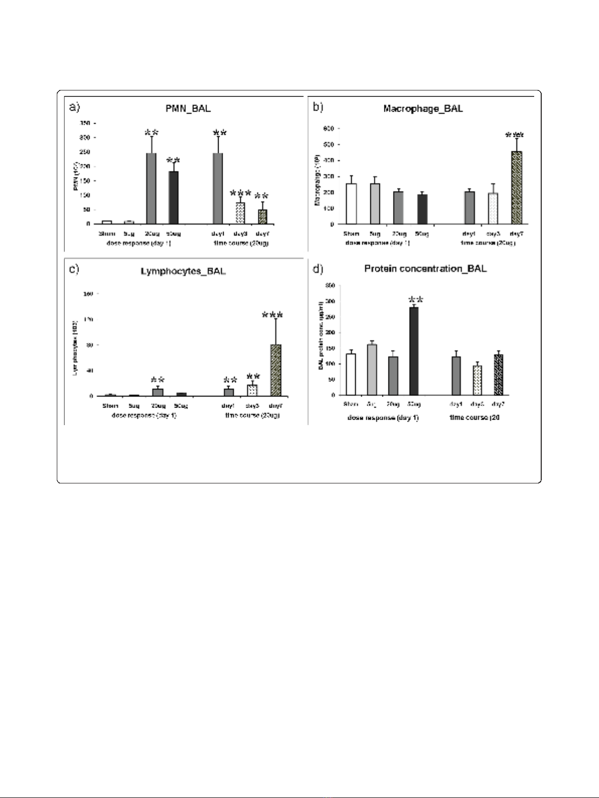
Dose and time response of BAL cells
The total number of retrieved BAL leucocytes was not
affected after i.t. instillation of 5 μg CNP (0.27 ± 0.04 ×
10E6 cells/lung versus 0.27 ± 0.05 × 10E6 cells/lung in
sham exposed mice) but increased two fold to 0.53 ±
0.06 × 10E6 cells/lung at day 1 (p < 0.05) after instillation
of 20 μg CNP (data not shown). The time course analysis
revealed an intermittent decline of BAL leucocytes almost
to control level on day 3 (0.31 ± 0.07 × 10E6 cells/lung,
n.s.) followed by an increase to 0.51 ± 0.11 × 10E6 cells/
lung on day 7, i.e. to 188% of that observed in sham
exposed mice.
PMN
As observed for the total cell count no significant induc-
tion of PMN was detected following i.t instillation of 5 μg
CNP (8.9 ± 2.3 × 10E3 PMNs) compared to control (9.7 ±
1.5 × 10E3 PMNs, Figure 1a). The increase of PMN num-
bers detected on day1 after 20 μgor50μg CNP instillation
already reached saturation at the 20 μg dosage. A 25-fold
induction of PMN was detected following 20 μg(246±58
× 10E3 PMNs) and a 19-fold after 50 μg (182 ± 31 × 10E3
PMNs) CNP instillation. Time course analysis of PMN
numbers revealed significantly reduced PMN counts after
3 days (73.9 ± 22.1 × 10E3 PMNs) and 7 days (48.5 ± 27.4
× 10E3 PMNs) compared to that of day1. However at day
3 and day 7 the PMN count remained 8 and 5 times
higher (< 0.05), respectively, than the baseline values indi-
cating incomplete resolution of the neutrophil influx
related inflammation following 20 μg of CNP instillation.
Macrophages
No dose dependent increase of macrophage numbers
was observed (Figure 1b). Strikingly, in the time course
analysis an obvious, 1.8-fold induction of macrophage
numbers was detected at day 7 (455 ± 83 × 10E3, vs.
sham 257 ± 48 × 10E3, p < 0.05).
Lymphocytes
The acute response of lymphocytes on day 1 is to some
extent comparable to that of PMNs showing the highest
influx with the 20 μgbutnotatthe50μg dosage (Fig-
ure 1c). Strikingly, in the time course analysis a moder-
ate induction of lymphocyte numbers in response to 20
μg CNP was detected on day 1 (4.9 times) and day3 (7.6
times) followed by a strong induction of lymphocyte
numbers being detected on day 7 (35.1 times). It is
interesting to note that the time course response of lym-
phocytes resembles that of the macrophages as both cell
types show a maximum influx at day 7.
BAL protein concentration
Compared to sham exposed mice, only the instillation of
the highest dose (50 μg/mouse) caused a significant (2.1
fold) increase in total BAL protein concentration (131.5
± 14.5 μg/ml versus 280.1 ± 8.3 μg/ml, p < 0.05) 1 day
after CNP instillation indicating alveolar barrier injury
with capillary leakage only at this concentration (Figure
1d). Time course investigation of 20 μginstilledlungs
revealed no changes of BAL protein concentrations
from day 1 to day 3 and day 7.
Histopathology
Histopathological analysis of paraffin embedded JF1 lung
sections (n = 4) showed a typical dose dependent accu-
mulation of particle laden macrophages on days 1, 3
and 7 (Figure 2a-e). In 50 μg/day1 samples inflammatory
cell infiltration (PMN) was clearly visible (Figure 2e)
whereas at 20 μg/day1 only slight PMN infiltration was
detectable (data not shown).
Molecular analysis for lung and BAL compartment
In the present study a panel of 62 protein markers was
applied to assess the CNP response in JF1 mice as
Ganguly et al.Respiratory Research 2011, 12:94
http://respiratory-research.com/content/12/1/94
Page 4 of 11
previously described for C3 mice [21]. A detailed list of
each of the 62 markers is supplied in Additional File 1,
Table S1.
Lung compartment
From the 62 markers 17 proteins did not exhibit any
response in lung and were therefore not considered for
analysis (CD40, CRP, EDN1, FGF9, F3, haptoglobin,
IgA, IL17, IL2, SAP, SCF, SGOT, TF, HO-1, GST-a,
myoglobin, VCAM1). Dose and time course responses
of the remaining 45 proteins are represented as a heat
mapinFigure3.IntheCNPdoseresponsemostpro-
teins showed a strong upregulation already at a dose of
20 μgandtheexpressionlevelof14ofthemdidnot
significantly increase further at a dose of 50 μg(F7,
FGA, GCP2/CXCL5, MCP1/CCL2, MCP3/CCL7,
MCP5/CCL12, IP10/CXCL10, KC/CXCL1, MDC/
CCL22, MIP1b/CCL4, MIP2/CXCL2, MIP1g/CCL9,
THPO and vWF). This observation can be associated
with the dose response of PMN numbers in the BAL as
these proteins include the major PMN recruiters
CXCL1, 2, 5 and 10. From the 45 proteins 13 proteins
were not significantly regulated at 20 μg/day1 but were
significantly induced at 50 μg/day1 (APOA1, CD40L,
EGF, FGF2, IFN-gamma, IL18, IL1B, IL3, IL4, IL5, M-
CSF/CSF1, MIP1a/CCL3, and RANTES/CCL5).
Forthetimecoursestudythemajorityofproteins
exhibited the expected response pattern of initial
increase (day1) and decline to baseline levels by day 7
following 20 μg CNP exposure. Among them were:
IL1B, MCP1/CCL2, MIP1/CCL4, MMP9, MCP3/CCL7,
IP10/CXCL10, MPO, MIP2/CXCL2, IL6, GM-CSF/
CSF2, MIP1-gamma/CCL9, GCP-2/CXCL5, TIMP1,
FGA, MCP-5/CCL12, KC/CXCL1. As evident from the
list, the major PMN recruiters CXCL1, 2, 5 and 10 were
down regulated at day 7 compared to day 1 reflecting
the phenotypic observation of an obvious decline in
PMN numbers until day 7 (Figure 3).
Figure 1 Bronchoalveolar lavage fluid (BAL) cell differentials and protein concentration. Dose dependent influx and time dependent
resolution of (a) polymorphonuclear leukocytes (PMN), (b) macrophages, and (c) lymphocytes in the BAL following intratracheal (i.t.) instillationof
carbon nanoparticles particles in the JF1/Msf (JF1) mice (n = 7 animals/experimental group). Total protein concentration is provided in Figure 1d.
(**) Significantly different with respect to (w.r.t) both sham control and 5 μg exposed; (***) significantly different w.r.t 20 μg exposed at day1,
sham control and 5 μg exposed; p ≤0.05.
Ganguly et al.Respiratory Research 2011, 12:94
http://respiratory-research.com/content/12/1/94
Page 5 of 11


