
Rosengren et al. Arthritis Research & Therapy 2010, 12:R65
http://arthritis-research.com/content/12/2/R65
Open Access
RESEARCH ARTICLE
BioMed Central
© 2010 Rosengren et al.; licensee BioMed Central Ltd. This is an open access article distributed under the terms of the Creative Com-
mons Attribution License (http://creativecommons.org/licenses/by/2.0), which permits unrestricted use, distribution, and reproduc-
tion in any medium, provided the original work is properly cited.
Research article
Platelet-derived growth factor and transforming
growth factor beta synergistically potentiate
inflammatory mediator synthesis by fibroblast-like
synoviocytes
Sanna Rosengren, Maripat Corr and David L Boyle*
Abstract
Introduction: The objective of this study was to model the effects of transforming growth factor beta (TGF-β) and
platelet-derived growth factor (PDGF), both present in rheumatoid arthritis (RA) synovia, on the behavior of fibroblast-
like synoviocytes (FLS) in response to pro-inflammatory cytokine (interleukin (IL)1β, tumor necrosis factor-alpha (TNFα))
challenge.
Methods: Gene and protein expression by fibroblast-like synoviocytes in vitro was studied by quantitative Polymerase
Chain Reaction (qPCR), ELISA and multiplex bead cytokine assays. Intracellular signaling pathway activation was
determined by Western blot for phospho-kinases and the use of specific inhibitors.
Results: In combination, TGF-β and PDGF (2GF) synergistically augmented TNFα- or IL1β-induced matrix
metalloproteinase 3 (MMP3), IL6, IL8, and macrophage inflammatory protein 1 alpha (MIP1α) secretion by FLS. Other
FLS-derived mediators remained unaffected. Individually, neither growth factor significantly potentiated TNFα or IL1β-
induced MMP3 secretion, and only slightly enhanced IL6. The effect of 2GF on TNFα-induced gene expression was
transcriptionally mediated; blocked by imatinib mesylate; and occurred even if 2GF was added as much as four hours
prior to TNFα. In addition, a 15-minute pulse of 2GF four hours prior to TNFα stimulation yielded a synergistic response.
The extracellular-signal-regulated kinase (ERK) and phosphoinositide 3-kinase (PI3K) signaling pathways were induced
for at least four hours by 2GF, as demonstrated by persistently upregulated levels of phospho-Akt and phospho-ERK.
However, pharmacologic inhibitor studies demonstrated that the potentiating action of 2GF was dependent on PI3
kinase only, and not on ERK.
Conclusions: The combination of PDGF and TGF-β dramatically potentiates FLS response to cytokines in a receptor-
mediated and PI3 kinase-dependent fashion. These data suggest that 2GF contribute to synovitis by directing synovial
fibroblasts toward a more aggressive phenotype in response to TNFα. Therefore, inhibition of growth factor signaling
may constitute a complementary therapeutic approach to cytokine-targeted treatments for RA.
Introduction
Expression of the regulatory peptides, platelet-derived
growth factor (PDGF) and transforming growth factor
beta (TGF-β) are increased in synovial tissue and fluid of
rheumatoid arthritis (RA) patients [1-4]. PDGF has been
implicated in RA pathogenesis, mainly through its func-
tion as a growth factor for fibroblast-like synoviocytes
(FLS) [3,5]. In contrast, the actions of TGF-β are more
complex. TGF-β plays a crucial role in maintaining
immunological tolerance through the inhibition of lym-
phocytes and macrophages [6]. On the other hand, it
recruits and activates naive monocytes [6], stimulates
proliferation [7] and induces aggrecanase synthesis [8] by
FLS. Systemic administration of TGF-β protects against
development of collagen arthritis in mice [9], whereas
direct injection of TGF-β into rat joints leads to pro-
nounced synovitis [10].
* Correspondence: dboyle@ucsd.edu
1 Division of Rheumatology, Allergy and Immunology, University of California at
San Diego School of Medicine, 9500 Gilman Drive, La Jolla, CA 92093-0656, USA
Full list of author information is available at the end of the article

Rosengren et al. Arthritis Research & Therapy 2010, 12:R65
http://arthritis-research.com/content/12/2/R65
Page 2 of 11
In addition to these growth factors, chronically
inflamed RA synovia contain a multitude of inflamma-
tory mediators that may act in concert with each other. In
this context, aggravating as well as mitigating effects of
growth factors and cytokines on FLS have been demon-
strated. For example, PDGF was reported to enhance
IL1β-induced prostaglandin E2 production, while inhibit-
ing collagenase synthesis [11]. Also, PDGF was shown to
induce synthesis of IL8 and MIP1α, along with IL1β, by
FLS [12], and also to synergize with TNFα to stimulate
IL1β secretion, although these results are somewhat con-
fusing since FLS are not typically considered a significant
source of IL1β. On the other hand, TGF-β was earlier
shown to inhibit TNFα-induced RANTES synthesis by
FLS [13]. A systematic study of the nature of the interac-
tion among these mediators was not undertaken to date.
Hence, the interplay between PDGF, TGF-β, and cytok-
ines such as TNFα and IL1β on the activation of FLS
remains unclear, albeit of potential significance consider-
ing the abundance of these proteins in the RA synovial
environment.
Consequently, we set out to systematically determine
the effect of PDGF and TGF-β, alone and in combination,
on inflammatory biomarker expression and secretion by
FLS. We describe significant potentiation by PDGF and
TGF-β of the production of certain cytokines, chemok-
ines, and matrix metalloproteinases (MMP) by FLS. This
synergy was mediated by tyrosine-kinase receptor activa-
tion and dependent on PI3K, both of which are receiving
attention as possible novel approaches to RA drug ther-
apy.
Materials and methods
Reagents
Cytokines and TGF-β were obtained from R&D Labora-
tories (Minneapolis, MN, USA). Imatinib mesylate (LC
Laboratories, Woburn, MA, USA) was dissolved in water.
All other reagents, including PDGF-BB, were from Sigma
(St. Louis, MO, USA) unless otherwise noted. Stock solu-
tions in DMSO (1000×) of PD98059 and LY294002 were
kept at -80°C.
Fibroblast-like synoviocytes (FLS)
FLS were cultured from the synovial tissues of RA
patients undergoing arthroplastic surgery, as previously
described [14], after obtaining informed consent under
approval from the University of California, San Diego
Institutional Review Board, and maintained in Dulbecco's
Modified Eagle Medium (DMEM) supplemented with
antibiotics, glutamine, and 10% fetal bovine serum. Pas-
sages 4 through 8 were used in experiments. Cells were
subjected to a two- to three-day reduced serum condition
(0.1% fetal bovine serum) prior to stimulation to mini-
mize baseline activity.
Secreted protein assays
FLS supernatants at 24 hours following stimulation were
assayed by ELISA for IL6 (eBioscience, San Diego, CA,
USA), MMP1, and MMP3 (GE Healthcare Life Sciences,
Piscataway, NJ, USA). Standard curves were constructed
by regression line fitting on log(absorbance) vs log(con-
centration). Levels of cytokines and chemokines in super-
natants were determined by Luminex multiplex analysis
(BioRad Bio-Plex assays, Hercules, CA, USA) from four-
parameter standard curve fits.
Gene expression assays
Messenger RNA for IL6, MIP1α, and MMP3 were quanti-
fied by real-time TaqMan quantitative Polymerase Chain
Reaction (qPCR), using FLS cDNA, with GAPDH used as
a housekeeper (all reagents from Applied Biosystems,
Foster City, CA, USA). Resulting threshold cycle (Ct) data
were normalized to standard curves constructed from
cDNA from IL1β-stimulated FLS [15], yielding cell equiv-
alents. The ratio between the specific cytokine and
GAPDH cell equivalents (relative expression units, REU)
is reported.
Western blot
FLS extracts were prepared in RIPA buffer with Complete
Protease Inhibitors (Roche Applied Science, Indianapolis,
IN, USA), denatured in sample buffer and 0.1 M
dithiotreitol, and fractioned on Invitrogen (Carlsbad, CA,
USA) NuPage 4 to 12% precast gels. Following blotting to
polyvinylidene fluoride (PVDF) membranes and blocking
with 5% dry milk, blots were probed with antibodies
against phospho- or total p38, JNK, Erk, or Akt, as well as
with secondary anti-rabbit-IgG-HRP (all Cell Signaling
Technologies, Danvers, MA, USA). GAPDH was used as
a gel loading control (antibody from Santa Cruz Biotech-
nology, Santa Cruz, CA, USA). Membranes were devel-
oped with Immun-Star WesternC ECL substrate (BioRad,
Hercules, CA, USA) and imaged on a VersaDoc imaging
system (BioRad), using QuantityOne software (Hercules,
CA, USA) for image capture and densitometry.
Statistical analysis
Data are reported as mean and standard error of the
mean (SEM). Protein secretion and gene expression data
in single time-point experiments were analyzed by one-
way ANOVA followed by Tukey-Kramer's post-hoc test
comparing all groups, or by Dunnett's post-hoc test com-
paring control to all others, as appropriate. Time course
data were analyzed by two-way ANOVA followed by con-
trast testing. Student's t-test was used to examine syner-
Rosengren et al. Arthritis Research & Therapy 2010, 12:R65
http://arthritis-research.com/content/12/2/R65
Page 3 of 11
gistic effects of growth factors and cytokines. Real-time
qPCR data were log-transformed prior to analysis.
Results
Effect of PDGF-BB and TGF-β on FLS secretion of
inflammatory mediators
Since PDGF and TGF-β are abundant in the rheumatoid
synovium, their effect on cytokine-induced inflammatory
mediator secretion by FLS was examined. TGF-β induced
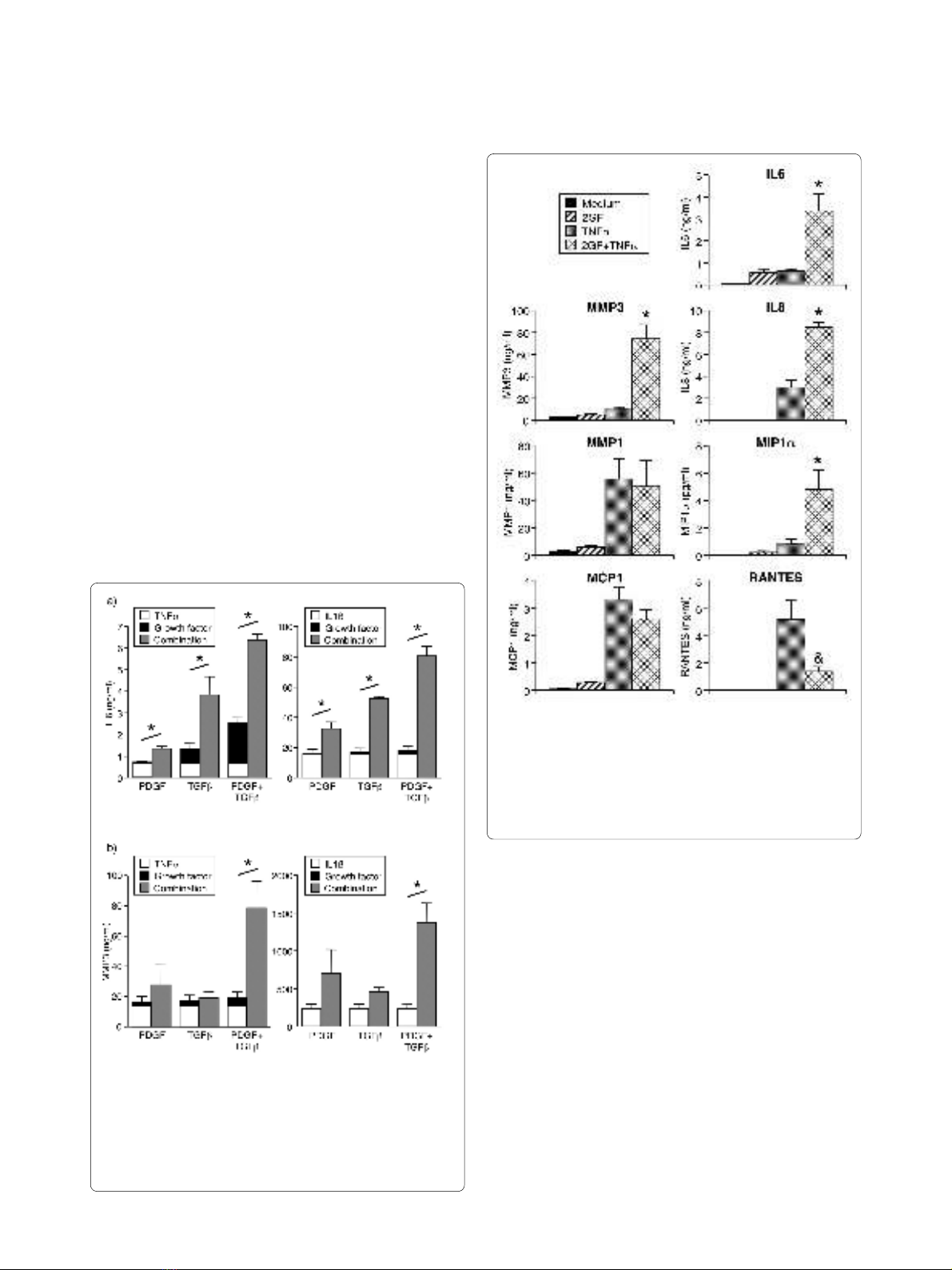
only a small amount of IL6 (Figure 1a), and no effect on
IL6 (Figure 1a) or MMP3 (Figure 1b) was observed by
PDGF-BB alone. PDGF and TGF-β in combination (2GF)
induced low-level secretion of IL6, but not MMPs or
chemokines (Figures 1 and 2). The amount of IL6
secreted after 2GF stimulation was comparable to that
observed with TNFα as the stimulant (Figure 2).
Surprisingly, the two growth factors in combination
potently augmented secretion of IL6 (Figure 1a) and
MMP3 (Figure 1b) in response to TNFα or IL1β. The
effect of 2GF was truly synergistic, in that the secretion
observed by 2GF and TNFα or IL1β in combination was
significantly higher than that obtained when adding the
values for 2GF alone and cytokine alone (Figure 1). When
PDGF-BB and TGF-β were examined individually, nei-
ther augmented TNF- or IL1β-induced MMP3 secretion,
and the effect on TNF- or IL1β-induced IL6 secretion
was smaller than that of the growth factor combination
(Figure 1). The potentiating effect of 2GF was not simply
due to a non-specific effect of cell activation, since the
secretion of some but not all mediators was affected.
TNFα-induced secretion of MMP1 and MCP1 was unal-
tered by addition of 2GF, and RANTES secretion was
inhibited, at the same time that IL8 and MIP1α secretion
was potentiated (Figure 2) along with that of IL6 and
MMP3.
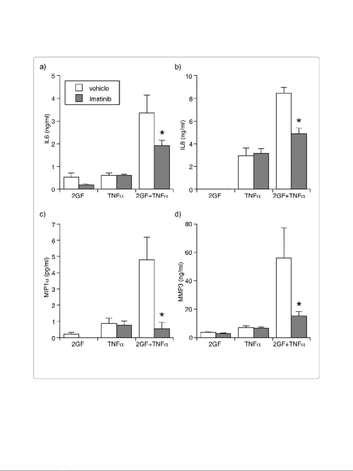
The effect of 2GF was mediated through activation of
growth factor receptors, since the receptor tyrosine
kinase inhibitor, imatinib mesylate significantly reversed
Figure 1 Potentiation by PDGF alone, TGF-β alone, or their com-
bination (2GF), of (a) IL6 and (b) MMP3 secretion from FLS. FLS
were cultured for 24 hours with TNFα (10 ng/ml) or IL1β (2 ng/ml), and/
or growth factors (10 ng/ml), and supernatants analyzed by ELISA.
Mean & SEM, n = 3 RA FLS lines. Asterisk indicates P < 0.05 between the
combination and the added values for TNF alone and growth factor
alone by Students' t-test.
Figure 2 Augmentation by 2GF of FLS secretion of particular cy-
tokines, chemokines and MMPs induced by TNFα. FLS were cul-
tured for 24 hours with TNFα and growth factors as in Figure 1, and
supernatants analyzed by ELISA (MMPs) or Luminex multiplex bead as-
say (all others). Mean & SEM, n = 3 to 6 RA FLS lines. Asterisk indicates P
< 0.05 to TNFα alone and 2GF alone, and ampersand indicates P < 0.05
to TNFα alone, by ANOVA/Tukey-Kramer's.
Rosengren et al. Arthritis Research & Therapy 2010, 12:R65
http://arthritis-research.com/content/12/2/R65
Page 4 of 11
the potentiating effect of 2GF on TNFα-induced secre-
tion of IL6, IL8, MIP1α, and MMP3 (Figure 3). Impor-
tantly, imatinib did not alter secretion of these mediators
in response to TNFα alone.
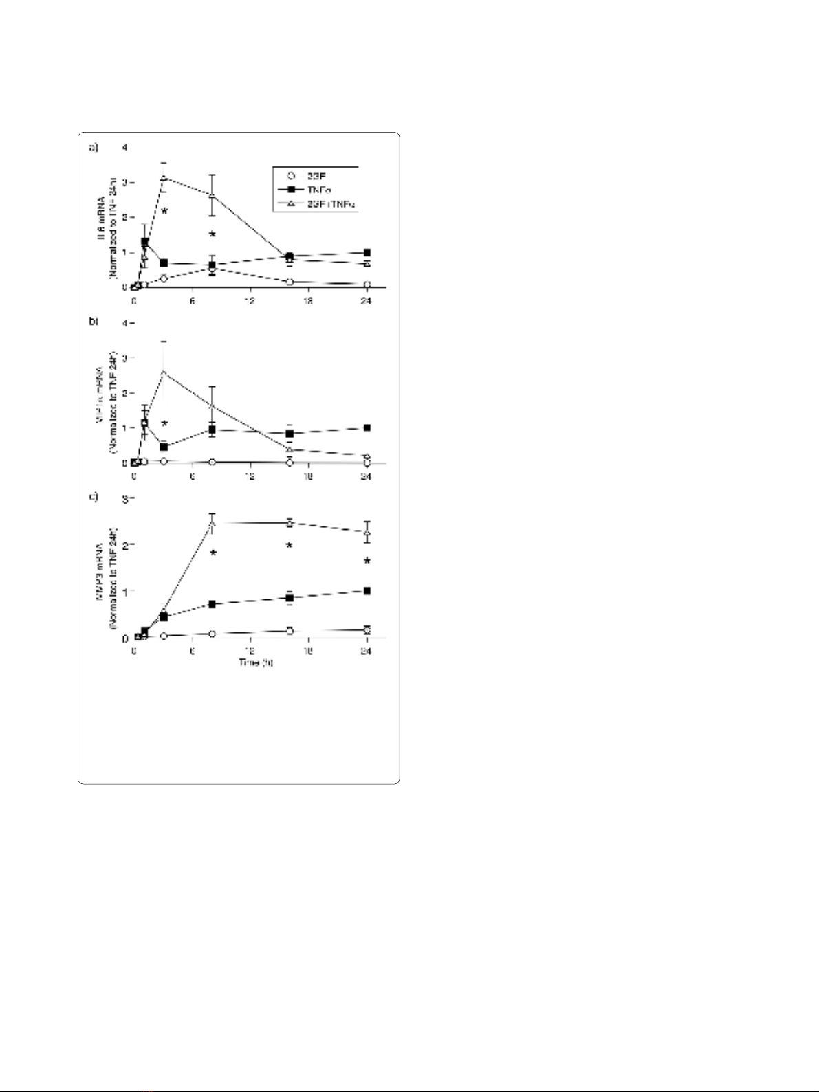
Effect of PDGF-BB and TGF-β on the time course of FLS
mRNA expression
In order to determine whether the effect of 2GF on FLS
protein secretion was observed at the mRNA expression
level, a time course experiment was conducted and the
expression of IL6, MIP1α, and MMP3 mRNA in FLS was
studied. TNFα caused a rapid rise in IL6 (Figure 4a) and
MIP1α (Figure 4b) mRNA expression, reaching a plateau
at one hour and maintaining significant expression until
the end of the experiment at 24 h. 2GF alone induced a
small amount of IL6 mRNA at three and eight hours, but
no MIP1α. When 2GF and TNFα was added in combina-
Figure 3 Reversal by imatinib (1 μM) of 2GF potentiation of TNFα-induced (a) IL6, (b) IL8, (c) MIP1α, and (d) MMP3 secretion. For culture con-
ditions and definitions, see legends for Figure 1 and 2. Supernatants were analyzed by ELISA or Luminex multiplex bead assay. Mean & SEM, n = 3 RA
FLS lines. Asterisk indicates P < 0.05 to vehicle by Students' t-test.
Rosengren et al. Arthritis Research & Therapy 2010, 12:R65
http://arthritis-research.com/content/12/2/R65
Page 5 of 11
tion, significantly elevated IL6 levels were observed at
three and eight hours (Figure 4a). For MIP1α (Figure 4b),
potentiation by 2GF of TNFα-induced chemokine was
only observed at three hours. Similar results were
obtained for IL8 expression (data not shown). In the case
of MMP3, TNFα alone induced a slow steady increase of
mRNA levels evident from three hours and lasting until
the end of the experiment at 24 h. The addition of 2GF in
combination with TNFα led to significantly elevated
MMP3 levels at 8, 16 and 24 h (Figure 4c). Thus, the syn-
ergistic effect of 2GF on TNFα-induced inflammatory
mediator production by FLS is evident at the transcrip-
tional level.
Effect of temporal separation of the addition of growth
factors and TNFα to FLS
Next, the addition of 2GF and TNFα was separated in
time to determine whether the potentiating effect of 2GF
would be maintained. PDGF and TGF-β were added at
various time points in relation to TNFα, which was in
turn allowed to stimulate the FLS for 24 h before super-
natants were analyzed for secreted proteins. Under these
conditions, 2GF was able to potentiate TNFα-induced
IL6, IL8 and MMP3 secretion when added at any time
between -2 h and +2 h in relation to a TNFα addition
(Figure 5a). The extent of the potentiating effect was sim-
ilar to that observed when 2GF and TNFα were added
simultaneously (crosshatched bars). For IL6 and MMP3
secretion, potentiation by 2GF was also observed when
added as much as six hours prior to TNFα (Figure 5a).
In similar experiments studying the gene mRNA
expression at three hours following TNFα addition, 2GF
synergistically potentiated TNFα-induced IL6 expression
when added between -4 h and +2 h in relation to TNFα
addition (Figure 5b). In separate experiments, FLS could
be exposed to 2GF for as little as 15 minutes, even when
added as early as four hours before TNFα, and signifi-
cantly elevated IL6 expression could still be noted (Figure
5c). This suggests that the synergistic effect does not
require continuous exposure to the 2GF, and that it
involves signaling pathways that are maintained over the
course of several hours.
Sustained activation of Erk and Akt in FLS by growth factors
For the purpose of elucidating the relevant signaling
pathways causing the synergistic effect, FLS were treated
with TNFα, 2GF, or a combination for 15 minutes to four
hours, and cell extracts analyzed by Western blot (Figure
6a). TNFα induced a short-lived peak of phosphorylation
of p38, JNK isoforms, and ERK isoforms (Figure 6b-e) but
had a marginal effect on Akt phosphorylation (Figure 6f).
In contrast, 2GF induced a different pattern: phosphory-
lation of ERK and Akt that lasted for the four hours stud-
ied (Figure 6e-f), no phosphorylation of p38 (Figure 6b)
nor JNK-p54 (Figure 6d), and a short-lived upregulation
of phospho-JNK-p46 (Figure 6c). In combination, 2GF
and TNFα generated phospho-protein levels similar to
those induced by the mediators added separately, with
the sole exception of phospho-JNK which was signifi-
cantly higher after 15 minutes of 2GF + TNFα than after
TNF alone or 2GF alone (Figure 6c, d). At the four-hour
time point, no synergistic effect of 2GF and TNFα was
noted on any phospho-protein studied. These studies
Figure 4 Time course of 2GF-induced potentiation of (a) IL6, (b)
MIP1α and (c) MMP3 RNA induced by TNFα. FLS were cultured for
indicated times with TNFα and growth factors, and mRNA levels quan-
tified by real-time qPCR using GAPDH as housekeeper. Data are nor-
malized to levels with TNFα alone at 24 h. Mean & SEM, n = 3 RA FLS
lines. Asterisk indicates P < 0.05 to TNF alone and 2GF alone by two-
way ANOVA and contrast testing on log-transformed data.


