
Open Access
Available online http://arthritis-research.com/content/9/3/R49
Page 1 of 10
(page number not for citation purposes)
Vol 9 No 3
Research article
Pro-apoptotic Bid is required for the resolution of the effector
phase of inflammatory arthritis
John C Scatizzi1, Jack Hutcheson1, Emily Bickel1, G Kenneth Haines III2 and Harris Perlman1,2
1Saint Louis University, School of Medicine, Department of Molecular Microbiology and Immunology, Saint Louis, MO 63104, USA
2Yale University, School of Medicine, Department of Pathology, New Haven CT 06510, USA
Corresponding author: Harris Perlman, perlmanh@slu.edu
Received: 12 Feb 2007 Revisions requested: 16 Mar 2007 Revisions received: 10 Apr 2007 Accepted: 17 May 2007 Published: 17 May 2007
Arthritis Research & Therapy 2007, 9:R49 (doi:10.1186/ar2204)
This article is online at: http://arthritis-research.com/content/9/3/R49
© 2007 Scatizzi et al.; licensee BioMed Central Ltd.
This is an open access article distributed under the terms of the Creative Commons Attribution License (http://creativecommons.org/licenses/by/2.0),
which permits unrestricted use, distribution, and reproduction in any medium, provided the original work is properly cited.
Abstract
Rheumatoid arthritis is an autoimmune disease characterized by
hyperplasia of the synovial lining and destruction of cartilage and
bone. Recent studies have suggested that a lack of apoptosis
contributes to the hyperplasia of the synovial lining and to the
failure in eliminating autoreactive cells. Mice lacking Fas or Bim,
two pro-apoptotic proteins that mediate the extrinsic and
intrinsic death cascades, respectively, develop enhanced K/BxN
serum transfer-induced arthritis. Since the pro-apoptotic protein
Bid functions as an intermediate between the extrinsic and
intrinsic apoptotic pathways, we examined the role that it plays
in inflammatory arthritis. Mice deficient in Bid (Bid-/-) show a
delay in the resolution of K/BxN serum transfer-induced arthritis.
Bid-/- mice display increased inflammation, bone destruction,
and pannus formation compared to wild-type mice. Furthermore,
Bid-/- mice have elevated levels of CXC chemokine and IL-1β in
serum, which are associated with more inflammatory cells
throughout the arthritic joint. In addition, there are fewer
apoptotic cells in the synovium of Bid-/- compared to Wt mice.
These data suggest that extrinsic and intrinsic apoptotic
pathways cooperate through Bid to limit development of
inflammatory arthritis.
Introduction
Rheumatoid arthritis (RA) is an autoimmune disease charac-
terized by hyperplasia of the synovial lining, inflammation, high
levels of circulating and local IL-1β and tumor necrosis factor
(TNF)α, and destruction of cartilage and bone. Antagonists to
IL-1β or TNFα lead to decreased joint destruction through an
unknown mechanism and also result in reduced numbers of
macrophages [1], one of the principal cell types that contrib-
ute to the pathogenesis of RA. Since the numbers of macro-
phages are associated with worse clinical outcome [2,3], one
prevailing hypothesis is that there is a failure to delete autore-
active cells, particularly macrophages, in the RA joint [4].
While the RA joint is replete with noxious molecules, including
reactive oxidative species and death ligand expressing cells,
histological evidence of apoptosis is rarely observed [5,6]. The
induction of synoviocyte apoptosis in animal models of inflam-
matory arthritis results in either amelioration of the disease or
reduction in joint inflammation and destruction [7-9]. Addition-
ally, patients with pauciarticular juvenile chronic arthritis dis-
play enhanced mononuclear cell apoptosis in synovial tissue
compared to patients with polyarticular arthritis [10]. These
data suggest that increasing the level of apoptosis in the joint
may be associated with improved clinical outcome. However,
the apoptotic factors that are essential to limit the inflammatory
response in RA remain elusive.
Apoptosis proceeds through two major pathways, an 'intrinsic'
pathway that signals through the mitochondria, and an 'extrin-
sic' pathway that transduces an apoptotic signal following the
aggregation of a death receptor to its ligand. The intrinsic
pathway is regulated by the Bcl-2 protein family, which are
divided into anti-apoptotic (Bcl-2, Bcl-xL, Mcl-1, A1/Bfl-1 and
Bcl-w) and pro-apoptotic (Bax, Bak, Bad, Bim/Bod, Bok/Mtd,
Bik/Blk/Nbk, Bid, Hrk/DP5, Bmf, Noxa, Puma/Bbc3) members
[11]. The pro-apoptotic family proteins are divided into two
additional groups based on the expression of the Bcl-2-hom-
ology (BH 1–4) domain: the multi-BH domain (BH1-3: for
example, Bak, Bax) and the BH3-only (for example, Bid, Bim)
proteins [12]. Recent studies have suggested that BH3-only
proteins are also subdivided into two categories based on
BH = Bcl-2-homology; Wt = wild-type; ELISA = enzyme-linked immunosorbent assay; FasL = Fas ligand; H&E = hematoxylin and eosin; IL = inter-
leukin; KC = CXC chemokine; MCP = monocyte chemoattractant protein; PMN = polymorphonuclear; RA = rheumatoid arthritis; TNF = tumor necro-
sis factor; TUNEL = terminal transferase dUTP nick end labeling.

Arthritis Research & Therapy Vol 9 No 3 Scatizzi et al.
Page 2 of 10
(page number not for citation purposes)
their ability to induce apoptosis [13-17]. Bid and Bim are suf-
ficient to sequester anti-apoptotic Bcl-2 family members,
induce oligomerization of Bak and Bax, permeabilization of
liposomes, and release of cytochrome C [13,14,17]. In con-
trast, Bad, Bmf, Hrk, Noxa, and Puma are sensitizers for apop-
tosis since they are able to bind only to the anti-apoptotic Bcl-
2 members and require Bid or Bim to induce the death
response [13-17]. During the intrinsic apoptotic death, cyto-
chrome C is released into the cytosol from the inner mitochon-
drial space, where it binds to Apaf1, forming the apoptosome.
This complex leads to the activation of initiator pro-caspase 9
[18,19]. Caspase 9 activates caspases 3 and 7 [20], which
then induce the downstream degradative events of apoptosis
[21]. These events are prevented by the overexpression of Bcl-
2/Bcl-xL or by the complete ablation of Bax and Bak [22].
The induction of apoptosis mediated by the extrinsic pathway
is initiated by binding of death ligands to their receptors, as in
the case of Fas ligand (FasL) binding to Fas. Oligomerization
of Fas upon FasL binding leads to the recruitment of both
FADD and pro-caspase 8 to the carboxyl terminus of Fas [23].
Aggregation or oligomerization of pro-caspase 8 results in its
autocatalysis and/or activation, and the induction of the degra-
dative phase of apoptosis through the activation of caspases
3 and 7 [23]. An additional pathway of death receptor-induced
cell death may proceed through the mitochondrial pathway by
activating the Bcl-2 pro-apoptotic protein Bid [24,25], which
is cleaved by caspase 8 following death receptor ligation.
Cleaved Bid is targeted to the mitochondria and ultimately
results in the induction of apoptosis mediated by the mito-
chondrial apoptotic pathway [26].
The vast majority of studies in RA have focused on the expres-
sion patterns of Bcl-2 family members and death receptor sig-
naling factors in the synovium. Recently, two studies have
demonstrated that Fas and Bim are required to limit the inflam-
matory response in a mouse model of the effector phase of
inflammatory arthritis [27,28]. These data suggest that a syn-
ergy between the extrinsic and intrinsic apoptotic pathways
may be required to prevent or reduce the development of
inflammatory arthritis. One potential factor that bridges the two
apoptotic pathways is the BH3-only protein Bid. To this end,
we examined the impact of deleting Bid (Bid-/-) on the devel-
opment of inflammatory arthritis in mice. Bid-/- mice show
increased ankle swelling accompanied by more articular
destruction and a delay in the resolution phase of arthritis. His-
tological examination of arthritic ankle sections reveals an
increase in infiltrating leukocytes, particularly macrophages
and neutrophils in Bid-/- mice compared to controls. Further-
more, there are fewer apoptotic cells in Bid-/- mice. Collec-
tively, these data suggest that the decreased apoptosis in Bid-
/- mice prolongs the inflammatory phase, leading to enhanced
joint destruction and a delay in the resolution phase.
Materials and methods
Mice
Bid-/- mice backcrossed for 12 generations onto C57BL/6
background were a kind gift from the late Dr Stanley Kors-
meyer (Dana-Farber Cancer Institute, Boston, MA, USA).
C57BL/6 mice (congenic control for Bid-/- mice) were pur-
chased from Jackson Laboratory (Bar Harbor, ME, USA). Non-
obese diabetes (NOD) mice were purchased from Taconic
(Germantown, NY, USA) and the homozygous KRN T-cell
receptor transgenic mice (C57BL/6 background) were a kind
gift from Drs D Mathis and C Benoist (Harvard Medical
School, Boston MA, USA, and the Institute de Gene-tique et
de Biologie Moleculaire et Cellulaire, Strasbourg, France). All
experiments on mice were approved by the Animal Care and
Use Committee at Saint Louis University.
K/BxN serum transfer-induced arthritis
The KRN T-cell receptor transgenic mouse was crossed with
the NOD mouse expressing the Ag7 MHC class II allele and all
progeny (K/BxN) developed spontaneous arthritis [29,30].
Serum from K/BxN mice may be transferred via intra-peritoneal
injection to allogeneic hosts regardless of the genetic back-
ground [31]. The host mice develop a transient inflammatory
joint disease that lasts for 7 to 14 days. Peripheral blood from
seven-week-old K/BxN mice was isolated, and serum were
collected and pooled. K/BxN serum (150 μl) was intraperito-
neally injected on each flank of 6-week old wild-type (Wt) and
Bid-/- mice as previously described [32]. At each time point
and prior to euthanasia, the degree of arthritis as indicated by
joint swelling was quantified by measuring two perpendicular
diameters of the ankles using a caliper (Lange Caliper: Cam-
bridge Scientific Industries, Cambridge, MA, USA). Joint cir-
cumference was calculated using the geometric formula of
ellipse circumference (2π × v(a2 + b2)/2) as previously
described [32]. Following euthanasia, ankle joints were
removed and either fixed in 10% neutral buffered formalin for
24 hours, decalcified in EDTA-decalcification buffer for two
weeks, embedded in paraffin, and sectioned, or placed in liq-
uid nitrogen, ground into a fine powder by mortar and pestle,
digested in protein lysis buffer (150 μM NaCl, 0.5% NP-40,
50 mM Tris, 2 mM EDTA) in the presence of phosphatase and
protease inhibitors, homogenized on ice for 20 s, and lysed
overnight at 4°C.
Immunohistochemistry
Paraffin embedded ankle sections were stained with hematox-
ylin and eosin (H&E) and Safranin O and methyl green. His-
topathological scoring was performed as previously described
in detail [28,33,34]. A pathologist blinded to the study (GKH)
evaluated ankle sections by examining at least 3 sections/
ankle and 3 fields/section at 1,000 × magnification. H&E ankle
sections were scored on a 0 to 5 scale for inflammation, with
0 = normal, 1 = minimal infiltration, 2 = mild infiltration, 3 =
moderate infiltration, 4 = marked infiltration, and 5 = severe
infiltration. Bone erosion was scored on a 0 to 5 scale by

Available online http://arthritis-research.com/content/9/3/R49
Page 3 of 10
(page number not for citation purposes)
viewing H&E ankle sections, with 0 = no or normal bone
resorption, 1 = small areas of resorption, 2 = more numerous
areas of resorption, 3 = obvious resorption, 4 = full thickness
defects in the bone without distortion of the profile, 5 = full
thickness defects in the bone with distortion of the profile.
H&E ankle stained sections were scored on a 0 to 5 scale for
pannus formation, with 0 = no pannus formation, 1 = minimal
pannus formation, 2 = mild pannus formation, 3 = moderate
pannus formation, 4 = marked pannus formation, and 5 =
severe pannus formation. Polymorphonuclear (PMN) leuko-
cyte infiltration: 0 = no PMNs, 1 = rare scattered PMNs, 2 =
more frequent scattered PMNs, 3 = small clusters of PMNs, 4
= larger clusters of PMNs, and 5 = sheets of PMNs (abscess).
Histopathological scoring was conducted on an Olympus
BX40 microscope (1,000 ×). Photographs were taken on a
Nikon (Tokyo, Japan) microscope equipped with the Nikon
digital camera DMX1200.
Macrophages were identified by the expression of F4/80 anti-
gen, a cell surface glycoprotein with homology to the G-pro-
tein linked transmembrane 7 hormone receptor family [35].
Previous studies have shown that F4/80 is expressed on all
macrophages [36,37] and that macrophages isolated from
mice lacking F4/80 do not stain for the F4/80 antigen [38,39].
To stain for F4/80-positivity, antigens were retrieved using the
Dako target retrieval solution (Dako, Carpinteria, CA). Follow-
ing antigen retrieval, sections were blocked in hydrogen per-
oxide, incubated with anti-F4/80 antibody (Clone BM8;
Invitrogen, Carlsbad, CA) or isotype control, and then incu-
bated with secondary biotinylated rabbit anti-rat antibody
(Dako). Sections were treated with streptavidin peroxidase
conjugate (Dako), color was visualized with diaminobenzidine,
and sections were counterstained with hematoxylin. All F4/80
antigen staining was performed on a DAKO autostainer
(Dako). Six fields of representative pannus and synovium
stained with anti-F4/80 antibody were viewed under oil emer-
sion at 1,000 × magnification, and the number of F4/80 posi-
tive cells was counted.
Immunophenotyping
Peripheral blood was isolated by cardiac sticks from Wt and
Bid-/- mice after euthanasia. Nonspecific staining was pre-
vented by incubation with anti-CD16/32 (24G2) antibody (BD
Biosciences, San Jose, CA). The blood was incubated with
fluorochrome-conjugated antibodies specific for CD3, CD4,
CD8, CD19, CD11b, CD45, CD62L, and Gr-1 (BD Bio-
sciences), or isotype controls for 30 minutes at 4°C. After
incubation with antibodies, red blood cells were lysed and the
samples were fixed by incubation in FACS Lyse (BD Bio-
sciences) for 10 minutes at room temperature. Samples were
collected on a BD FACS Calibur at the St Louis University
Flow Cytometry Core Facility, and the data were analyzed in
FlowJo (TreeStar, Inc. Ashland, OR). Total peripheral blood
leukocyte numbers were determined on the automated hema-
tology analyzer ABX Pentra 60.
ELISA
For detection of mouse CXC chemokine (KC), monocyte che-
moattractant protein (MCP-1/CCL2), TNFα, and IL-1β in ankle
extracts, sandwich ELISAs were performed according to the
manufacturer's instructions (R & D Systems, Minneapolis, MN,
USA). The sensitivity of TNFα and MCP-1 ELISAs was 7.8 pg/
ml, while the sensitivity of IL-1β and KC ELISAs was 15.6 pg/
ml. ELISAs were quantified by absorbance at 450 nm on a
microplate reader (BioRad, Hercules, CA, USA). Data
obtained using ELISA on ankle extracts (pg/ml) were normal-
ized by the total protein concentration (μg/μl) for each individ-
ual ankle extract. The levels of cytokines and chemokines in
serum were determined using a Luminex based assay accord-
ing to manufacturer's specifications (Linco Research, Earth
City, MO).
TUNEL analysis
Paraffin embedded ankle sections (5 μm) were deparaffinized,
rehydrated, and permeabilized with 20 μg/ml of proteinase K
for 15 minutes. Mouse thymus was used as a positive control
for TUNEL (data not shown). TdT enzyme and dUTP conju-
gated to a fluorescein cocktail were added to sections accord-
ing to the manufacturer's specifications (in situ death
detection kit; Roche Biochemical, Indianapolis, IN, USA).
Nuclei were stained with Hoechst 33258 (Invitrogen). Slides
were mounted with glass coverslips using mounting medium
for fluorescence (Kirkegaard and Perry Laboratories Inc.,
Gaithersburg, MD, USA). Three different areas per joint of
TUNEL positive cells were identified at 400 × power. The
number of TUNEL positive cells were counted and then
divided by the total number of cells in the field as determined
by Hoechst staining. The percent of TUNEL positive cells per
field was averaged with two other fields identified from differ-
ent areas of the joint. Photographs were taken on a Nikon
microscope equipped with the Nikon digital camera
DMX1200.
Statistical analysis
Results were expressed as the mean ± standard error. Differ-
ences between groups were analyzed using Student's t test.
Results
Bid-/- mice have a delay in the resolution of
inflammatory arthritis following transfer of K/BxN serum
Previous studies have implicated the extrinsic and intrinsic
apoptotic pathways in preventing or limiting the effector phase
of inflammatory arthritis [27,28]. Since the pro-apoptotic pro-
tein Bid links the extrinsic to the intrinsic pathway, we exam-
ined the affect of inducing experimental inflammatory arthritis
in mice lacking Bid (Bid-/- mice). We used the K/BxN serum
transfer-induced arthritis model, which is widely used to
assess factors that mediate the effector phase of RA. Unlike
the collagen-induced arthritis model, the K/BxN model may be
used in mice on a C57BL/6 background [31]. This model
shares many common features with human RA, including
Arthritis Research & Therapy Vol 9 No 3 Scatizzi et al.
Page 4 of 10
(page number not for citation purposes)
invasion of leukocytes, proliferation of synoviocytes resulting in
the thickening of the synovial lining, formation of pannus, and
erosion of cartilage and bone [40]. The K/BxN serum transfer
model is independent of T and B lymphocytes [30], but
requires Fc receptors [41,42] and the alternative pathway of
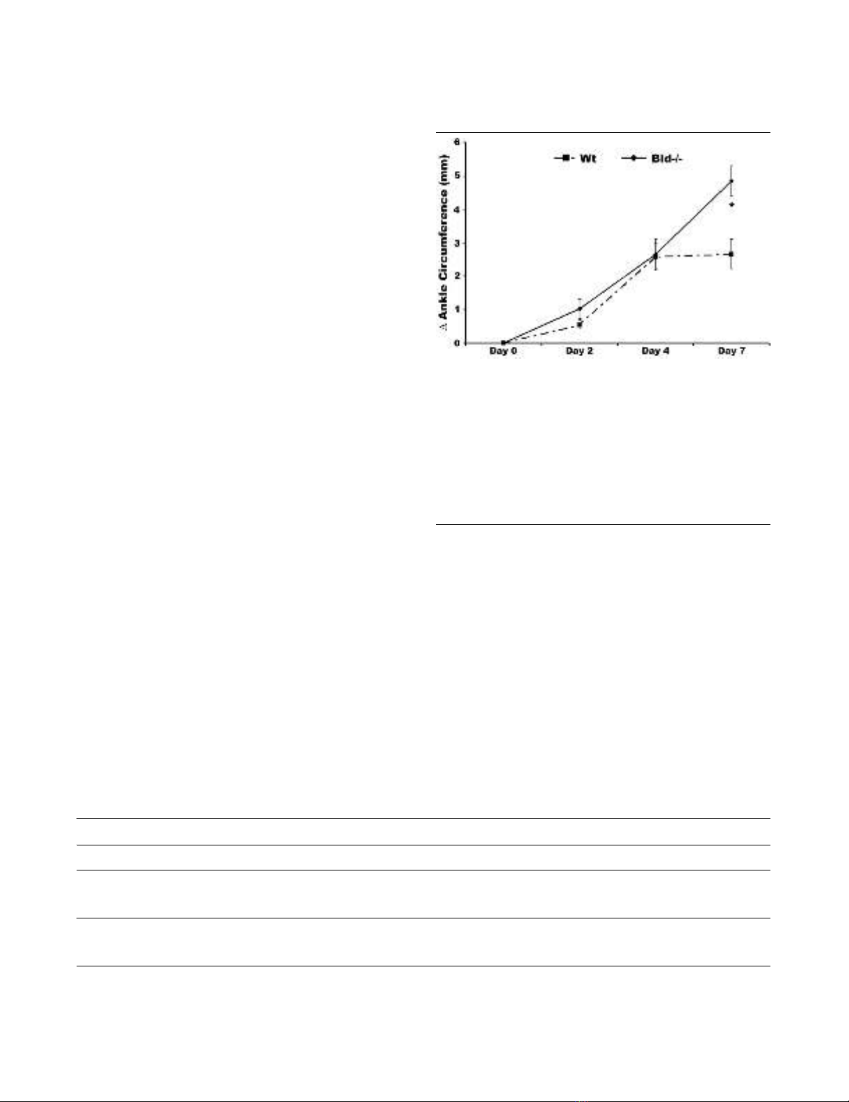
complement [31,42]. There was no difference in edema of the
ankle joint in Bid-/- compared to Wt mice at days two and four
post-serum transfer as indicated by a change in ankle circum-
ference (Figure 1). However, ankle circumference increased
by 2.0-fold (p < 0.002) in Bid-/- compared to Wt mice at day
seven. There was no change in ankle swelling in Wt mice
between days four and seven. These data suggest that the
loss of Bid causes impairment in the resolution of K/BxN
serum transfer-induced arthritis.
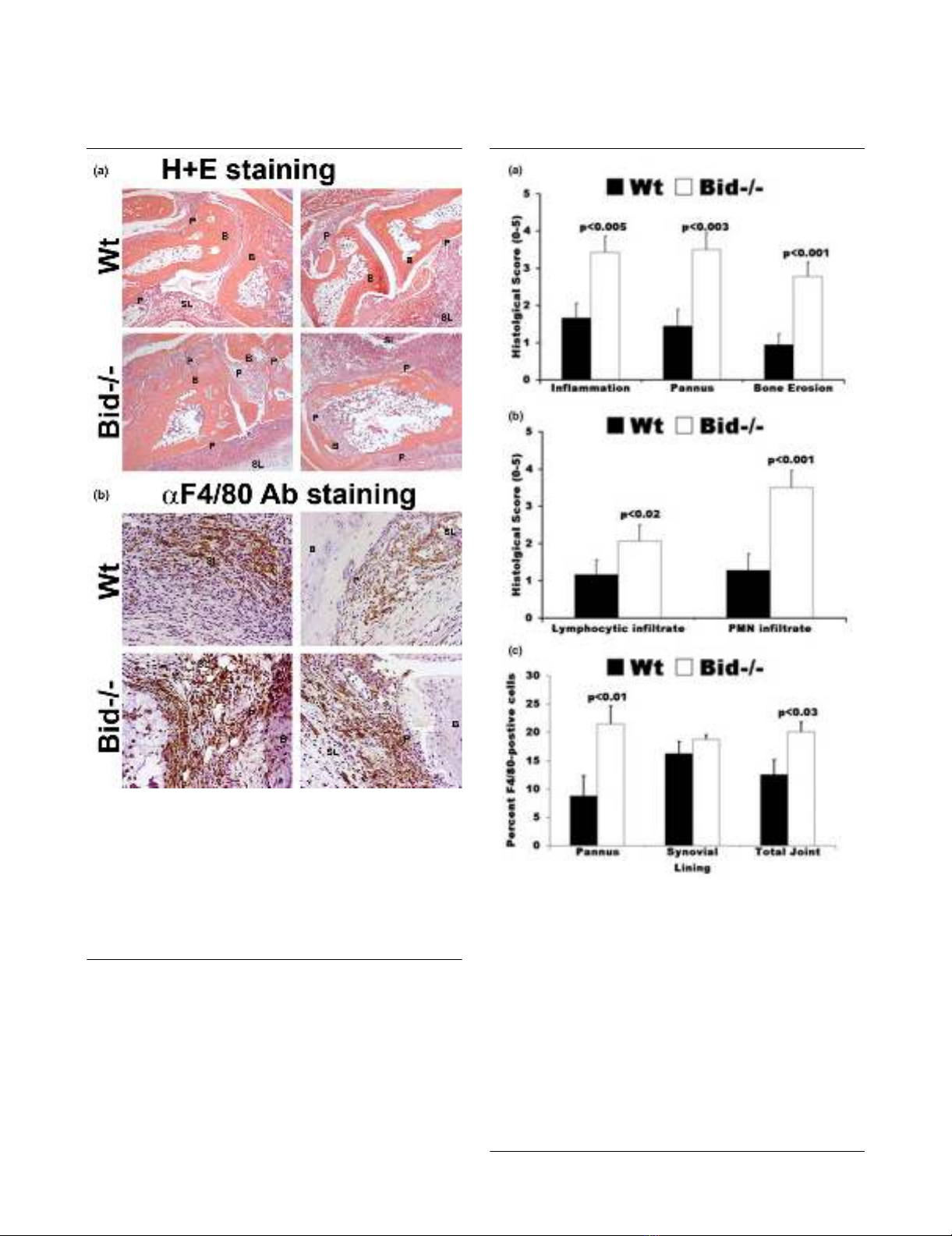
Arthritic Bid-/- mice display increased histopathological
scores
Ankle sections were examined using a histopathological scor-
ing system to further identify differences in Bid-/- compared to
Wt mice following the induction of arthritis. There was a 2.0-
fold increase (p < 0.005) in inflammation score, a 2.5-fold
increase (p < 0.003) in pannus formation score, and a 3.0-fold
increase in bone erosion score (p < 0.001) in Bid-/- compared
to Wt ankles (Figures 2a and 3a). No difference in cartilage
destruction was detected in Wt and Bid-/- mice (data not
shown). Analysis of the cells infiltrating the joints (Figure 3b)
showed a 2.7-fold increase (p < 0.002) in polymorphonuclear
cells and a 2.0-fold increase (p < 0.02) in lymphocytes in Bid-
/- compared to Wt joints. Bid-/- ankles had a 1.6-fold increase
(p < 0.03) in the number of macrophages compared to Wt
ankles. More specifically, there was a 2.5-fold increase (p <
0.02) in the number of macrophages in the pannus of Bid-/-
compared to Wt joints. There was no statistical difference in
average number of macrophages in the synovial lining in Bid-/
- compared to Wt mice. There were no differences in total
numbers of lymphocytes, neutrophils, or monocytes circulat-
ing in peripheral blood in Wt and Bid-/- mice (Table 1). These
data suggest that the increase in the numbers of inflammatory
cells in the joints of Bid-/- mice may not be attributed to an ele-
vation in circulating leukocytes. Since the K/BxN serum trans-
fer model has been shown to be independent of T and B cells
[30], these data suggest that the impairment in the resolution
of arthritis in Bid-/- mice may be due to an inability to clear infil-
trating leukocytes, specifically neutrophils and macrophages.
Expression of pro-inflammatory factors is similar in Wt
and Bid-/- mice following serum transfer
The cytokine and chemokine milieu of the joint is necessary for
the initiation and the perpetuation of inflammatory arthritis. Pre-
vious studies have shown that lpr and Bim-/- mice display
increased levels of pro-inflammatory factors in the joint and in
serum [27,28]. There were no differences in TNFα, IL-1β, KC,
or MCP-1 levels in Bid-/- and Wt untreated ankle joints and in
ankle joints isolated at days 3, 5, or 7 post transfer of serum
(Figure 4). However, there was a 2.0-fold increase in circulat-
ing levels of IL-1β (p > 0.09) and KC (p < 0.01) in Bid-/- com-
pared to Wt serum at day 3 post-serum transfer (Table 2).
Figure 1
Bid-deficient mice develop a sustained and prolonged edema of the ankles following transfer of K/BxN serumBid-deficient mice develop a sustained and prolonged edema of the
ankles following transfer of K/BxN serum. Pooled serum (300 μl) from
K/BxN mice was injected intra-peritoneally (IP) into Bid-/- (n = 32) and
wild-type (Wt) (n = 42) mice. Ankle joints were examined for arthritis by
measuring two perpendicular diameters of both joints (anterior-poste-
rior; medio-lateral) by calipers. The change in (Δ) ankle circumference
at each time point is defined as the difference between the ankle cir-
cumference and the measurement at day 0. values represent the mean
± standard error of ankles/time point, which were compared by Stu-
dent's t-test to Wt mice under parallel conditions. The asterisk denotes
p < 0.002 compared to Wt under parallel conditions.
Table 1
Wt and Bid-/- mice have similar numbers of leukocyte subpopulations in peripheral blood
CD19+ CD3+ CD3+ CD3+ CD11b+ CD11b+ CD11b+
CD4+ CD8+ Gr-1- Gr-1+ Gr-1++
CD62L - CD62L+
Wt (n = 20) 46.5 ± 0.8 22.5 ± 0.6 12.1 ± 0.4 7.4 ± 0.3 3.5 ± 0.2 2.6 ± 0.2 6.2 ± 0.8
Bid-/- (n = 15) 40.2 ± 3.8 21.1 ± 1.4 12.2 ± 1.0 8.5 ± 1.1 4.1 ± 0.7 2.6 ± 0.6 5.7 ± 0.5
Quantitative analysis of peripheral blood leukocyte numbers (expressed as 1 × E5/ml) in blood isolated from wild-type (Wt) and Bid-/- mice. All
data expressed as means ± standard error.
Available online http://arthritis-research.com/content/9/3/R49
Page 5 of 10
(page number not for citation purposes)
There were no differences in untreated serum samples from
Wt and Bid-/- mice (Table 2). These data suggest that while
the local inflammatory milieu remains similar in Bid-/- and Wt
mice, the systemic levels of IL-1β and KC are increased. These
elevated levels of IL-1β and/or KC may lead to the increased
numbers of neutrophils and macrophages in Bid-/- mice.
Figure 2
Increased inflammation and destruction of the joint is associated with more macrophages in Bid-/- compared to wild-type (Wt) mice following transfer of serumIncreased inflammation and destruction of the joint is associated with
more macrophages in Bid-/- compared to wild-type (Wt) mice following
transfer of serum. Mice (Wt, n = 9; Bid-/-, n = 7) underwent K/BxN
serum transfer as described in Figure 1 and were euthanized at seven
days post-serum transfer. Both ankles from each mouse were har-
vested, fixed, embedded in paraffin, sectioned, and stained with either
(a) hematoxylin (blue) and eosin (pink) (H&E) or (b) F4/80 antigen
(macrophage specific marker). Shown are representative photomicro-
graphs of the synovium and pannus formation from Wt and Bid-/- mice.
B, bone; SL, synovial lining; P, pannus.
Figure 3
Histological scores of ankle sections from wild-type (Wt) and Bid-/- miceHistological scores of ankle sections from wild-type (Wt) and Bid-/-
mice. (a) Bid-/- mice have increased inflammation and joint destruction
compared to Wt mice. Ankles isolated from mice (Wt, n = 9; Bid-/- n =
7) were prepared as described in Figure 2. Ankle sections were evalu-
ated and scored by a pathologist blinded to the study as described in
the Materials and methods section. Values represent the mean ± stand-
ard error of ankles/time point, which were compared by Student's t-
test. (b) Increased numbers of lymphocytes and polymorphonuclear
(PMNs) cells in inflamed Bid-/- joints. Ankles were prepared as
described above. Values represent the mean ± standard error of
ankles/time point, which were compared by Student's t-test. (c)
Arthritic Bid-/- mice have more macrophages in the pannus and in the
whole joint. Ankles were examined for F4/80 antigen as described in
Materials and methods. The number of positive cells for F4/80 in pan-
nus, synovial lining, and whole joint was determined by a pathologist
blinded to the study. Values represent the mean ± standard error of
ankles/time point, which were compared by Student's t-test.


