
Open Access
Available online http://arthritis-research.com/content/8/2/R52
Page 1 of 11
(page number not for citation purposes)
Vol 8 No 2
Research article
Ultrasonography of the metacarpophalangeal and proximal
interphalangeal joints in rheumatoid arthritis: a comparison with
magnetic resonance imaging, conventional radiography and
clinical examination
Marcin Szkudlarek1, Mette Klarlund2, Eva Narvestad3, Michel Court-Payen3, Charlotte Strandberg3,
Karl E Jensen3, Henrik S Thomsen4 and Mikkel Østergaard1
1Department of Rheumatology, University of Copenhagen Hvidovre Hospital, Kettegård Allé 30, 2650 Hvidovre, Denmark
2Magnetic Resonance Research Centre, University of Copenhagen Hvidovre Hospital, Kettegård Allé 30, 2650 Hvidovre, Denmark
3Department of Radiology, University of Copenhagen Hvidovre Hospital, Kettegård Allé 30, 2650 Hvidovre, Denmark
4Department of Radiology, University of Copenhagen Herlev Hospital, Herlev Ringvej 75, 2730 Herlev, Denmark
Corresponding author: Marcin Szkudlarek, marcin@dadlnet.dk
Received: 16 Aug 2005 Revisions requested: 26 Sep 2005 Revisions received: 22 Dec 2005 Accepted: 26 Jan 2006 Published: 6 Mar 2006
Arthritis Research & Therapy 2006, 8:R52 (doi:10.1186/ar1904)
This article is online at: http://arthritis-research.com/content/8/2/R52
© 2006 Szkudlarek et al.; licensee BioMed Central Ltd.
This is an open access article distributed under the terms of the Creative Commons Attribution License (http://creativecommons.org/licenses/by/2.0),
which permits unrestricted use, distribution, and reproduction in any medium, provided the original work is properly cited.
Abstract
Signs of inflammation and destruction in the finger joints are the
principal features of rheumatoid arthritis (RA). There are few
studies assessing the sensitivity and specificity of
ultrasonography in detecting these signs. The objective of the
present study was to investigate whether ultrasonography can
provide information on signs of inflammation and destruction in
RA finger joints that are not available with conventional
radiography and clinical examination, and comparable to the
information provided by magnetic resonance imaging (MRI). The
second to fifth metacarpophalangeal and proximal
interphalangeal joints of 40 RA patients and 20 control persons
were assessed with ultrasonography, clinical examination,
radiography and MRI. With MRI as the reference method, the
sensitivity, specificity and accuracy of ultrasonography in
detecting bone erosions in the finger joints were 0.59, 0.98 and
0.96, respectively; they were 0.42, 0.99 and 0.95 for
radiography. The sensitivity, specificity and accuracy of
ultrasonography, with signs of inflammation on T1-weighted MRI
sequences as the reference method, were 0.70, 0.78 and 0.76,
respectively; they were 0.40, 0.85 and 0.72 for the clinical
examination. With MRI as the reference method,
ultrasonography had higher sensitivity and accuracy in detecting
signs of inflammation and destruction in RA finger joints than did
clinical and radiographic examinations, without loss of
specificity. This study shows that ultrasonography has the
potential to improve assessment of patients with RA.
Introduction
New aggressive and powerful treatments that permit fast and
effective suppression of inflammation in rheumatoid arthritis
(RA) demand sensitive and specific methods for detecting dis-
ease signs and monitoring disease activity. Finger joints are
frequently the first to be involved in RA, and therefore methods
of assessment of these joints are of particular importance at
the onset of disease. The methods currently used, including
clinical examination and conventional radiography, are not sen-
sitive, especially in the evaluation of early stages of RA. In
recent years magnetic resonance imaging (MRI) has been rig-
orously tested in patients with RA, and its value has been con-
firmed both in studies of large joints (for example, knee joints
[1,2]) and in finger joints [3] compared with histological evalu-
ation of biopsy specimens acquired at microarthroscopy. Thus
far, because of the expensive equipment required and the
need for highly qualified personnel, it has not become widely
used as a joint assessment tool in RA. However, its benefits of
high sensitivity and specificity in the evaluation of RA joints [4-
6] make it a worthy surrogate 'gold standard' in settings where
FoV = field of view; Gd-DTPA = gadolinium-diethylenetriamine penta-acetic acid; ICC = intraclass correlation coefficient; MCP = metacarpophalan-
geal; MRI = magnetic resonance imaging; PIP = proximal interphalangeal; RA = rheumatoid arthritis; ST = slice thickness; TE = echo time; TR =
repetition time.
Arthritis Research & Therapy Vol 8 No 2 Szkudlarek et al.
Page 2 of 11
(page number not for citation purposes)
acquiring histological specimens is difficult (for example, fin-
ger joints).
Ultrasonography is an imaging technique that has attracted
much interest in the field of rheumatology in recent years [7,8].
As a result of technological improvements and wide availabil-
ity, ultrasonography has the potential to facilitate diagnosis of
RA and improve the assessment of disease activity, and its use
by rheumatologists may soon become routine. Few studies
have compared ultrasonography with other imaging modalities
with respect to their ability to detect signs of destruction and
inflammation; furthermore, data are seldom gathered from
homogenous populations and studies rarely include control
persons. Despite of appearance in the literature of reports pre-
senting the results of longitudinal studies of ultrasonographic
assessment of RA, the more basic issues of agreement, sensi-
tivity and specificity of ultrasonography in detecting RA pathol-
ogy remain to be addressed.
We therefore planned a systematic study in order to investi-
gate whether ultrasonography can provide information on RA
finger joints that is not available with conventional radiography
and clinical examination and comparable to the information
provided by MRI.
Materials and methods
Patients
We examined a total of 158 second to fifth metacarpophalan-
geal (MCP) joints and 140 second to fifth proximal inter-
phalangeal (PIP) joints of 40 patients with RA (fulfilling
American College of Rheumatology 1987 criteria) and 80 sec-
ond to fifth MCP joints and 80 second to fifth PIP joints of 20
healthy control persons. In the first part of the study we
attempted to evaluate the wrists of RA patients, but after we
had examined the first five patients the evaluation was omitted
because of poor accessibility of most bone surfaces. The
median age of the RA patients was 58 (range 23–79) years
and that of the control persons was 52 (27–79) years. The
female/male ratio was 4:1 both in the RA group and in the con-
trol group. The median disease duration in RA patients was 5
(range 0–20) years.
Twenty patients in the series had a disease duration in excess
of 2 years (established disease). Their median age and dis-
ease duration were 64 (range 23–79) years and 8 (2–20)
years, respectively. A further 20 patients had a disease dura-
tion of under 2 years (early disease). Their median and disease
duration were 53 (range 23–72) years and 1 (0–1) year,
respectively. All patients with established RA and 15 patients
with early RA were being treated with disease-modifying
antirheumatic drugs. The healthy control individuals had nei-
ther history of previous nor any current joint complaints.
The patients were recruited from two outpatient hospital-
based arthritis clinics. The study was conducted in accord-
ance with the Declaration of Helsinki and was approved by the
local ethics committee. Signed informed consent was
obtained from each participant. The inclusion criteria for RA
patients were swelling or tenderness of at least three finger
joints (MCP and/or PIP joints). The exclusion criteria were
severe deformity of MCP or PIP joint and contraindications to
MRI.
Ultrasonographic, clinical, laboratory and MRI examinations of
each patient were conducted on the same day.
Ultrasonography
Ultrasonography was performed using a General Electric
LOGIQ 500 unit (General Electric, Solingen, Germany) using
a 7–13 MHz linear array transducer. Ultrasonography was
conducted in the accessible aspects of the second to fifth
MCP joints and the second to fifth PIP joints of the dominant
hand: the dorsal, radial and palmar aspects of the second
MCP joint; the dorsal and palmar aspects of the third and
fourth MCP joints; the dorsal, ulnar and palmar aspects the
fifth MCP joint; and the dorsal, palmar, radial and ulnar aspects
of all PIP joints. Ultrasonographic examination from the dorsal
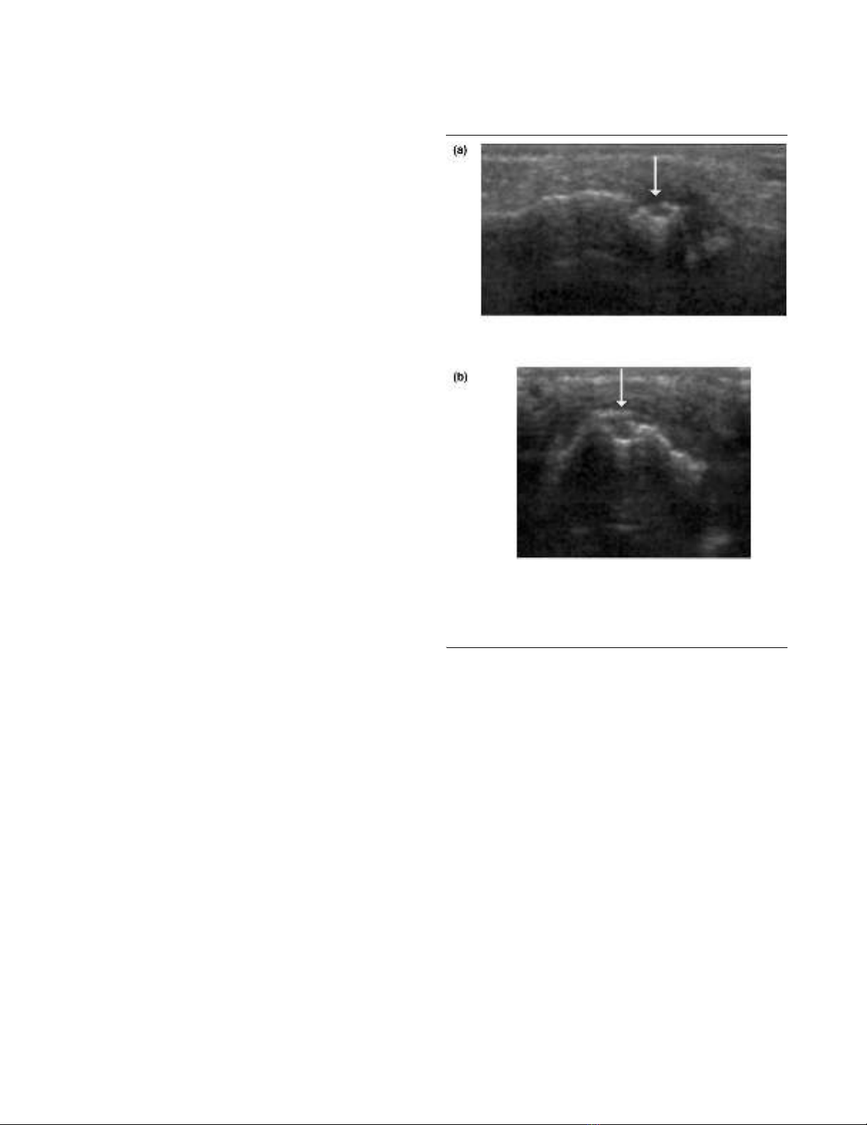
Figure 1
Signs of destruction on ultrasonography in the fourth proximal inter-phalangeal joint: early RASigns of destruction on ultrasonography in the fourth proximal inter-
phalangeal joint: early RA. MRI and conventional radiography revealed
no signs of destruction in the joint. A bone erosion (arrow) is visualized
with ultrasonography in (a) the longitudinal and (b) the transverse
planes. MRI, magnetic resonance imaging; RA, rheumatoid arthritis.
Available online http://arthritis-research.com/content/8/2/R52
Page 3 of 11
(page number not for citation purposes)
aspect was performed both in the neutral position and at about
70° of flexion. Each joint was assessed by quadrant for the
presence or absence of bone erosions (Figures 1 and 2) and
each joint was assessed for the presence or absence of signs
of inflammation (joint effusion and synovitis; Figures 2 and 3).
The following definitions of ultrasonographic changes were
employed: bone erosion = break in bone cortex in the area
adjacent to the joint, visualized in two planes; joint effusion =
compressible anechoic intracapsular area; and synovitis =
uncompressible hypoechoic intracapsular area. The ultrasono-
graphic changes were scored according to a semiquantitative
scoring system (grades 0–3) introduced in an earlier report
[9]. In relation to the original system, scoring of synovitis was
widened to include grade 4, defined as a hypoechoic area
bulging out of the joint and stretching over both bone diaphy-
ses of the joint.
Ultrasonographic examinations were performed by two radiol-
ogists with expertise in musculoskeletal ultrasonography and
a rheumatologist with training in the examination of the small
joints of the extremities. Ultrasonography was performed with-
out knowledge of the clinician's assessment or MRI data. The
interobserver variation between one of the radiologists and the
rheumatologist was presented in an earlier report [9].
Clinical examination
Prior to ultrasonography, clinical disease activity (presence or
absence of swelling and/or tenderness) in the MCP and PIP
joints was evaluated in all patients by the consultant rheuma-
tologist on duty. The number and localization of swollen and/
or tender joints was determined.
Conventional radiography
Radiography of the dominant hand was performed using
standard postero-anterior and oblique (Nørgaard's) views
within four weeks of the other examinations. The films were
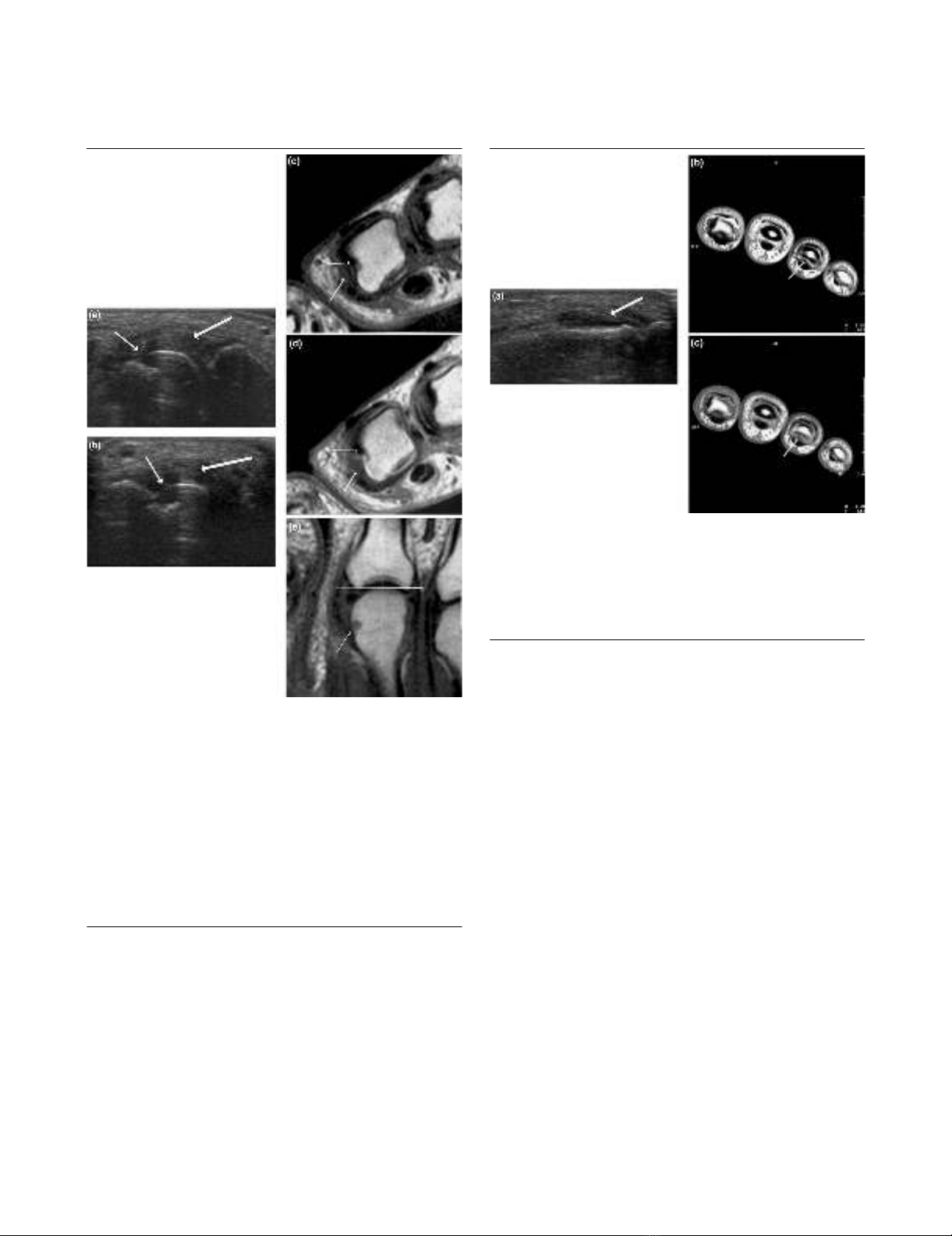
Figure 2
Signs of destruction and inflammation on ultrasonography and MRI in second metacarpophalangeal joint: established RASigns of destruction and inflammation on ultrasonography and MRI in
second metacarpophalangeal joint: established RA. Thin arrows indi-
cate an erosive change; thick arrows indicate synovitis. Ultrasonogra-
phy in the (a) longitudinal and (b) the transverse planes shows both
signs of destruction (grade 2) and inflammation (grade 3). Axial T1-
weighted magnetic resonance images were obtained (c) before and
(d) after contrast administration (grade 3 synovitis). Additionally, a
coronal T1-weighted magnetic resonance image (e) before contrast
administration visualizes the same bone erosion as shown in panels c
and d. The coronal magnetic resonance image of the second metacar-
pophalangeal joint (panel e) is additionally covered by a grid illustrating
division of the assessed joints into quadrants: proximal radial, proximal
ulnar, distal radial and distal ulnar. MRI, magnetic resonance imaging;
RA, rheumatoid arthritis.
Figure 3
Signs of synovitis on ultrasonography and MRI in fourth proximal inter-phalangeal joint: early RASigns of synovitis on ultrasonography and MRI in fourth proximal inter-
phalangeal joint: early RA. Arrows indicate an area with synovitis. Ultra-
sonography in (a) the longitudinal plane from the dorsal aspect shows
signs of synovitis (grade 4). Axial T1-weighted magnetic resonance
images were obtained (b) before and (c) after contrast administration
(grade 3 synovitis). MRI, magnetic resonance imaging; RA, rheumatoid
arthritis.

Arthritis Research & Therapy Vol 8 No 2 Szkudlarek et al.
Page 4 of 11
(page number not for citation purposes)
Table 1
Number of quadrants with bone erosions in finger joints, stratified by imaging modality and combinations thereof
Joint Quadrants with erosions Quadrants
with no
erosions on
US, MRI or
CR
Agreement Sensitivity Specificity
US + MRI
+ CR
US +
MRI
MRI +
RAD
US +
CR
US
only
MRI
only
CR
only
US versus
MRI (%)
CR versus
MRI (%)
US CR US CR
MCP 2nd 10 10 2 1 2 7 3 205 225(94) 217(90) 0.64 0.41 0.98 0.98
Est. 9 8 2 1 1 2 2 55 72(90) 66(82)
Early 1 2 0 0 1 2 0 74 77(96) 75(94)
Control 0 0 0 0 0 3 1 76 76(95) 76(95)
MCP 3rd 7 7 4 1 4 12 2 230 217(90) 214 (89) 0.47 0.37 0.97 0.98
Est. 6 6 4 1 3 6 2 52 64(80) 62 (78)
Early 1 1 0 0 1 2 0 75 77(96) 76 (95)
Control 0 0 0 0 0 4 0 76 76(95) 76 (95)
MCP 4th 5 2 0 1 1 5 0 222 229 (97) 227 (96) 0.58 0.42 0.99 0.99
Est. 4 1 0 1 1 5 0 64 69 (91) 68 (89)
Early 1 1 0 0 0 0 0 78 80 (100) 79 (99)
Control 0 0 0 0 0 0 0 80 80 (100) 80 (100)
MCP 5th 7 1 0 1 3 2 1 221 229 (97) 228 (97) 0.80 0.70 0.98 0.99
Est. 6 0 0 1 2 0 0 67 73 (96) 73 (96)
Early 1 1 0 0 1 0 1 76 78 (97) 77 (96)
Control 0 0 0 0 0 2 0 78 78 (97) 78 (97)
PIP 2nd 0 0 0 0 6 1 1 212 212 (96) 212 (96) - - 0.97 0.99
Est. 0 0 0 0 5 0 1 54 54 (90) 54 (90)
Early 0 0 0 0 1 0 0 79 79 (99) 79 (99)
Control 0 0 0 0 0 1 0 79 79 (99) 79 (99)
PIP 3rd 1 0 0 1 7 1 0 210 211 (96) 211 (96) 0.50 0.50 0.96 0.99
Est. 1 0 0 1 6 1 0 51 52 (87) 52 (87)
Early 0 0 0 0 1 0 0 79 79 (99) 79 (99)
Control 0 0 0 0 0 0 0 80 80 (100) 80 (100)
PIP 4th 0 0 0 0 2 1 1 216 216 (98) 216(98) - - 0.99 0.99
Est. 0 0 0 0 2 0 1 57 57 (95) 57 (95)
Early 0 0 0 0 0 1 0 79 79 (99) 79 (99)
Control 0 0 0 0 0 0 0 80 80 (100) 80 (100)
PIP 5th 0 0 0 0 1 0 4 215 215 (98) 215(98) - - 0.99 0.98
Est. 0 0 0 0 0 0 2 58 58 (97) 58(97)
Early 0 0 0 0 1 0 2 77 77 (96) 77(96)
Control 0 0 0 0 0 0 0 80 80 (100) 80 (100)
Total 30 20 6 5 26 29 12 1,704 1,754 (96) 1,740 (95) 0.59 0.42 0.98 0.99
Est. 26 15 6 5 20 14 8 458 499 (90) 490 (89)
Early 4 5 0 0 6 5 3 617 626 (98) 621 (97)
Control 0 0 0 0 0 10 1 629 629 (98) 629 (98)
The following numbers of joints were evaluated (1,832 in total): 240 MCP second, 240 MCP third, 236 MCP fourth, 236 MCP sixth, 220 PIP
second, 220 PIP third, 220 PIP fourth, and 220 PIP fifth. All study participants included. CR, conventional radiography; early, early rheumatoid
arthritis; Est., established rheumatoid arthritis; MCP, metacarpophalangeal joint; MRI, magnetic resonance imaging; PIP, proximal interphalangeal
joint; US, ultrasonography.

Available online http://arthritis-research.com/content/8/2/R52
Page 5 of 11
(page number not for citation purposes)
assessed by quadrant for the presence or absence of bone
erosions in the second to fifth MCP joints and the second to
fifth PIP joints by an experienced radiologist, who was una-
ware of the findings of the other examinations.
Magnetic resonance imaging
Later in the day on which ultrasonography was performed,
continuous axial and coronal pre-Gd-DTPA (gadolinium-dieth-
ylenetriamine penta-acetic acid) and post-Gd-DTPA T1-
weighted spin-echo magnetic resonance sequences of the
second to fifth MCP and second to fifth PIP joints of the dom-
inant hand were performed. This MRI assessment employed a
1.0 T Siemens Impact MR unit (Siemens, Erlangen, Germany)
equipped with a receive-only, wrap-around flex coil, and was
conducted in the group with established disease, three
patients with early disease and five control persons. The Gd-
DTPA (0.1 mmol/kg body weight) was injected intravenously
between repeated T1-weighted spin-echo magnetic reso-
nance sequences. The patients and control persons were in
the supine position with the hand in the coil along the femur.
The parameters of the applied sequences were as follows for
coronal sequences: repetition time (TR) 600 ms, echo time
(TE) 15 ms, slice thickness (ST) 3 mm, field of view (FoV) 140
mm, and matrix 192 × 256. For axial sequences the parame-
ters were as follows: TR 700 ms, TE 15 ms, ST 3 mm, FoV 120
mm, and matrix 192 × 256.
An extremity coil was used in 17 patients with early RA and 15
control persons. The use of different coils was necessary
because technical problems meant that the wrap-around flex
coil was unavailable for a lengthy period. The persons under-
going MRI were in supine position with the hand stretched
above the head ('Superman' position). The parameters of the
applied sequences for coronal sequences were as follows: TR
600 ms, TE 15 ms, ST 3 mm, FoV 145 mm, and matrix 192 ×
256. For axial sequences the parameters were as follows: TR
600 ms, TE 15 ms, ST 3 mm, FoV 120 mm, and matrix 192 ×
256.
The definitions of the applied MRI RA pathologies were in
accordance with OMERACT recommendations [10].
The examinations were assessed by quadrant for the presence
or absence of bone erosions (Figure 2) and by joint for the
presence or absence of signs of inflammation (joint effusion
and synovitis; Figures 2 and 3). Synovitis was scored accord-
ing to the semiquantitative system (grades 0–4) introduced by
Klarlund and coworkers [11]. The MRI observer was blinded
to clinical and ultrasonographical data.
The numbers of finger joints assessed using ultrasonography/
clinical examination and MRI were different (480 versus 433)
because the MRI data for 47 joints were not available: 20 PIP
joints were not visualized in the five patients in whom MRI of
wrists and MCP joints was performed, and the MRIs of six
MCP and 21 PIP joints were not assessable because the
patients moved between pre- and post-contrast MRI
sequences.
Statistical analysis
The agreement between imaging methods and compared with
clinical examination is reported as the overall agreement,
defined as the proportion of exact agreements to the overall
number of trials (expressed as a percentage). Furthermore,
agreement was expressed as means of sensitivity and specifi-
city. The correlation between ultrasonographic and MRI syno-
vitis scores was estimated using calculations of intraclass
correlation coefficients (ICCs; two-way mixed effects model,
consistency definition).
Results
Signs of bone destruction
A total of 1,832 quadrants of second to fifth MCP joints (952
quadrants) and PIP joints (880 quadrants) from 40 RA
patients and 20 healthy control individuals were examined
using ultrasonography, MRI and radiography (Table 1).
In MCP joints, at least one modality detected bone erosions in
101 of 952 examined quadrants (11%). Agreement between
all modalities on the presence of erosions was found in 29 out
of 101 quadrants (29%), whereas ultrasonography and MRI
agreed in 49 quadrants (49%). In 10 (11%) quadrants only
ultrasonography and in 26 (26%) quadrants only MRI identi-
fied bone erosions. Half of the ultrasonographic erosions in RA
patients that were not visualized by MRI were located in sec-
ond and fifth MCP joints (7 out of 14), whereas MRI quadrants
with erosions in RA patients not visualized with ultrasonogra-
phy were located predominantly in third to fourth MCP joints
(17 out of 23).
In PIP joints, at least one modality detected bone erosions in
27 of 880 quadrants (3%). Of these 27, only one quadrant
(4%) was identified as erosive with all modalities. In 16 (59 %)
quadrants only ultrasonography and in three (11 %) quadrants
only MRI detected bone erosions. Ultrasonographic bone ero-
sions, not visualized with other modalities, were distributed
between all examined PIP joints, but most of them were
located in the second and third PIP joints (15 out of 18). Radi-
ography detected six (22%) quadrants with erosions in PIP
joints that were not detected with other modalities.
Ten of the MRI quadrants with bone erosions in MCP joints
were detected in healthy control persons (10 erosions in 238
MCP joints; frequency 4.2%), which is in contrast to none with
ultrasonography and one with radiography. Ultrasonography
and radiography detected no erosions in PIP joints of the
healthy persons examined; one quadrant with erosions was
found with MRI.


