
Open Access
Available online http://arthritis-research.com/content/9/4/R64
Page 1 of 9
(page number not for citation purposes)
Vol 9 No 4
Research article
Reduction of urate crystal-induced inflammation by root extracts
from traditional oriental medicinal plants: elevation of
prostaglandin D2 levels
Sung Mun Jung1,3,5, H Ralph Schumacher1,3, Hocheol Kim5, Miyeon Kim5, Seoung Hoon Lee2 and
Frank Pessler4
1Division of Rheumatology, University of Pennsylvania, 3600 Spruce St, Philadelphia, PA 19104, USA
2Department of Pathology and Laboratory Medicine, 3400 Spruce St, University of Pennsylvania, Philadelphia, PA 19104, USA
3Division of Rheumatology, Veteran Affairs Medical Center, University and Woodland Avenues, Philadelphia, PA 19104, USA
4Division of Rheumatology, The Children's Hospital of Philadelphia, 3405 Civic Center Blvd, Philadelphia, PA 19104, USA
5Faculty of Oriental Medicine, Department of Herbal Pharmacology, Kyung Hee University College of Oriental Medicine, 1 Hoekidong,
Dongdaemoonku, Seoul 130-701, Korea
Corresponding author: Frank Pessler, pessler@email.chop.edu
Received: 5 Feb 2007 Revisions requested: 26 Feb 2007 Revisions received: 18 Apr 2007 Accepted: 5 Jul 2007 Published: 5 Jul 2007
Arthritis Research & Therapy 2007, 9:R64 (doi:10.1186/ar2222)
This article is online at: http://arthritis-research.com/content/9/4/R64
© 2007 Jung et al.; licensee BioMed Central Ltd.
This is an open access article distributed under the terms of the Creative Commons Attribution License (http://creativecommons.org/licenses/by/2.0),
which permits unrestricted use, distribution, and reproduction in any medium, provided the original work is properly cited.
Abstract
Dried roots of the plants Acanthopanax senticosus, Angelica
sinensis and Scutellaria baicalensis are used in traditional
oriental medicine and reportedly possess anti-inflammatory
properties. Using the murine air pouch model of inflammation,
we investigated the efficacy and mode of action of an extract
from these three plants in crystal-induced inflammation. Air
pouches were raised on the backs of 8-week-old BALB/c mice.
Mice were fed 100 mg/kg body weight of root extracts (A.
senticosus:A. sinensis:S. baicalensis mixed in a ratio of 5:4:1 by
weight) or vehicle only on days 3–6. Inflammation was elicited
on day 6 by injecting 2 mg of monosodium urate (MSU) crystals
into the pouch. Neutrophil density and IL-6 and TNF-α mRNA
levels were determined in the pouch membrane, and the
leukocyte count and IL-6, prostaglandin E2 (PGE2) and
prostaglandin D2 (PGD2) levels were determined in the pouch
exudate. Treatment with the root extracts led to a reduction in all
inflammatory parameters: the leukocyte count in the pouch
exudate decreased by 82%; the neutrophil density in the pouch
membrane decreased by 68%; IL-6 and TNF-α mRNA levels in
the pouch membrane decreased by 100%; the IL-6
concentration in the pouch fluid decreased by 50%; and the
PGE2 concentration in the pouch fluid decreased by 69%.
Remarkably, the concentration of the potentially anti-
inflammatory PGD2 rose 5.2-fold in the pouch exudate (p <
0.005), which led to a normalization of the PGD2:PGE2 ratio. A
3.7-fold rise in hematopoietic PGD synthase (h-PGDS) mRNA
paralleled this rise in PGD2 (p = 0.01).
Thus, the root extracts diminished MSU crystal-induced
inflammation by reducing neutrophil recruitment and expression
of pro-inflammatory factors and increasing the level of the
potentially anti-inflammatory PGD2. These results support a
need for further studies of the efficacy of these extracts in the
treatment of inflammatory arthropathies and suggest elevation of
PGD2 levels as a novel mechanism for an anti-inflammatory
agent.
Introduction
Powderized dried roots of the plants Acanthopanax sentico-
sus (Siberian ginseng), Angelica sinensis (Dong Quai) and
Scutellaria baicalensis (Baikal Skullcap) are commonly used
in oriental medicine for a variety of indications based on tradi-
tional concepts. A. senticosus is used as a general tonic to
stimulate Qi forces [1]. A. sinensis is used, for instance, to
treat blood deficiency with wind–damp painful obstruction
[2,3], and S. baicalensis is used to clear heat, remove toxins
and restrain bleeding [4,5]. All three plants are contained in
COX = cyclo-oxygenase; Ct = threshold cycle; ELISA = enzyme-linked immunosorbent assay; GAPDH = glyceraldehyde 3-phosphate dehydroge-
nase; H&E = hematoxylin and eosin; h-PGDS = hematopoietic prostaglandin D synthase; HPLC = high-performance liquid chromatography; IL
= interleukin; MSU = monosodium urate; NSAID = nonsteroidal anti-inflammatory drug; PBS = phosphate-buffered saline; PGD2= prostaglandin D2;
PGE2= prostaglandin E2; PGJ2= 15-deoxy-Δ12,14-prostaglandin J2; ΔRn = reporter-dye signals; RT-PCR = reverse transcriptase polymerase chain
reaction; TGF = transforming growth factor; TNF = tumor necrosis factor.

Arthritis Research & Therapy Vol 9 No 4 Jung et al.
Page 2 of 9
(page number not for citation purposes)
herbal mixtures used for the treatment of chronic inflammatory
disorders, including arthritis [6]. Pharmacologic studies in ani-
mals have documented the anti-inflammatory effects of all
three plants. A. senticosus has been shown to reduce the
expression of cyclo-oxygenase (COX)-2 and complement type
3 receptor (a marker for microglia in the central nervous
system) in cerebral ischemia [7] and to inhibit mast cell-
dependent anaphylaxis [8]. A. sinensis root polysaccharides
inhibited neutrophil migration in ethanol-induced gastrointesti-
nal inflammation in rats [9] and reduced expression of pro-
inflammatory factors in experimental colitis in rats [10]. The fla-
vonoids baicalein, which binds to chemokine ligands and
inhibits leukotriene C4 synthesis, and wogonin have been
implicated as the principal anti-inflammatory active ingredients
of S. baicalensis [11,12].
Considering their anti-inflammatory properties, extracts or mix-
tures of extracts from these plants might be suitable for the
treatment or prevention of inflammatory arthropathies. Mix-
tures of medicinal herbs containing root preparations from
these three herbs are indeed used in traditional oriental medi-
cine for this purpose [6], and there is anecdotal evidence from
clinical experience in traditional oriental medicine that these
herbs might be effective in treating musculoskeletal pain and
arthritis (H.C. Kim, S.M. Jung, unpublished data). However,
these herbs have not been validated for the treatment of acute
or chronic synovitis in clinical studies or animal models of
arthritis. As a first step, we therefore wanted to investigate the
efficacy and mode of action of a mixture of standardized root
extracts from the three plants in a simple animal model that
resembles acute synovitis in humans.
The murine air pouch model represents an easily manipulable
animal model of acute inflammation that has been used exten-
sively in studies of a variety of anti-inflammatory agents. In con-
trast to animal models of chronic arthritis, the murine air pouch
model lends itself well to the study of orally administered
agents because it does not require prolonged gavage feed-
ings of test substances to the animals. The air pouch is a newly
formed, bursa-like tissue that grows around subcutaneously
injected air and resembles the human synovial lining [13]. For
the purposes of definition, we shall refer to this newly formed
tissue as the 'pouch membrane'. Depending on the pro-inflam-
matory agent instilled into the pouch, distinct forms of inflam-
mation can be elicited [14]. Injection of monosodium urate
(MSU) crystals results in transient neutrophilic inflammation
that resembles acute gouty arthritis in humans [15,16] and
induces major pro-inflammatory cytokines that are active in
chronic inflammatory arthropathies, such as TNF-α and IL-1
and -6 [17-19]. Here, we show that the root extracts strongly
inhibit inflammation in this model by decreasing neutrophil
immigration into the pouch membrane, reducing expression of
pro-inflammatory factors, including prostaglandin E2 (PGE2),
and raising the level of the potentially anti-inflammatory pros-
taglandin D2 (PGD2), thereby normalizing the PGD2:PGE2
ratio. These findings suggest elevation of PGD2 levels as a
novel mechanism of action for an anti-inflammatory agent.
Materials and methods
Air pouches
Air pouches were raised on the backs of 8-week-old female
BALB/c mice (Taconic, Germantown, NY, USA) by subcuta-
neous injection of 3 cc of filtered air. MSU crystals were pre-
pared as described by McCarty and Faires [20]. On day 6, 2
mg of sterile crystals in 1 ml of PBS or 1 ml of PBS alone was
injected into the pouch space. After 9 hours (the peak of neu-
trophil accumulation in the pouch lumen), the animals were
sacrificed by asphyxiation with carbon dioxide (Figure 1a). The
dorsal skin and underlying dorsal pouch membrane were then
punctured and opened with a small cruciform incision, and the
pouch exudates were lavaged out of the pouch under direct
visualization, using a small pipette and 2 ml of PBS. The leuko-
cyte count in the lavage fluid was determined manually using
a hemocytometer. In this protocol, erythrocytes are lysed in
hypotonic buffer and thus do not interfere with determination
of the leukocyte count [21]. For immunoassay analysis, lav-
aged pouch exudates were flash-frozen in liquid nitrogen, with-
out prior centrifugation, and kept at -70°C until further
analysis; thus, levels of the test substances in both cells and
extracellular fluid were assayed without differentiating
between their synthesis and their secretion into the extracellu-
lar environment. Exudate IL-6, PGE2 and PGD2 levels were
determined by commercially available immunoassays (eBio-
science, San Diego, CA, USA (IL-6) and Cayman Chemical,
Ann Arbor, MI, USA (PGE2 and PGD2)).
RNA extraction and analysis of gene expression
Air pouch membranes were carefully dissected free of adja-
cent subcutaneous and paraspinal tissues by a method
recently developed in our laboratory [18]. Briefly, the pouch
membrane was meticulously separated from the adjacent sub-
cutaneous tissue by blunt dissection using curved scissors,
and the base of the membrane was then cut from the dorsal
fascia using straight surgical scissors. Using a rotatory tissue
homogenizer and disposable tips (Omni International, Warren-
ton, VA, USA), pouch membranes were homogenized in TRIzol
medium (Invitrogen, Carlsbad, CA, USA) immediately after dis-
section. Total RNA was extracted using RNeasy minicolumns
(Qiagen, Valencia, CA, USA) and tested for integrity and quan-
tity on an Agilent 2100 Bioanalyzer (Agilent Technologies,
Palo Alto, CA, USA). After enzymatic digestion of DNA by
DNase 1, aliquots of the RNA were reverse transcribed into
cDNA according to standard methods. Target-gene expres-
sion was then analyzed by real-time RT-PCR using an ABI
Prism 7000 sequence detector (Applied Biosystems, Foster
City, CA, USA) and the SYBR Green system (Applied Biosys-
tems). The house-keeping gene glyceraldehyde 3-phosphate
dehydrogenase (GAPDH) was co-amplified as an internal con-
trol. Artifacts from primer-dimer formation were excluded by
dissociation analysis. Sequences of the primers used are sum-
Available online http://arthritis-research.com/content/9/4/R64
Page 3 of 9
(page number not for citation purposes)
marized in Table 1. cDNA was synthesized from 5 μg of total
RNA in 80 μl reaction mixtures. For real-time RT-PCR, sense
and antisense primer pairs specific for the murine genes
encoding IL-6, TNF-α and hematopoietic PGD synthase (h-
PGDS) were reconstituted at a concentration of 4 μM. Reac-
tions were performed in a final volume of 25 μl, containing
12.5 μl of 2 × SYBR Green PCR Master Mix (Applied Biosys-
tems), 1 μl of each target primer (2 μl in total), 2 μl of cDNA
and 8.5 μl of distilled water. Forty cycles were performed at
95°C for 15 seconds and 60°C for 1 minute. The values of the
threshold cycle (Ct) at which a statistically significant increase
in reporter-dye signals (ΔRn) was first detected were imported
into Microsoft Excel software (Microsoft Corporation, Red-
mond, WA, USA) and then used to calculate relative expres-
sion of the target genes. All results were normalized to the Ct
value of GAPDH. The mean Ct value of target gene expression
from control pouches was assigned the reference value 1. The
relative target-gene expression values of the samples were cal-
culated according to the relative ΔCt method, as defined in
[22].
Histology and immunohistochemistry
Full-thickness tissue pieces, containing skin and pouch mem-
brane and measuring approximately 2 × 2 cm, were excised
from the lateral aspects of the pouch (the same location was
used in all cases). They were then fixed in formalin for 24–48
hours, embedded in paraffin and sectioned. H&E stains were
performed according to standard laboratory procedures. The
neutrophil density was counted independently by two observ-
ers (S.M. Jung, F. Pessler) in one section from each of two tis-
sue pieces per animal. As recommended previously [23], five
representative high-power fields (× 600 magnification), con-
taining intact pouch membrane and adjacent subcutaneous
tissues, were evaluated per section. Fields containing large
blood vessels or follicular inflammatory aggregates were
excluded. In all analyses, statistical significance was deter-
mined using the Student's t test.
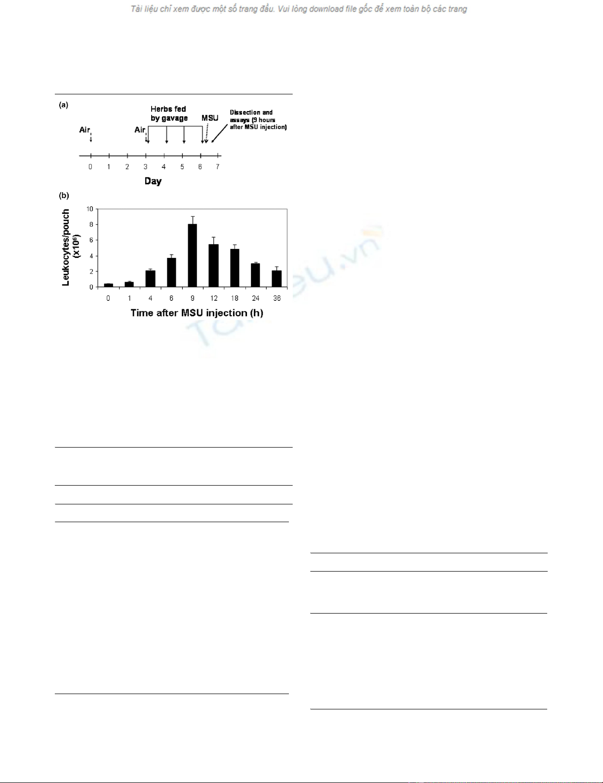
Figure 1
Sequence of events in the murine air pouch model (a) Outline of a typi-cal experimentSequence of events in the murine air pouch model (a) Outline of a typi-
cal experiment. Air is injected subcutaneously on day 0 and repeated
on day 3, as needed, to keep the pouch inflated. The root extracts or
water are gavage-fed once daily on days 3–6. A suspension of MSU
crystals in PBS (or PBS only) is injected into the pouch cavity on day 6
after the last gavage feeding. Pouch exudate and tissue are obtained
for analysis 9 hours after crystal injection. (b) Determination of the time
of maximal inflammation. The MSU crystal suspension was injected into
the pouch at 0 hours. Leukocyte counts in the pouch exudate were
determined by manual cell counting at the indicated time points (n = 4
mice for each time point). MSU, monosodium urate; PBS, phosphate-
buffered saline.
Table 1
Sequences of PCR primers used
Target gene Sequence
GAPDH forward 5'TGCAGTGGCAAAGTGGAGATT3'
GAPDH reverse 5'ATTTGCCGTGAGTGGAGTCAT3'
IL-6 forward 5'GGAGAGGAGACTTCACAG3'
IL-6 reverse 5'GCCATTGCACAACTCTTTTC3'
TNF-α forward 5'CATCTTCTCAAAATTCGAGTGACAA3'
TNF-α reverse 5'TGGGAGTAGACAAGGTACAACCC3'
h-PGDS forward 5'ATCCAAGGCTGGTGACTTTACG3'
h-PGDS reverse 5'TGAAGGCAACATGGATCAGCTA3'
GAPDH, glyceraldehyde 3-phosphate dehydrogenase; h-PGDS,
hematopoietic prostaglandin D synthase; IL, interleukin; PCR,
polymerase chain reaction; TNF, tumor necrosis factor.
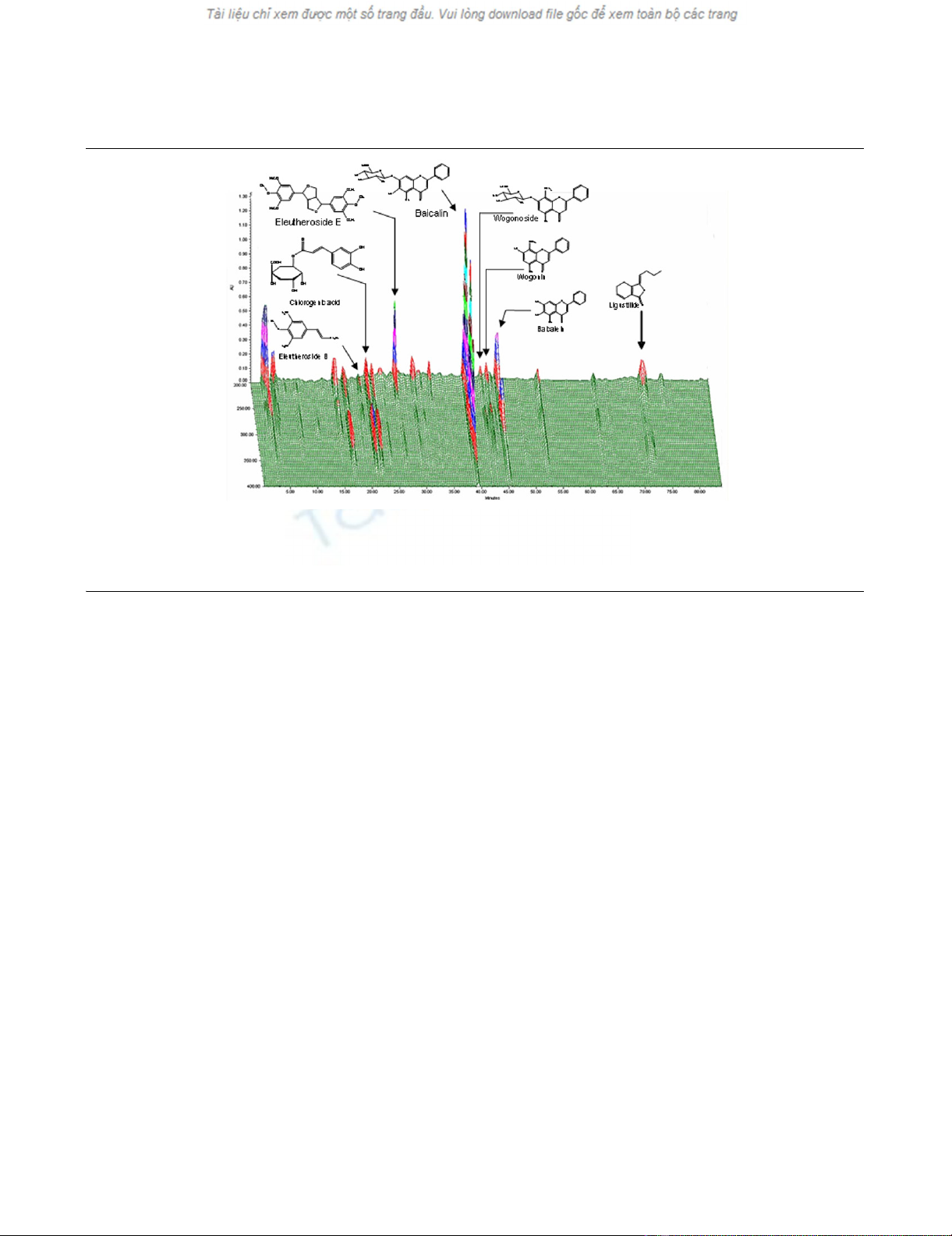
Table 2
Authentication of the extracts by HPLC
Botanical source Concentration ratio Final concentration of
compound used for
standardization (mg/100
g)
Acanthopanax
senticosus 15:1 Eleutheroside B, 0.081
Eleutheroside D, 0.44
Scutellaria
baicalensis 8:1 Baicalein, 22.8
Wogonin, 9.3
Angelica sinensis 7:1 Lingustilide, 8.64
HPLC, high-performance liquid chromatography.
Arthritis Research & Therapy Vol 9 No 4 Jung et al.
Page 4 of 9
(page number not for citation purposes)
Treatment with the root extracts
Plant materials were imported from China: A. senticosus
(Araliaceae) was from Heilongjiang Province, A. sinensis
(Umbelliferae) was from Gan'su Province, and S. baicalensis
(Labiatae) was from Shan'xi Province. The identities of the
plant materials were verified by one of the authors (H. Kim) and
voucher specimens were deposited in the Department of
Herbal Pharmacology, College of Oriental Medicine, Kyung
Hee University, Korea. The roots were heat-dried, ground and
extracted for several hours with 70% ethanol solution. The
resulting extracts were then concentrated using a rotatory
evaporator and freeze-dried. The results of quantitative
authentication of the extracts by HPLC are summarized in
Table 2. The corresponding chromatogram is shown in Figure
2, in which details of the HPLC procedure are also outlined.
Freeze-dried plant extracts were combined (A. senticosus:A.
sinensis:S. baicalensis in a ratio of 5:4:1 by weight) and then
dissolved in distilled water, to a final concentration of 2 mg/ml.
These proportions were chosen according to previous prelim-
inary results in a mouse model of cerebral reperfusion injury,
which has a strong inflammatory component (H. Kim, unpub-
lished data). Using a 22-gauge, 1.5-inch rigid feeding tube
(Ejay International, Glendora, CA, USA) mice were gavage-fed
1 ml of this solution (corresponding to 100 mg of freeze-dried
extracts/kg body weight) or 1 ml of water once daily, as out-
lined in Figure 1. There were no deaths or illnesses among the
mice.
Results
Validating the time of maximal inflammation in this
model
The leukocyte count of the pouch exudate is the commonly
used end point in the air pouch model. A time-course experi-
ment showed that the leukocyte density of the pouch exudate
peaked 9 hours after instillation of MSU crystals and then sub-
sided gradually over the following 27 hours (Figure 1b). The 9-
hour time point, which reflected a 24-fold increase in the leu-
kocyte count of the exudate, was thus chosen for all subse-
quent experiments.
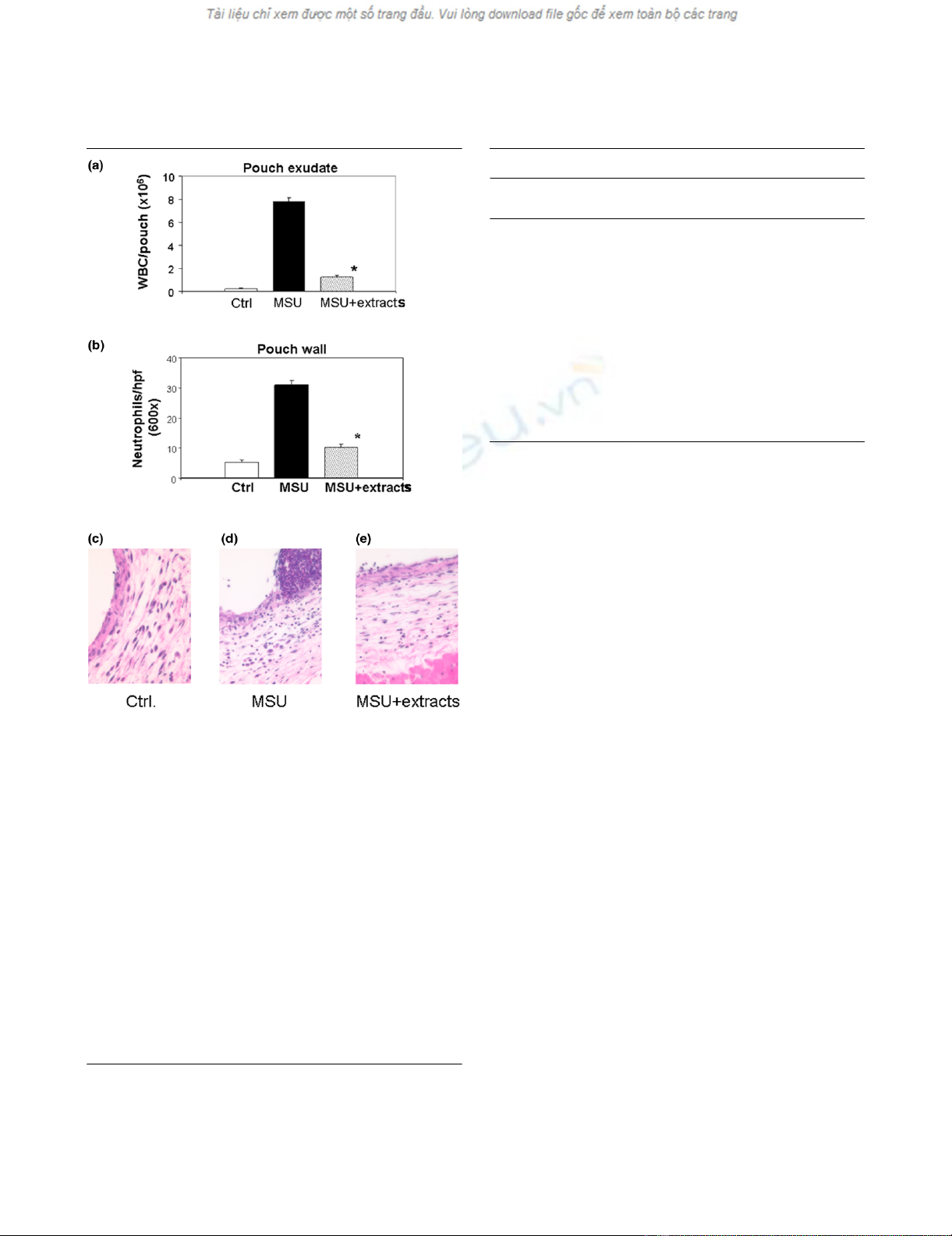
Reduction of inflammation and inflammatory mediators
by treatment with the root extracts
In a first experiment into the ability of the root extracts to
reduce inflammation, we assessed their effect on the leuko-
cyte count in the pouch exudate at the 9-hour time point. The
expected vigorous neutrophilic inflammation was observed in
the MSU-stimulated pouches from mice fed water, as
reflected in a 26-fold rise in the leukocyte count of the pouch
fluid (Figure 3a). As expected, the neutrophil density within the
pouch membrane also increased, but to a lesser extent
(approximately sixfold; Figure 3b). Treatment with the root
extracts blunted both parameters significantly: the MSU-asso-
Figure 2
Standardization of the root extracts (high-performance liquid chromatography (HPLC) chromatogram)Standardization of the root extracts (high-performance liquid chromatography (HPLC) chromatogram). Compounds were detected with a photodi-
ode array. X-axis, retention time; Y-axis, wavelength; and Z-axis, absorbance unit. The analytic conditions were as follows: column, C18 Φ 4 × 250
mm; mobile phase, 1% phosphoric acid (H3PO4; solvent A) and acetonitrile (CH3CN; solvent B); flow rate, 1 ml/min; and eluting gradient, 5% to
50% of solvent B in A (during minutes 1–60), followed by standing 70% of solvent B in A (during minutes 61–85).
Available online http://arthritis-research.com/content/9/4/R64
Page 5 of 9
(page number not for citation purposes)
ciated increases in the leukocyte count of the pouch fluid and
neutrophil density of the pouch membrane were 87% and
68% lower, respectively, in the treatment group (Figure 3a,b).
Table 3 summarizes the percentage changes detected in this
and all subsequent experiments. H&E stained histologic
sections of pouch walls from representative control, MSU and
MSU + extracts mice are shown in Figure 3c–e.
We next assessed changes in the expression of pro-inflamma-
tory factors in the pouch membrane and exudate (Figure 4).
MSU crystals led to a 55-fold rise in the level of IL-6 mRNA and
17-fold rise in the level of TNF-α mRNA in the membrane.
Treatment with the root extracts prevented this MSU-depend-
ent increase in mRNA levels for both factors (Figure 4a,b). In
the exudate, the level of IL-6 protein rose 8.7-fold in response
to MSU crystals (Figure 4c) and the level of PGE2 protein
increased 11.3-fold (Figure 4d). The increase in IL-6 was 50%
lower and that of PGE2 was 69% lower in the mice treated
with the root extracts (Figure 4c,d). Treatment with the root
extracts thus decreased inflammation in this model by reduc-
ing neutrophil migration into the pouch wall and fluid and
reducing the synthesis of pro-inflammatory factors.
Increase in the level of prostaglandin D2 by treatment
with the root extracts
PGD2 is a pleiotropic prostaglandin that has been associated
with anti-inflammatory properties and the resolution of
inflammation [24,25], and it is the precursor of the anti-inflam-
matory prostaglandin 15-deoxy-Δ12,14-prostaglandin J2
(PGJ2) [24]. We hypothesized that the root mixture might func-
tion partially by increasing the level of this potentially anti-
inflammatory substance. At the 9-hour time point, a modest
rise in the PGD2 level was seen in the MSU-treated pouches
Figure 3
Treatment with root extracts reduces leukocyte recruitment into the pouch wall and their accumulation in the pouch exudateTreatment with root extracts reduces leukocyte recruitment into the
pouch wall and their accumulation in the pouch exudate. The experi-
mental groups in these and subsequent experiments were as follows:
(1) Ctrl (gavage feeding with water and intrapouch injection of PBS);
(2) MSU (gavage feeding with water and intrapouch injection of MSU
crystals in PBS); and (3) MSU + extracts (gavage feeding with extracts
and intrapouch injection of MSU crystals in PBS). (a) Leukocyte count
in the pouch exudate, expressed as leukocytes per pouch. The numeri-
cal values (all × 106 ± standard error of the mean) were as follows: Ctrl,
0.26 ± 0.03; MSU, 7.80 ± 0.33; and MSU + extracts, 1.24 ± 0.18. The
percentage changes detected in this and all other experiments are
summarized in Table 3. (b) Polymorphonuclear cell density in the pouch
wall (cells per × 600 field ± SEM): Ctrl, 5.30 ± 0.78; MSU, 31.02 ±
1.55; MSU + extracts, 10.08 ± 1.12. (c–e) H&E stains of representa-
tive sections from pouch walls obtained from control (c), MSU (d) and
extract treatment (e) groups. Higher magnification revealed that the
control wall contained mostly fibroblasts and mononuclear cells. Abun-
dant polymorphonuclear cells were seen in the MSU-stimulated pouch
wall (d), the number of which was decreased by treatment with the root
extracts (e). Ctrl, control; H&E, hematoxylin and eosin; Hpf, high-power
field (× 600); MSU, monosodium urate; PBS, phosphate-buffered
saline; WBC, white blood cell count.
Table 3
Summary of effects of the root extracts*
Parameter Assay Change No. of mice
per group
Leukocyte count, exudate Cell count -87% 10
Neutrophil density, membrane Cell count -68% 4
IL-6 protein, exudate ELISA -50% 7
IL-6 mRNA, membrane qRT-PCR -100% 4 + 4**
TNF-α mRNA, membrane qRT-PCR -100% 4 + 4**
PGE2, exudate ELISA -69% 7
PGD2, exudate ELISA +5.2-fold 7
Ratio of PGD2:PGE2ELISA +9.0-fold 7
h-PGDS mRNA, membrane qRT-PCR +3.7-fold 5
* Compared with MSU-stimulated pouches from mice fed water. All
percentage differences were significant at p < 0.05.
**Duplicate experiments. ELISA, enzyme-linked immunosorbent
assay; h-PGDS, hematopoietic prostaglandin D synthase; IL,
interleukin; MSU, monosodium urate; PGD2, prostaglandin D2;
PGE2, prostaglandin E2; qRT-PCR, relative quantitative reverse
transcriptase polymerase chain reaction; TNF, tumor necrosis factor.


