
Open Access
Available online http://arthritis-research.com/content/7/4/R825
R825
Vol 7 No 4
Research article
Characterization of histopathology and gene-expression profiles
of synovitis in early rheumatoid arthritis using targeted biopsy
specimens
Takahito Tsubaki1, Norimasa Arita1, Takuma Kawakami2, Takayuki Shiratsuchi2,
Haruyasu Yamamoto1, Nobuo Takubo3, Kazuhito Yamada3, Sanpei Nakata3, Sumiki Yamamoto3
and Masato Nose1
1Ehime University School of Medicine, Ehime, Japan
2Otsuka Pharmaceutical Co Ltd, Tokushima, Japan
3Center for Rheumatic Diseases, Matsuyama Red Cross Hospital, Ehime, Japan
Corresponding author: Masato Nose, masanose@m.ehime-u.ac.jp
Received: 30 Sep 2004 Revisions requested: 27 Oct 2004 Revisions received: 17 Mar 2005 Accepted: 29 Mar 2005 Published: 25 Apr 2005
Arthritis Research & Therapy 2005, 7:R825-R836 (DOI 10.1186/ar1751)
This article is online at: http://arthritis-research.com/content/7/4/R825
© 2005 Tsubaki et al.; licensee BioMed Central Ltd.
This is an Open Access article distributed under the terms of the Creative Commons Attribution License (http://creativecommons.org/licenses/by/
2.0), which permits unrestricted use, distribution, and reproduction in any medium, provided the original work is properly cited.
Abstract
The disease category of early rheumatoid arthritis (RA) has been
limited with respect to clinical criteria. Pathological
manifestations of synovitis in patients whose disease is clinically
classified as early RA seem to be heterogeneous, with regular
variations. To clarify the relation between the molecular and
histopathological features of the synovitis, we analyzed gene-
expression profiles in the synovial lining tissues to correlate them
with histopathological features. Synovial tissues were obtained
from knee joints of 12 patients with early RA by targeted biopsy
under arthroscopy. Surgical specimens of long-standing RA
(from four patients) were examined as positive controls. Each
histopathological parameter characteristic of rheumatoid
synovitis in synovial tissues was scored under light microscopy.
Total RNAs from synovial lining tissues were obtained from the
specimens selected by laser capture microdissection and the
mRNAs were amplified by bacteriophage T7 RNA polymerase.
Their cDNAs were analyzed in a cDNA microarray with 23,040
cDNAs, and the levels of gene expression in multilayered lining
tissues, compared with those of normal-like lining tissues in
specimens from the same person, were determined to estimate
gene-expression profiles characteristic of the synovial
proliferative lesions in each case. Based on cluster analysis of all
cases, gene-expression profiles in the lesions in early RA fell into
two groups. The groups had different expression levels of genes
critical for proliferative inflammation, including those encoding
cytokines, adhesion molecules, and extracellular matrices. One
group resembled synovitis in long-standing RA and had high
scores for some histopathological features – involving
accumulations of lymphocytes and plasma cells – but not for
other features. Possible differences in the histopathogenesis
and prognosis of synovitis between the two groups are
discussed in relation to the candidate genes and
histopathology.
Introduction
Synovial lesions in rheumatoid arthritis (RA) show complex his-
topathological manifestations, involving several diagnostic
hallmarks such as multilayered synovial lining tissues associ-
ated with a palisading structure of the intimal lining cells and
the presence of non-foreign-body-type giant cells, formation of
lymphoid follicles, and massive accumulation of plasma cells
and macrophages [1]. Mesenchymoid transformation and fibri-
noid degeneration are definite histopathological features of
RA [2]. These lesions are specific to the synovium in the pro-
gression stage of RA and their developmental processes
remain unclear.
'Early RA' is a clinical term referring to the early stage of RA
used to predict the eventual progression stage of RA. The
American College of Rheumatology (ACR) 1987 classification
criteria for RA [3] have often been used as a diagnostic tool in
patients with recent-onset arthritis. However, these criteria
ACR = American College of Rheumatology; IFN = interferon; IL = interleukin; LCM = laser capture microdissection; OA = osteoarthritis; RA = rheu-
matoid arthritis; SAM = significance analysis of microarrays; SSC = saline sodium citrate; TNF = tumor necrosis factor.

Arthritis Research & Therapy Vol 7 No 4 Tsubaki et al.
R826
were developed in a population of patients selected according
to their disease status to classify rather than to diagnose RA.
Thus, the diagnostic usefullness of these criteria in early arthri-
tis is probably not optimal. Likewise, previous histopathologi-
cal studies have been inconclusive with respect to elucidating
histological features typical of early RA [4-6]. Therefore, stud-
ies of potential molecular changes in the synovium of patients
with early RA may improve our understanding of this disease
entity and aid diagnosis in the future.
Biopsy targeting of articular lesions in synovial tissues should
be a powerful tool for clarifying the initial events of synovitis in
RA. Immunohistochemical analyses of synovitis in RA using
targeted biopsy specimens have shown that the histopatho-
logical features of synovium in early RA are representative of
those in long-standing RA [7,8], suggesting quantitative rather
than qualitative differences between various forms of synovitis
in RA [9,10]. Laser capture microdissection (LCM) and extrac-
tion of total RNA followed by a cDNA microarray are tech-
niques that have been developed mainly in molecular oncology
and are used for clarifying molecular markers that have the
potential to predict metastasis, sensitivity to drugs, and prog-
nosis [11,12]. The use of these techniques to study the his-
topathogenesis of the initial step of synovitis in RA and its
progression should improve our understanding at the molecu-
lar level.
In this study, we focused on the analysis of gene-expression
profiles characteristic of proliferative lesions in the synovial lin-
ing tissues, which are one of the initial histopathological
events of synovitis in early RA. That is, we prepared synovial
specimens from early RA by targeted biopsy under arthros-
copy, and analyzed gene-expression profiles in the synovial lin-
ing tissues selected by LCM in a cDNA microarray by
comparing those in multilayered lining tissues with those in
normal-like lining tissues in each case. On the basis of a clus-
ter analysis, we propose that the synovial proliferative lesions
in early RA can be classified into at least two groups. We dis-
cuss the histopathological manifestations characteristic of
rheumatoid synovitis in these two groups and also the possible
differences in pathogenesis and prognosis of synovitis
between them.
Materials and methods
Patients and tissue samples
We studied 12 patients with early RA (duration of less than 1
year before the diagnosis), and 4 with long-standing RA (dura-
tion of more than 3 years before the diagnosis). Not all patients
with early RA could be accurately diagnosed at the time of tar-
geted biopsy, although diagnosis was possible with follow-up
assessments. All patients had arthritis of the knee and fulfilled
the ACR criteria for RA [3] except E-09 (early RA case no. 9)
(see Table 1). Written, informed consent was obtained from
each patient before they were entered into the study.
Synovial specimens in early RA were obtained from knee joints
by targeted biopsy under arthroscopy, and specimens from
long-standing RA were obtained by total knee arthroplasty at
the Center for Rheumatic Disease, Matsuyama Red Cross
Hospital. The number of specimens obtained from each
patient and the macroscopic signs of synovitis with the maxi-
mum inflammatory activity at biopsy sites are shown in Table
1. For intraindividual comparison, normal-like synovial speci-
mens that were macroscopically thin and translucent and con-
tained only a few vessels were also obtained from each patient
[13].
Histopathology
One-half of each synovial specimen was used for histopatho-
logical analysis. The tissue specimens were fixed with 10%
formalin in 0.01 mol/l phosphate buffer, pH 7.2, and embed-
ded in paraffin wax. They were stained with hematoxylin and
eosin for examination by light microscopy. Histopathological
parameters of synovitis were evaluated in accordance with
established criteria [14], with modifications involving the
degree of proliferation of synovial cells, typical palisading of
synovial cells in the intimal lining layers, non-foreign-body-type
giant cells in the lining regions, lymphoid and plasma cell infil-
tration, neovascularization, mesenchymoid transformation, and
fibrinoid necrosis in synovium. Of these features, the degree of
proliferation of synovial cells was scored as follows: fewer than
three layers (0), three to four layers (1), five to six layers (2), or
more than six layers (3). Lymphoid cell infiltration was scored
as follows: none to diffuse infiltration (0), lymphoid cell aggre-
gates (1), lymphoid follicles (2), or lymphoid follicles with ger-
minal center formation (3). The other features were evaluated
using a quantitative grading system consisting of a 4-point
scale: none (0), mild (1), moderate (2), or severe (3). The max-
imum score with this system was 24. The results of scoring of
each histopathological feature are presented as the highest
score among all the specimens for the patient. The remaining
half of the synovial specimen showing the highest score in the
feature 'proliferation of synovial cells' was used as multilayered
lining tissue for LCM. Nearly normal synovial tissues from the
same patient that had no inflammatory lesions and received a
score of 0 for all of the histopathological features were used
as 'normal-like lining tissue' for LCM.
Laser capture microdissection
The tissue samples were placed in embedding medium (Tis-
sue-Tek OCT Compound, Sakura Finetechnical, Tokyo, Japan)
and immediately snap frozen in acetone/dry ice in the operat-
ing room before transport to the laboratory. All cryoblocks
were stored at -80°C until 7-µm-thick cryosections were pre-
pared and mounted on a 1.35-µm-thick polyethylene mem-
brane (PALM, Wolfratshausen, Germany). The sections were
immediately fixed for 3 min with acetone and for 1 min with
70% ethanol and then stained rapidly for 1 min with His-
toGene™ staining solution (Arctrus, BM Equipment Co Ltd,
Tokyo, Japan). They were washed with distilled water and

Available online http://arthritis-research.com/content/7/4/R825
R827
were then dehydrated with 100% ethanol and air-dried with a
fan for 3 min.
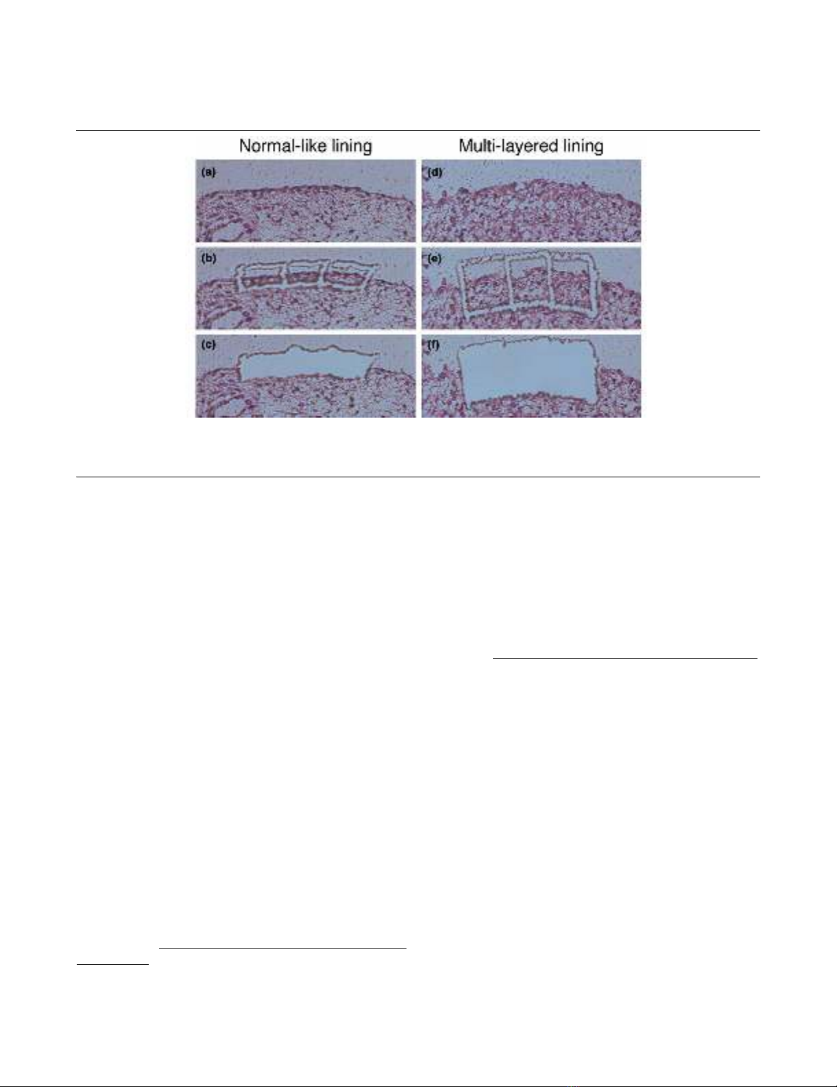
LCM was done to collect small regions from a specimen using
a Robot-Microbeam (PALM) and an inverted microscope (Carl
Zeiss, Oberkochem, Germany) [15]. In brief, the specimen
was set on a computer-controlled microscope stage and
observed from the upper side with a charged-coupling device
(CCD) camera. The image was displayed, and the multilayered
lining tissue and the normal-like lining tissue of the same case
were selected using the computer mouse (Fig. 1a,d). We
traced around the lining and then dissected it to the bottom of
the specimen together with the thin membrane, using a laser
microbeam through the objective lens (Fig. 1b,e). The selected
tissue was then catapulted with a single laser shot into a
microcentrifuge cap (0.6 ml), which was held by the microma-
nipulator (Fig. 1c,f). More than 5,000 cells in each specimen
were dissected and pooled for RNA extraction.
RNA extraction and T7-based RNA amplification
Total RNA was extracted from the samples collected by LCM
using an RNeasy spin column purification kit (Qiagen, Hilden,
Germany) in accordance with the manufacturer's procedure.
To remove possible genomic DNA contamination, RNase-free
DNase (Qiagen) was used during the RNA purification steps.
Messenger RNA was then amplified by bacteriophage T7
RNA polymerase using a RiboAmp™RNA amplification kit
(Arctrus). Two or three rounds of in vitro amplification were
done with the samples. The amplified RNAs from each multi-
layered lining tissue and normal-like lining tissue of each case
were reverse-transcribed using the SuperScript preamplifica-
tion system (Life Technologies, Rockville, MD, USA) with ran-
dom hexamers in the presence of Cy5-dCTP and Cy3-dCTP
(Amersham Biosciences Co, Piscataway, NJ, USA),
respectively.
cDNA microarray
A cDNA microarray was fabricated with 23,040 cDNAs
selected from the UniGene database of the National Center
for Biotechnology http://www.ncbi.nlm.nih.gov/. The cDNAs
were amplified by RT-PCR using poly(A) + RNAs isolated from
various human organs as templates. The PCR products were
spotted in duplicate on type VII glass slides (Amersham Bio-
Table 1
Characteristics of studied patients with early (E) and long-standing (L) rheumatoid arthritis (RA)
Patient Age Sex Disease duration ACR criterion nos.
fulfilledaNumber of samples Macroscopic signs
of synovitis
With early RA
E-01 51 F 11 months 1, 2, 3, 4 13 Vi, Ve
E-02 50 F 2 months 1, 2, 3, 4, 6 8 Vi, Ve
E-03 34 F 4 months 1, 2, 3, 4, 7 8 Vi, Ve
E-04 34 F 3 months 1, 2, 3, 4, 6, 7 13 Vi, Ve
E-05 77 F 2 months 1, 2, 3, 4, 7 11 Vi
E-06 50 M 4 months 1, 2, 3, 4 11 Vi, Ve
E-07 37 F 7 months 1, 2, 3, 4, 6 6 Ve
E-08 61 F 2 months 1, 2, 3, 4 7 Vi
E-09 75 F 4 months 1, 4, 6 12 Vi, Ve, Gr
E-10 25 F 12 months 1, 2, 3, 4 12 Vi, Ve, Gr
E-11 54 M 12 months 1, 2, 3, 4, 6 11 Ve
E-12 60 F 4 months 1, 2, 3, 4, 6 13 Vi, Ve, Gr
With long-standing RA
L-01 54 M 9 years 1, 2, 3, 4, 6, 7 6 Vi, Ve, Gr
L-02 77 M 5 years 1, 2, 3, 4, 5, 6, 7 8 Vi, Ve, Gr
L-03 54 F 7 years 1, 2, 3, 4, 6, 7 6 Vi, Ve
L-04 55 F 3 years 1, 2, 3, 4, 6, 7 11 Vi, Ve, Gr
aACR (American College of Rheumatology) criteria: 1, morning stiffness; 2, arthritis of three or more joint areas; 3, arthritis of hand joints; 4,
symmetric arthritis; 5, rheumatoid nodules; 6, serum rheumatoid factor; 7, radiographic changes. F, female; Gr, granulation; M, male; Ve, increased
number of vessels; Vi, villi.
Arthritis Research & Therapy Vol 7 No 4 Tsubaki et al.
R828
sciences) with a Microarray Spotter Generation III (Amersham
Biosciences).
Labeled probes were mixed with Microarray Hybridization
Solution Version 2 (Amersham Biosciences) and formamide
(Sigma Chemical Co, St Louis, MO, USA) to a final concentra-
tion of 50%. After hybridization for 14 to 16 hours at 42°C, the
slides were washed for 10 min at 55°C in 2 X saline sodium
citrate (SSC) and 1% SDS, for 10 min at 55°C in 0.2 X SSC
and 0.1% SDS, and for 1 min at room temperature in 0.1 X
SSC. They were then scanned using an Array Scanner Gener-
ation III (Amersham Biosciences). The fluorescence intensities
of Cy5 and Cy3 for each target spot were evaluated photomet-
rically by the ArrayVision computer program (Amersham Bio-
sciences). Since data derived from low signal intensities are
less reliable, a cutoff value for signal intensities of 10,000 was
used.
Cluster analysis
To obtain reproducible clusters for classifying the 16 samples,
we selected 1,035 genes for which valid expression data were
obtained in all the experiments, and which included an up-reg-
ulated (Cy5/Cy3 >2) or down-regulated gene (Cy5/Cy3 <0.5)
in at least two of all samples. The analysis was performed
using Cluster 3.0 and TreeView software written by M Eisen
and updated by Michiel de Hoon, and available on the World
Wide Web http://genome-www5.stanford.edu/resources/
restech.shtml. Before the clustering algorithm was applied, the
fluorescence ratio for each spot was log-transformed (base 2).
Then the data were median-centered and normalized for each
sample, to remove experimental biases.
Statistical analysis
Euclidean distance was used to determine the differences
between expression levels of individual genes. Statistical anal-
ysis on microarray data was performed using the significance
analysis of microarrays (SAM) method, available on the World
Wide Web http://www-stat.stanford.edu/~tibs/SAM/faq.html.
The fold change in expression was calculated for each gene
between groups, and significance levels were indicated by the
Q value. A Q value less than 5% was considered significant.
A t-test was used to confirm the results by SAM. A P value less
than 0.05 was considered significant. The Mann–Whitney U
test was used to test for differences in histological scores and
disease duration between groups.
Results
Histopathological features of synovitis with variations
The histopathology of the early RA specimens showed regular
variations. The histological score for each lesion is summa-
rized in Table 2. For example, as shown in Fig. 2, in E-02 the
proliferation of synovial lining cells resulted in fewer than four
layers (score 1), and a typical palisading structure of the lining
cells was not clear (score 1); there was diffuse infiltration of
lymphocytes in the sublining regions (score 0). In E-07, the
proliferative lining contained fewer than four layers (score 1)
but showed a typical palisading structure (score 2).
Figure 1
Laser capture microdissection of synovial lining regions with normal-like lining or multilayered liningLaser capture microdissection of synovial lining regions with normal-like lining or multilayered lining. (a,d)before microdissection; (b,e) after
tracing around the lining regions together with the intimal lining layer, using a laser microbeam; (c,f) catapulted into a microcentrifuge tube by the
micromanipulator with a single, precisely aimed laser shot.

Available online http://arthritis-research.com/content/7/4/R825
R829
Some cases of early RA manifested synovitis, in which the his-
topathological features were similar to those of long-standing
RA such as L-01. In E-12, the specimen showed proliferation
of synovial lining cells, forming 5 to 6 layers (score 2), associ-
ated with a typical palisading structure (score 2), and there
were foci of lymphocyte aggregates in the sublining regions,
resembling lymphoid follicles but lacking germinal centers
(score 1). Many plasma cells were involved in these lesions
(score 3) (Fig. 2). Partial fibrinoid necrosis was also present
(score 1).
Gene-expression profiles and clustering
As shown in Fig. 3, 18 samples from 16 cases were clustered
into two major groups based on their gene-expression profiles.
The dendrogram shown at the top of Fig. 3 represents similar-
ities in expression patterns among individual cases, with
shorter branches indicating greater similarities. Two cases (E-
07 and E-08), which were examined with two and three rounds
of amplification, were clustered most closely, supporting the
reliability of our RNA amplification procedures. Of the 16
cases, ten (L-01, L-04, L-02, E-01, E-10, E-04, L-03, E-06, E-
12, and E-09) clustered into one group (I) and the other six (E-
03, E-02, E-08, E-07, E-05, and E-11) clustered into another
group (II). The clustering analysis of only the cases with early
RA, not including those with long-standing RA, gave results
similar to those shown in Fig. 3. (The result is attached as
Additional file 1). Moreover, there was no significant difference
in disease duration of the cases with early RA in groups I and
II (P = 0.34 on the Mann–Whitney test). Each group appeared
to have a specific gene-expression profile that should explain
the molecular nature of their etiological differences.
Candidate gene profiles in each group
Using the SAM software, we examined 1,035 genes to find
which were expressed significantly differently in groups I and
II. We found that the expression of 180 genes was significantly
increased and that of 235 was significantly decreased in
group II versus group I (Q value <5%). From these genes, we
Table 2
Histological scores in patients with early (E) and long-standing (L) rheumatoid arthritis (RA)
Group I Group II
Histological
feature
L-01 L-04 L-02 E-01 E-10 E-04 L-03 E-06 E-12 E-09 E-03 E-02 E-08 E-07 E-05 E-11
Proliferation of
synovial cells
3211122222112121
1.80 ± 0.63 (1.67 ± 0.52) 1.33 ± 0.52
Typical palisading3332222123112220
2.30 ± 0.68*(2.00 ± 0.63) 1.33 ± 0.82
Non-foreign-body
giant cells
2331211121131200
1.70 ± 0.82 (1.33 ± 0.52) 1.17 ± 0.48
Lymphoid cell
infiltration
3130212112000000
1.60 ± 0.97† (1.17 ± 0.75*) 0.00 ± 0.00
Plasma cell
infiltration
3330323133001000
2.40 ± 1.08† (2.00 ± 1.27*) 0.17 ± 0.41
Neovascularizatio
n
2222223223332213
2.20 ± 0.42 (2.17 ± 0.41) 2.33 ± 0.82
Mesenchymoid
transformation
1120001003000000
0.80 ± 1.03 (0.50 ± 1.23) 0.00 ± 0.00
Fibrinoid necrosis1320001012001010
1.00 ± 1.05 (0.50 ± 0.84) 0.33 ± 0.52
Total 18 18 19 6 12 10 15 8 13 19 6 8 9 7 6 4
13.80 ± 4.76† (11.33 ± 4.37*) 6.67 ± 1.75
The value in the upper row is the histological score of each case. More than 6 samples were taken from each patient for the feature studied. The
value in the lower row is the mean ± standard deviation for the group. Values in parentheses (group I) are those for only the patients with early RA.
†P <0.01, *P <0.05 versus group II on the Mann–Whitney test. ACR, American College of Rheumatology.


