
Open Access
Available online http://arthritis-research.com/content/7/5/R1091
R1091
Vol 7 No 5
Research article
The protective effect of licofelone on experimental osteoarthritis
is correlated with the downregulation of gene expression and
protein synthesis of several major cartilage catabolic factors:
MMP-13, cathepsin K and aggrecanases
Jean-Pierre Pelletier1, Christelle Boileau1, Martin Boily1, Julie Brunet1, François Mineau1,
Changshen Geng1, Pascal Reboul1, Stefan Laufer2, Daniel Lajeunesse1 and Johanne Martel-
Pelletier1
1Osteoarthritis Research Unit, University of Montreal Hospital Centre, Notre-Dame Hospital, Montreal, Quebec, Canada
2Department of Pharmaceutical Chemistry/Medicinal Chemistry, Eberhard-Karls-University Tübingen, Institute of Pharmacy, Tübingen, Germany
Corresponding author: Jean-Pierre Pelletier, dr@jppelletier.ca
Received: 22 Dec 2004 Revisions requested: 3 Feb 2005 Revisions received: 6 Jun 2005 Accepted: 17 Jun 2005 Published: 19 Jul 2005
Arthritis Research & Therapy 2005, 7:R1091-R1102 (DOI 10.1186/ar1788)
This article is online at: http://arthritis-research.com/content/7/5/R1091
© 2005 Pelletier et al.; licensee BioMed Central Ltd.
This is an Open Access article distributed under the terms of the Creative Commons Attribution License (http://creativecommons.org/licenses/by/
2.0), which permits unrestricted use, distribution, and reproduction in any medium, provided the original work is properly cited.
Abstract
This study sought to evaluate the levels of mRNA expression
and protein synthesis of MMP-13, cathepsin K, aggrecanase-1
(ADAMTS-4), aggrecanase-2 (ADAMTS-5) and 5-lipoxygenase
(5-LOX) in cartilage in the experimental anterior cruciate
ligament (ACL) dog model of osteoarthritis (OA), and to examine
the effects of treatment with licofelone, a 5-lipoxygenase (LOX)/
cyclooxygenase (COX) inhibitor, on the levels of these catabolic
factors. Sectioning of the ACL of the right knee was performed
in three experimental groups: group 1 received no active
treatment (placebo group); and groups 2 and 3 received
therapeutic concentrations of licofelone (2.5 or 5.0 mg/kg/day
orally, respectively) for 8 weeks, beginning the day following
surgery. A fourth group consisted of untreated dogs that were
used as normal controls. Specimens of cartilage were selected
from lesional areas of OA femoral condyles and tibial plateaus,
and were processed for real-time quantitative PCR and
immunohistochemical analyses. The levels of MMP-13,
cathepsin K, ADAMTS-4, ADAMTS-5 and 5-LOX were found to
be significantly increased in OA cartilage. Licofelone treatment
decreased the levels of both mRNA expression and protein
synthesis of the factors studied. Of note was the marked
reduction in the level of 5-LOX gene expression. The effects of
the drug were about the same at both tested dosages. In vivo
treatment with therapeutic dosages of licofelone has been found
to reduce the degradation of OA cartilage in experimental OA.
This, coupled with the results of the present study, indicates that
the effects of licofelone are mediated by the inhibition of the
major cartilage catabolic pathways involved in the destruction of
cartilage matrix macromolecules. Moreover, our findings also
indicate the possible auto-regulation of 5-LOX gene expression
by licofelone in OA cartilage.
Introduction
Along with the graying of the world's population, osteoarthritis
(OA), the most common form of arthritis, is becoming an
increasingly significant medical and financial burden. In this
context, the clear need for a better understanding of the dis-
ease process has rendered undeniable the importance of find-
ing drugs that can reduce or stop its progression.
Recent studies have revealed new and interesting information
regarding the role played by eicosanoids in the pathophysiol-
ogy of arthritic diseases, including OA [1-6]. For instance, leu-
kotriene-B4 (LTB4) has proven to be an important regulating
factor in the synthesis of IL-1β by OA synovium [6-8]. Both in
vitro and in vivo studies have demonstrated that the excess
production of IL-1β in OA tissue is a key factor in its destruc-
tion and in the progression of the disease itself [1,9]. The
ABC = avidin-biotin complex; ACL = anterior cruciate ligament; ADAMTS = a disintegrin and metalloproteinase with thrombospondin motifs; COX =
cyclooxygenase; Ct = threshold cycle; GAPDH = glyceraldehyde-3-phosphate dehydrogenase; IL = interleukin; LOX = lipoxygenase; LTB4 =
leukotriene-B4; MMP = matrix metalloproteinase; NSAID = non-steroidal anti-inflammatory drug; OA = osteoarthritis; PBS = phosphate buffered

Arthritis Research & Therapy Vol 7 No 5 Pelletier et al.
R1092
endogenous production of LTB4 in OA synovium is a crucial
element in the upregulation of IL-1β synthesis in this tissue [8].
The synthesis of LTB4, and subsequently of IL-1β, can be sig-
nificantly increased by non-steroidal anti-inflammatory drugs
(NSAIDs) [10,11]. It has been hypothesized that this could be
related to a 'shunt' of the arachidonic acid cascade from the
cyclooxygenase (COX) to the lipoxygenase (LOX) pathway
[2]. These findings could help explain how some NSAIDs
accelerate the progression of clinical OA [12]. A recent study
from our laboratory has demonstrated that, in in vivo experi-
mental OA, licofelone, a drug that can inhibit both the COX
and 5-LOX pathways, was capable of reducing the develop-
ment of OA structural changes while simultaneously reducing
the synthesis of LTB4 and IL-1β by the OA synovium [6]. These
findings are in strong support of the in situ role played by LTB4
in the structural changes that occur in OA.
The progression of the structural changes that occur during
the course of the disease is related to a number of complex
pathways and mechanisms, among which the excess produc-
tion of proteolytic enzymes that can degrade the cartilage
matrix and soft tissues surrounding the joint is believed to be
of particular importance [1]. The degradation of the OA carti-
lage matrix has been shown to be related to the excess synthe-
sis of a large number of proteases and, more particularly, to
that of the matrix metalloproteinases (MMPs) and thiol-
dependent families. Among the MMPs, two collagenases,
MMP-1 and MMP-13, have been the subject of extensive
investigation and were found likely to be the primary enzymes
involved in the breakdown of type II collagen in OA cartilage
[13]. Cathepsin K, a thiol-dependent enzyme that works pref-
erentially under acidic pH conditions, has also been demon-
strated to be synthesized by OA chondrocytes and is likewise
believed to play an important role in the breakdown of the OA
cartilage collagen network [14] as well as the aggrecans, and
thus likely involved in degrading the cartilage extracellular
matrix. The mechanisms involved in the degradation of the
aggrecans in OA cartilage have also been extensively explored
and studied, which has led to the identification of a number of
proteolytic enzymes that can specifically degrade aggrecans
[15]. Comprehensive investigation has indicated that the
MMPs, including MMP-13, aggrecanase-1 (a disintegrin and
metalloproteinase with thrombospondin motifs (ADAMTS)-4)
and aggrecanase-2 (ADAMTS-5), are the proteolytic enzymes
that seem the most likely to be involved in the degradation of
aggrecans in OA cartilage [16,17].
The present study is an extension of previous ones that inves-
tigated the mechanisms by which licofelone, a dual inhibitor of
5-LOX and COXs, can reduce the development of experimen-
tal OA. This study focuses on the in situ effect of licofelone on
the gene expression and protein synthesis of the major colla-
genolytic enzymes (MMP-13 and cathepsin K) and aggrecan-
degrading proteases (ADAMTS-4 and ADAMTS-5) in OA car-
tilage using the experimental anterior cruciate ligament (ACL)
model in dogs. The level of 5-LOX in OA cartilage as well as
the drug treatment effects were also explored.
Materials and methods
Experimental groups
Specimens were obtained from different experimental groups,
including some that had been included in previous studies
[6,18]. Adult crossbred dogs of 2 to 3 years of age, weighing
20 to 25 kg each, were used in the study. The surgical section-
ing of the ACL of the right knee was performed through a stab
wound, as previously described [6]. Prior to surgery, the ani-
mals were intravenously anesthetized with pentobarbital
sodium (25 mg/kg) and intubated. After surgery, the dogs
were kept in animal care facilities for one week, and were then
sent to a housing farm. Dogs were housed in a large pen in
which they could exercise ad libitum under supervision to
ensure that they were bearing weight on the operated knee.
The University of Montreal Hospital Centre Research Ethics
Committee at the Notre-Dame Hospital approved the protocol.
The dogs were separated into four experimental groups: group
1 (n = 7) consisted of OA operated dogs that received the pla-
cebo (encapsulated methylcellulose); group 2 (n = 7) of OA
operated dogs that received encapsulated licofelone (2.5 mg/
kg/day orally) (Merckle GmbH, Ulm, Germany); group 3 (n =
7) of OA operated dogs that received encapsulated licofelone
(5.0 mg/kg/day orally); and group 4 (n = 6) of normal unoper-
ated dogs (n = 6) that received no treatment. All treatments
began the day after surgery. The dosages were selected on
the basis of those given to patients for the treatment of symp-
tomatic OA [6]. Licofelone was administered twice daily (at 8
a.m. and 4 p.m.) with food to a total dosage of 2.5 or 5.0 mg/
kg. All dogs were sacrificed 8 weeks after surgery, including
group 4, which was used as a control group. Morphologic
changes in OA dogs have already been reported [6].
Specimen selection and preparation
As previously described [6,19], a full-thickness section of
articular cartilage was removed from the lesional areas of the
femoral condyles and tibial plateaus of the placebo-treated OA
dogs, and from the OA dogs treated with 2.5 mg/kg/day or 5.0
mg/kg/day of licofelone. Specimens were also obtained from
equivalent anatomical sites in the normal dogs. The specimens
were embedded in paraffin and processed for immunohisto-
logical studies.
Histologic grading
Histologic evaluation was performed on sagittal sections of
cartilage from the lesional areas of femoral condyles and tibial
plateaus as described [6]. Specimens were fixed in TissuFix
#2 (Chaptec Inc., Montreal, QC, Canada) for 24 h, then
embedded in paraffin. Serial sections (5 µm) of paraffin-
embedded specimens were stained with safranin-O. The
severity of the OA lesions was graded on a scale of 0–14 by
two independent observers using the histologic/histochemical

Available online http://arthritis-research.com/content/7/5/R1091
R1093
scale of Mankin et al. [20]. The scale evaluates the loss of
safranin-O staining (scale 0–4), cellular changes (scale 0–3),
invasion of the tide mark by blood vessels (scale 0–1) and
structural changes (scale 0–6, where 0 = normal cartilage
structure and 6 = erosion of the cartilage down to the
subchondral bone). Scoring was based on the most severe
histologic changes within each cartilage section.
Immunohistochemistry
Cartilage specimens from femoral condyles and tibial plateaus
(n = 5 per group) were processed for immunohistochemical
analysis, as previously described [6,18,19]. Specimens were
fixed in TissuFix #2 (Chaptec Inc.) for 24 h, then embedded in
paraffin. Sections (5 µm) of paraffin-embedded specimens
were placed on Superfrost Plus slides (Fisher Scientific,
Nepean, ON, Canada), deparaffinized in xylene, rehydrated in
a reverse-graded series of ethanol, and preincubated with
chondroitinase ABC 0.25 units/ml (Sigma-Aldrich Canada,
Oakville, ON, Canada) in PBS pH 8.0 for 60 minutes at 37°C.
The specimens were subsequently washed in PBS, incubated
in 0.3% Triton X-100/PBS for 30 minutes, and then placed in
3% hydrogen peroxide/PBS for 15 minutes. Slides were fur-
ther incubated with a blocking serum (Vectastain ABC kit;
Vector Laboratories Inc., Burlingame, CA, USA) for 60 min-
utes, after which they were blotted and then overlaid with the
primary polyclonal goat antibody against collagenase-3 (MMP-
13) (15 µg/ml; R&D Systems, Minneapolis, MN, USA); poly-
clonal goat antibody against cathepsin K (1 µg/ml; Santa Cruz,
Santa Cruz, CA, USA); polyclonal rabbit antibody against
ADAMTS-4 (RP1ADAMTS-4) or ADAMTS-5 (RP1ADAMTS-
5) (10 µg/ml; Triple Point Biologics Inc., Forest Grove, OR,
USA); or rabbit antiserum against 5-LOX (dilution 1:50; Cay-
man Chemical, Ann Arbor, MI, USA) for 18 h at 4°C in a humid-
ified chamber. The antibodies against MMP-13, ADAMTS-4
and ADAMTS-5 recognized both the pro- and active forms of
the enzyme. Each slide was washed three times in PBS (pH
7.4) and stained using the avidin-biotin complex method
(Vectastain ABC kit), which entails incubation in the presence
of the biotin-conjugated secondary antibody for 45 minutes at
room temperature, followed by the addition of the avidin-biotin-
peroxidase complex for 45 minutes. All incubations were car-
ried out in a humidified chamber at room temperature and the
colour was developed with 3,3'-diaminobenzidine (Vector Lab-
oratories, Inc.) containing hydrogen peroxide. Slides were
counterstained with eosin.
To determine the specificity of staining, different control pro-
cedures were employed according to the same experimental
protocol: first, the use of adsorbed immune serum (1 h, 37°C)
with a 20-fold excess of human recombinant for MMP-13 pro-
tein (R&D Systems) and for 5-LOX protein (Cayman Chemi-
cal), or human blocking peptide for cathepsin K (Santa Cruz)
and ADAMTS-4 (Triple Point Biologics Inc.) (the peptide for
ADAMTS-5 was not commercially available); second, omis-
sion of the primary antibody; and third, substitution of the pri-
mary antibody with an autologous pre-immune serum. The
results of control experiments for MMP-13 and cathepsin K
have already been published [18] and showed only back-
ground staining.
Immunohistomorphometric analysis
Several sections were made from each block of cartilage, and
three non-consecutive representative sections from each
specimen were processed for immunohistochemical analysis.
Each section was examined under a light microscope (Leitz
Orthoplan; Wild Leitz, St. Laurent, QC, Canada) and photo-
graphed with a CoolSNAP cf Photometrics camera (Roper
Scientific, Rochester, NY, USA). The different antigen levels
were quantified using a method modified from our previously
published studies [6,21]. by determining the number (percent-
age) of chondrocytes that stained positive. Each section was
divided into six macroscopic fields (three in superficial and
three in the deep zones of cartilage) (×40; Leitz Diaplan). The
superficial zone of cartilage corresponds to the superficial and
to the upper intermediate layers. The deep zone of cartilage
corresponds to the lower intermediate and the deep layers.
The results from the six fields were averaged for each section.
The total number of cells and the number of cells that stained
positive for the specific antigen were determined. The results
were expressed as the percentage of cells that stained posi-
tive for the antigen (cell score), with the maximum score being
100%. Each slide was subjected to a double-blind evaluation,
which resulted in a variation of less than 5%. For the purposes
of statistical analysis, the data obtained for each specimen
(mean score of three sections) were considered independent.
Real-time quantitative PCR analysis
Extraction of total RNA from cartilage
Total RNA was extracted directly from the cartilage. The carti-
lage from the condyles and the plateaus (0.5–1.0 g) was
pooled to allow for the processing of a sufficient amount of tis-
sue for RNA extraction. Cartilage was suspended in a TRIzol
buffer (Invitrogen; Life Technologies, Burlington, ON, Canada)
and processed as previously described [22]. The purified RNA
was quantified by spectrophotometry.
PCR analysis
The quantification of gene expression for MMP-13, cathepsin
K, 5-LOX, ADAMTS-4, and ADAMTS-5 was determined by
real-time quantitative PCR with the GeneAmp® 5700
Sequence Detection System (Applied Biosystems, Foster
City, CA, USA) using the Quantitect Sybr Green PCR kit (Qia-
gen Inc., Mississauga, ON, Canada), as previously described
[23].
The oligonucleotides used for PCR studies are described in
Table 1. The data were collected and processed with Gene-
Amp® 5700 SDS software and given as a threshold cycle (Ct).
Plasmid DNA containing the target gene sequences was used
to generate standard curves. A DNA standard curve for each

Arthritis Research & Therapy Vol 7 No 5 Pelletier et al.
R1094
gene was prepared and used in quantitative PCR reactions.
The Ct was then converted to a number of molecules, and the
value for each sample was calculated as the ratio of the
number of molecules of the target gene to the number of mol-
ecules of glyceraldehyde-3-phosphate dehydrogenase
(GAPDH) gene. The primer efficiencies for the test genes
were the same as those for the GAPDH gene.
Statistical analysis
Unless otherwise specified, values are expressed as the
median with the range in parentheses. Statistical analysis was
performed using the Mann-Whitney U test. Correlations
between the histologic grade and the cell score were analyzed
using a linear regression test. Statistical analysis was per-
formed using the parametric (Pearson) linear correlation test.
P-values ≤ 0.05 were considered significant.
Results
Histologic analysis
Cartilage from normal controls had normal microscopic
appearance. Specimens from the OA group presented typical
OA changes with a Mankin score of 5.1 (3–11) and a safranin-
O score of 1 (0–4). Specimens from licofelone-treated groups
had a Mankin score of 3.5 (0–10) and a safranin-O score of
0.4 (0–3) with the 2.5 mg dosage and a Mankin score of 4.2
(1.5–6.5) and a safranin-O score of 0.3 (0–1.5) with the 5.0
mg dosage.
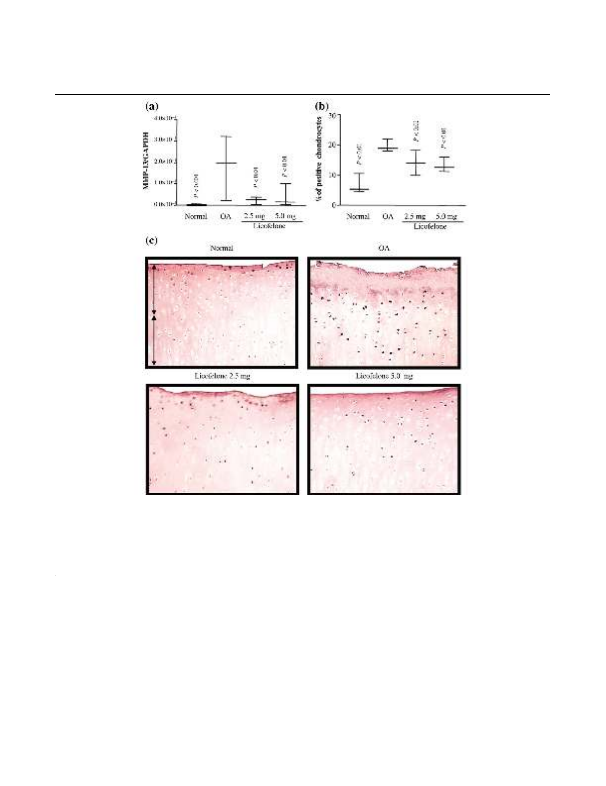
MMP-13 gene expression and protein synthesis
PCR analysis found a marked and significant increase in the
expression of mRNA for MMP-13 in OA cartilage compared to
normal (Fig. 1). Immunohistochemical analysis revealed that
the increased synthesis of MMP-13 was mainly found through-
out the tissue, as previously reported [18]; the controls were
negative (data not shown). A good correlation exists between
the mRNA and protein levels. At the two dosages tested, the
licofelone treatment significantly reduced the levels of both
MMP-13 mRNA expression and the protein to an approxi-
mately similar extent.
Cathepsin K gene expression and protein synthesis
The levels of both the gene expression and the protein of
cathepsin K were significantly increased in OA cartilage, com-
pared to normal cartilage (Fig. 2). These two levels were also
well correlated. Immunohistochemical staining showed that
the enzyme was found to be preferentially located in the super-
ficial zone of the OA cartilage, as previously reported [18]. The
controls were found to be negative (data not shown). Treat-
ment with licofelone at both concentrations reduced the levels
of mRNA expression and protein synthesis of cathepsin K. The
effect was similar at both of the tested dosages for gene
expression and more pronounced at the highest dosage
tested for the level of the enzyme per se.
ADAMTS-4 and ADAMTS-5 gene expression and protein
synthesis
The level of gene expression of ADAMTS-5 in OA cartilage
determined by PCR analysis was highly variable and, although
sometimes higher than that in normal cartilage, the differences
did not reach statistical significance (Fig. 3). The results were
somewhat similar with regards to the immunohistochemical
analysis. The staining showed that the enzyme was in the
chondrocytes mainly located in the superficial zone; some
matrix staining was also observed. The protein level of
ADAMTS-5 in OA cartilage was found to be significantly
higher than normal; the controls were found to be negative and
showed only background staining. Treatment with licofelone
had little effect on the level of its gene expression or on the
level of protein. In contrast, the level of expression of mRNA for
ADAMTS-4 was found to be significantly increased in OA car-
tilage compared to normal (Fig. 4). This was also reflected in
the immunohistochemical analysis, in which an increased level
Table 1
Primer design for quantitative RT-PCR analysis
mRNA Primersa
MMP-13 Fw: 5'-TTGGTCAGATGTGACACCTC
Rv: 5'-ATCGGGAAGCATAAAGTGGC
Cathepsin K Fw: 5'-AGGTGGATGAAATCTCTCGG
Rv: 5'-TTCTTGAGTTGGCCCTCCAG
5-LOX Fw: 5'-TGCGTTCCAGTGACTTCCAC
Rv: 5'-CTCTGCACCATCTGCACGTG
ADAMTS-4 Fw: 5'-TACTACTATGTGCTGGAGCC
Rv: 5'-AGTGACCACATTGTTGTATCC
ADAMTS-5 Fw: 5'-GGCATCATTCATGTGACAC
Rv: 5'-GCATCGTAGGTCTGTCCTG
GAPDH Fw: 5'-AGGCTGTGGGCAAGGTCATC
Rv: 5'-AAGGTGGAAGAGTGGGTGTC
aFw, forward; Rv, reverse. GAPDH, glyseraldehyde-3-phosphate dehydrogenase; MMP, matrix metalloproteinase.
Available online http://arthritis-research.com/content/7/5/R1091
R1095
of the enzyme was found more particularly in the superficial
layers. The level of the enzyme was found to be significantly
decreased by licofelone treatment at both the tested dosages.
5-LOX gene expression and protein synthesis
Although the level of gene expression of 5-LOX in normal car-
tilage was very low, as demonstrated by quantitative PCR
analysis (Fig. 5), it showed a marked and significant increase
in OA cartilage. There was a good correlation between these
results and those from immunohistochemistry, which also
showed a marked and significant increase in the level of the
enzyme that was mainly located in the superficial zone of OA
cartilage. The controls were negative. At both of the tested
dosages, licofelone treatment significantly reduced the level of
gene expression and protein synthesis of the enzyme to a sim-
ilar extent. There was also a correlation between the reduction
in the mRNA and protein levels.
Correlation analysis: Mankin score, safranin-O and cell
score
In specimens from OA dogs, a positive and significant correla-
tion was found between the Mankin score or the safranin-O
staining score and the chondrocyte cell score for ADAMTS-4
(r = 0.50, p = 0.005 for the Mankin score, and r = 0.59, p =
Figure 1
MMP-13 gene expression and protein synthesisMMP-13 gene expression and protein synthesis. (a) mRNA levels, as determined by real-time quantitative PCR analysis as described in Materials
and methods. (b) Morphometric analysis of MMP-13 immunostaining. (a, b) Data are expressed as median and range and are presented as box
plots, where the boxes represent the 1st and 3rd quartiles, the line within the box represents the median, and the lines outside the box represent the
spread of values. P-values were compared to the placebo group (OA) using the Mann-Whitney U test. (c) Representative MMP-13 immunohisto-
chemical sections of tibial plateaus. Superficial (superfical and upper intermediate layers) and deep (lower intermediate and deep layers) zones of
cartilage are indicated on the picture with arrows. No specific staining was detected in the OA cartilage with immunoabsorbed serum (data not
shown) (original magnification × 250). GAPDH, glyceraldehyde-3-phosphate dehydrogenase; MMP, matrix metalloproteinase.


