
In the previous issue of Arthritis Research and Th erapy,
Ibanez and colleagues [1] report on the ‘rational’ use of
gluco corticoids (GCs) in the management of early arthritis.
Th is article concludes that GCs cause minimal varia tion in
bone mineral density (BMD) at multiple skeletal sites, and
in fact may increase BMD at the ultra distal forearm, a
juxta-articular site. Although this article is notable for
examining the eff ect of GCs on BMD at fi ve anatomic
sites, a ‘rational’ use of GCs for rheumatoid arthritis (RA)
is still elusive. To grasp the complex relation ship between
GCs and both localized (juxta-articular, bony erosions)
and systemic (osteoporosis) bone loss in RA, we need to
fi rst step back and appreciate the interplay of the immune
system and bone metabolism.
Th e osteoclast plays a central role at the site of infl amed
joints and is critical in the pathogenesis of joint erosions
in RA [2]. Receptor activator of nuclear factor-kappa B
ligand (RANKL), expressed by TH1 and TH17 T cell
subsets, is a potent inducer of osteoclast diff erentiation.
Additionally, an array of pro-infl ammatory cytokines
such as TNF, IL-1, IL-6 and IL-17 can stimulate RANKL
expression [3] (Figure 1). GCs, in turn, directly aff ect
both osteoblast and osteoclast activity, and indirectly
exert many eff ects on bone metabolism, leading to an
increased fracture risk [4] (Figure 2).
How should the relationship between the bone biology
in RA and the eff ects of GCs translate into the use of GCs
in clinical practice? Th e COBRA trial provides a rationale
for the use of GCs in combination with other disease
modifying anti-rheumatic drugs (DMARDs) to signifi -
cantly reduce RA disease activity [5]. A subsequent
review demonstrates that GCs (mean cumulative dose of
2,300 mg prednisone equivalent over the fi rst year), when
used in combination with traditional DMARD therapy,
can decrease the rate of radiographic progression in RA
[6]. Th e eff ect of GCs on bone mass, among non-RA
patients, has been evaluated in a small randomized,
placebo-controlled trial demonstrating that serum
markers of bone formation are rapidly decreased among
healthy post-menopausal women treated with just 5 mg
of prednisone daily for 6 weeks [7]. In early RA patients
treated with prednisolone 7.5 mg per day and traditional
DMARD therapy (compared to traditional DMARD
therapy alone), markers of bone formation, markers of
bone resorption, and lumber spine BMD, but not femoral
BMD, were decreased [8]. However, in a randomized,
placebo-controlled trial of 95 early RA patients, GCs
decreased the degree of localized hand bone loss [9].
Th ese studies suggest that low dose GCs may reduce
markers of bone formation leading to generalized osteo-
porosis, but they also counteract RA-associated infl am-
ma tion and slow the rate of bone loss proximal to sites of
active disease. Other factors, such as GC resistance and
genetic polymorphisms that predispose to either GC
sensitivity or resistance, may play a role in the BMD
variation seen at various anatomic sites [10,11].
Despite the disease-modifying properties of GCs seen
in RA patients, the risk of GC-induced osteoporosis and
its associated morbidity often give the rheumatologist
pause when determining whether to use GCs, in what
dosing, for how long, at what time in the disease process,
and in which types of RA patients (seropositive versus
sero negative). Ibanez and colleagues have begun to
advance our knowledge of GC use in RA. In this cohort
of early RA patients, they found a signifi cant decrease in
BMD at all sites except the ultradistal and distal forearm.
In the multivariate analysis there was no signifi cant
relation ship between cumulative GC use and BMD
variation at multiple sites, except at the ultradistal
(increased BMD) and mid-forearm (decreased BMD) [1].
Abstract
The relationship between glucocorticoids and bone
mineral density in rheumatoid arthritis is complex.
Further study into the optimal dosing, timing and
duration of glucocorticoid use in rheumatoid arthritis is
necessary.
© 2010 BioMed Central Ltd
The use of glucocorticoids in rheumatoid arthritis -
no ‘rational’ approach yet
Sonali P Desai* and Daniel H Solomon
See related research by Ibanez et al., http://arthritis-research.com/content/12/2/R50
EDITORIAL
*Correspondence: sdesai5@partners.org
Brigham and Women’s Hospital, Division of Rheumatology, Immunology, and
Allergy, 75 Francis Street, Boston, MA 02115, USA
Desai and Solomon Arthritis Research & Therapy 2010, 12:127
http://arthritis-research.com/content/12/3/127
© 2010 BioMed Central Ltd
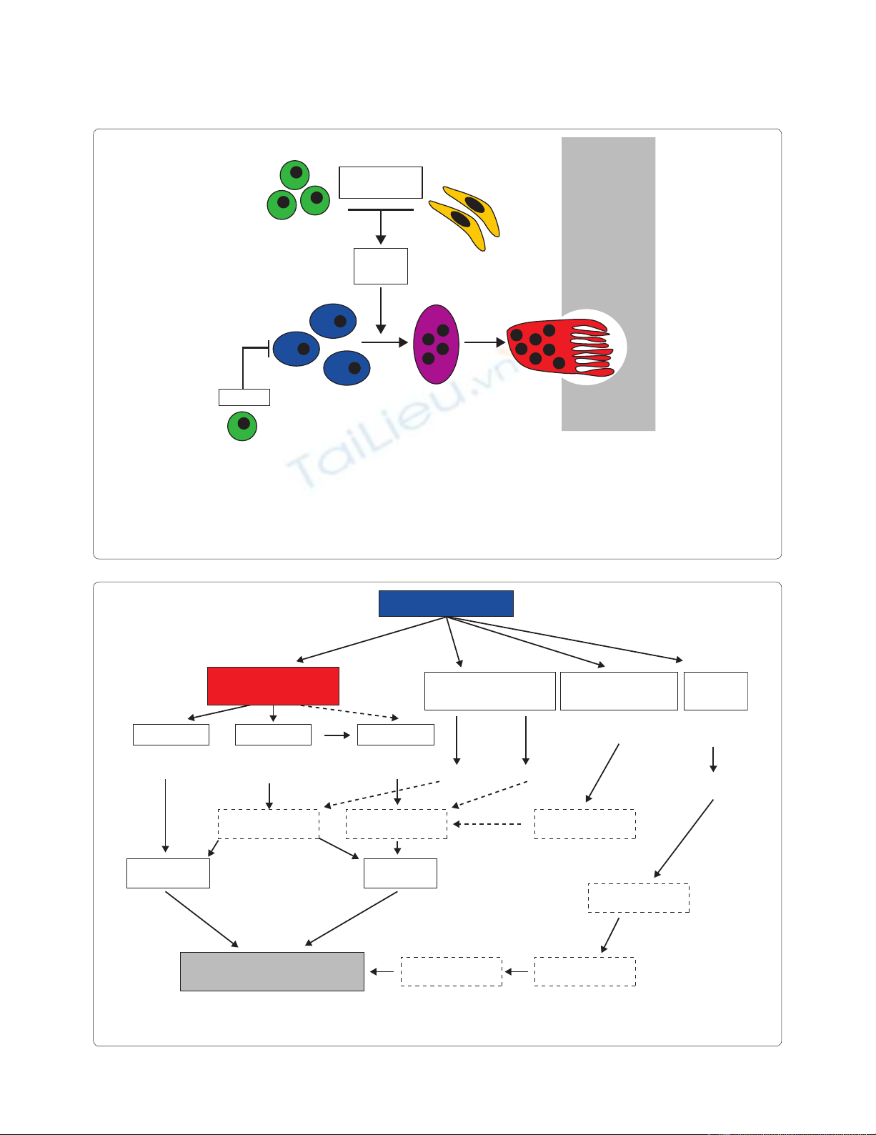
Figure 1. Osteoclast formation in the joint. Monocytic cells in the synovium serve as osteoclast precursors. Upon exposure to macrophage colony-
stimulating factor (MCSF) and Receptor activator of nuclear factor-kappa B ligand (RANKL) synthesized by T cells and synovial fi broblasts, osteoclasts
fuse to polykaryons termed preosteoclasts, which then undergo further diff erentiation into mature osteoclasts, acquiring specifi c features such as the
ruffl ed membrane. Infl ammatory cytokines such as TNF and IL-1, IL-6, and IL-17 increase the expression of RANKL and thus support osteoclastogenesis
in the joint. In contrast, regulatory T cells (Tregs) block osteoclast formation via Cytotoxic T-lymphocyte antigen 4 (CTLA4). Figure obtained with
permission from [3]. The full colour version of this fi gure is available online at http://arthritis-research.com/content/12/3/127
TH1 and TH17
Fibroblasts
TNF, IL-6
IL-1, IL-17
MCSF
RANKL
Treg
Preosteoclast
Osteoclast
Osteoclast
precursors
CTLA4
Bone
Figure 2. The direct and indirect eff ects of glucocorticoids on bone leading to glucocorticoid-induced osteoporosis and fractures. Figure
obtained with permission from [4]. CSF, colony stimulating factor; GH, growth hormone; IGF, insulin-like growth factor; RANKL, Receptor activator of
nuclear factor-kappa B ligand. The full colour version of this fi gure is available online at http://arthritis-research.com/content/12/3/127
Osteocytes Osteoblasts Osteoclasts
Neuroendocrine system Calcium metabolism Muscle
↓ Function
↑ Apoptosis
↓ Differentation
↓ Function
↑ Apoptosis
↑ Genesis
↓ Apoptosis ↓ GH/IGF-1 ↓ Sex steroids
↓ Intestinal absorption
↑ Renal excretion
Proteoolysis
of myofibrils
↓ Fibrils
↓ Bone quality
↓ Bone formation ↑ Bone resorption
↑ Risk of falls
Negative calcium
balance
↓ Bone mass
Myopathy
Increased risk of fracture Muscle weakness
RANKL
CSF
Bone
Glucocorticoids
Desai and Solomon Arthritis Research & Therapy 2010, 12:127
http://arthritis-research.com/content/12/3/127
Page 2 of 3

At fi rst glance, the seemingly simple lack of BMD
variation among a cohort of early RA patients treated
with GCs is a compelling argument for GC use - but in
this small 2-year study of early RA, the dosing, duration
and long-term eff ects remain unknown. Th e apparent
increase in ultradistal forearm BMD among patients
treated with GCs warrants further exploration. However,
this study of early RA examines only 116 patients treated
with a median cumulative GC dose of 22 mg/month and
45 mg/month among those who actually received GCs. Is
it the relatively small cumulative dose of GCs used in this
study that explains the lack of signifi cant BMD variation
or the fact that only 67% of the cohort actually received
GCs, with 17.3% of patients on GC therapy at the end of
the study?
Conclusion
While Ibanez and colleagues have furthered our
understanding of the eff ects of GCs on BMD variation
through their detailed analysis of fi ve skeletal sites, we are
far from a ‘rational’ use of GCs in the management of RA.
Abbreviations
BMD = bone mineral density; DMARD = disease modifying anti-rheumatic
drug; GC = glucocorticoid; IL = interleukin; RA = rheumatoid arthritis; RANKL
= Receptor activator of nuclear factor-kappa B ligand; TNF = tumor necrosis
factor.
Competing interests
The authors declare that they have no competing interests.
Acknowledgements
SPD’s eff ort is supported by the American College of Rheumatology Research
and Education Fund Physician Scientist Development Award. DHS’s eff ort is
supported by NIH grants (AR 055989 and AR 047782).
Published: 25 June 2010
References
1. Ibanez M, Ortiz AM, Castrejon I, Garcia-Vadillo A, Carvajal I, Castaneda S,
Gonzalez-Alvaro I: A rational use of glucocorticoids in patients with early
arthritis has a minimal impact on bone mass. Arthritis Res Ther 12:R50.
2. Gravallese EM, Harada Y, Wang JT, Gorn AH, Thornhill TS, Goldring SR:
Identifi cation of cell types responsible for bone resorption in rheumatoid
arthritis and juvenile rheumatoid arthritis. Am J Pathol 1998, 152:943-951.
3. Schett G: Osteoimmunology in rheumatic diseases. Arthritis Res Ther 2009,
11:210.
4. Canalis E, Mazziotti G, Giustina A, Bilezikian JP: Glucocorticoid-induced
osteoporosis: pathophysiology and therapy. Osteoporos Int 2007,
18:1319-1328.
5. Maurice MM, Nakamura H, van der Voort EA, van Vliet AI, Staal FJ, Tak PP,
Breedveld FC, Boers M, Verhoeven AC, Markusse HM, van de Laar MA,
Westhovens R, van Denderen JC, van Zeben D, Dijkmans BA, Peeters AJ,
Jacobs P, van den Brink HR, Schouten HJ, van der Heijde DM, Boonen A, van
der Linden S: Randomised comparison of combined step-down
prednisolone, methotrexate and sulphasalazine with sulphasalazine alone
in early rheumatoid arthritis. Lancet 1997, 350:309-318.
6. Kirwan JR, Bijlsma JW, Boers M, Shea BJ: Eff ects of glucocorticoids on
radiological progression in rheumatoid arthritis. Cochrane Database Syst
Rev 2007:CD006356.
7. Ton FN, Gunawardene SC, Lee H, Neer RM: Eff ects of low-dose prednisone
on bone metabolism. J Bone Miner Res 2005, 20:464-470.
8. Engvall IL, Svensson B, Tengstrand B, Brismar K, Hafstrom I: Impact of low-
dose prednisolone on bone synthesis and resorption in early rheumatoid
arthritis: experiences from a two-year randomized study. Arthritis Res Ther
2008, 10:R128.
9. Haugeberg G, Strand A, Kvien TK, Kirwan JR: Reduced loss of hand bone
density with prednisolone in early rheumatoid arthritis: results from a
randomized placebo-controlled trial. Arch Int Med 2005, 165:1293-1297.
10. Sliwinska-Stanczyk P, Pazdur J, Ziolkowska M, Jaworski J, Kaminska-
Tchorzewska E, Lacki JK: The eff ect of methylprednisolone on proliferation
of PBMCs obtained from steroid-sensitive and steroid-resistant
rheumatoid arthritis patients. Scand J Rheumatol 2007, 36:167-171.
11. Manenschijn L, van den Akker EL, Lamberts SW, van Rossum EF: Clinical
features associated with glucocorticoid receptor polymorphisms. An
overview. Ann N Y Acad Sci 2009, 1179:179-198.
doi:10.1186/ar3035
Cite this article as: Desai SP, Solomon DH: The use of glucocorticoids in
rheumatoid arthritis - no ‘rational’ approach yet. Arthritis Research & Therapy
2010, 12:127.
Desai and Solomon Arthritis Research & Therapy 2010, 12:127
http://arthritis-research.com/content/12/3/127
Page 3 of 3


