
BioMed Central
Page 1 of 6
(page number not for citation purposes)
Journal of Immune Based Therapies
and Vaccines
Open Access
Review
The effects of chemotherapeutics on cellular metabolism and
consequent immune recognition
M Karen Newell*1, Robert Melamede1, Elizabeth Villalobos-Menuey1,
Douglas Swartzendruber2, Richard Trauger3, Robert E Camley4 and
William Crisp5
Address: 1Department of Biology, University of Colorado at Colorado Springs, Colorado Springs, CO 80933-7150, USA, 2Natural Sciences
Division, Seaver College, Pepperdine University, Malibu, CA 90263, USA, 3Hollis Eden Pharmaceuticals, San Diego, CA 92121, USA, 4Department
of Physics, University of Colorado at Colorado Springs, Colorado Ssprings, CO 80933-7150, USA and 5Cancer Research Institute, Arizona State
University, Tempe, AZ 85287, USA
Email: M Karen Newell* - mnewell@uccs.edu; Robert Melamede - rmelamed@uccs.edu; Elizabeth Villalobos-Menuey - emvillal@uccs.edu;
Douglas Swartzendruber - douglas.swartzendruber@pepperdine.edu; Richard Trauger - rtrauger@holliseden.com;
Robert E Camley - rcamley@uccs.edu; William Crisp - adcrc1@getnet.net
* Corresponding author
chemotherapyimmune recognitionapoptosisFas (CD95)metabolism
Abstract
A widely held view is that oncolytic agents induce death of tumor cells directly. In this report we
review and discuss the apoptosis-inducing effects of chemotherapeutics, the effects of
chemotherapeutics on metabolic function, and the consequent effects of metabolic function on
immune recognition. Finally, we propose that effective chemotherapeutic and/or apoptosis-
inducing agents, at concentrations that can be achieved physiologically, do not kill tumor cells
directly. Rather, we suggest that effective oncolytic agents sensitize immunologically altered tumor
cells to immune recognition and immune-directed cell death.
Review
Do drugs kill tumor cells directly?
Our laboratories have been investigating the conse-
quences of chemotherapeutic agents on cell surface
expression of immunologically important molecules,
including Major Histocompatibility Complex (MHC)
encoded molecules (both MHC class I and II), B7.1
(CD80), B7.2 (CD86), Fas (CD95), and Fas Ligand
(CD95L) [1]. T cell activation requires recognition of anti-
gens associated with MHC molecules [2] and a second sig-
nal provided by co-stimulation [3] provided by the
interaction of molecules including B7.1 or B7.2 or Fas
(CD95) on the cell being recognized and CD28 or CTLA-
4 or Fas Ligand on the T cell. We, and others, have
reported that changes in the cell surface occur in drug-
treated cells [4-10]. First, we observe changes and
increases in cell surface expression of the B7 family mem-
bers, CD80 and CD86, on drug-treated (adriamycin, 5-
fluorouracil, or methotrexate-treataed) tumor cells. These
cell surface molecules have been extensively studied and
are now widely accepted as important in promoting the
immunogenicity of tumor cells by providing costimula-
tion for T cells [5]. Second, we, and others, have observed
that most of the drugs we have used increase cell surface
expression of Fas (CD95) and sensitize the Fas-bearing
tumor to Fas-induced death [1,7,9]. In the present report,
Published: 02 February 2004
Journal of Immune Based Therapies and Vaccines 2004, 2:3
Received: 29 December 2003
Accepted: 02 February 2004
This article is available from: http://www.jibtherapies.com/content/2/1/3
© 2004 Newell et al; licensee BioMed Central Ltd. This is an Open Access article: verbatim copying and redistribution of this article are permitted in all
media for any purpose, provided this notice is preserved along with the article's original URL.

Journal of Immune Based Therapies and Vaccines 2004, 2http://www.jibtherapies.com/content/2/1/3
Page 2 of 6
(page number not for citation purposes)
we discuss our working model that the concert of meta-
bolic interference with the ability of the tumor to be more
readily "seen" by the immune system may be the basis for
effectiveness of many currently effective strategies or the
basis for developing novel therapeutic approaches to
treating cancers.
We first explore one of the relevant immunological cell
surface receptors, Fas (CD95). Fas is a member of the
tumor necrosis receptor (TNFR) family. The cytoplasmic
tail of Fas contains a death domain able to trigger intrac-
ellular caspase cascades that culminate in apoptotic cell
death [11-13]. Fas can induce apoptosis when ligated by
its cognate ligand (FasL, CD95L) in Fas sensitive cells
[11,12]. Paradoxically, Fas, like other members of its fam-
ily, can transduce growth-enhancing signals as well as
death signals [14-18]. In chemo-sensitive leukemia and
solid tumors, anti-cancer drugs have been shown to
induce apoptosis and for many tumors the pathways
involved include, but are not limited to, Fas and FasL [19-
21].
In an attempt to reflect in vitro the concentrations of drugs
that can be achieved physiologically in vivo, we were sur-
prised to observe that tumor cells from many tissue ori-
gins were not dead at such concentrations. However, we
found (and continue to find with a broad spectrum of
agents) that the drugs have several important conse-
quences. Our results have shown that chemotherapeutic
agents sensitize Fas-bearing, Fas-insensitive tumors to Fas-
susceptibility and Fas-induced death [1]. Consistent with
these observations, cross-resistance to Fas/FasL and onco-
lytic agents has been reported by our group and others
[1,8,10,22]. While much of our work has involved Fas and
FasL, other members of "death inducing" receptor-ligand
pairs likely perform similarly in the presence of effective
oncolytic agents [23].
Together these data indicated that an important mecha-
nism of chemotherapeutic agents may be to sensitize
tumor cells to immune-directed death. Implied by these
results is the importance of identifying and preserving
(from death by high dose chemotherapy) the FasL (or
other ligand)-bearing cells to facilitate immunological
destruction of drug-treated tumor cells.
How do chemotherapeutic agents sensitize the tumor cells
to immune-mediated death?
Our efforts at understanding the molecular mechanisms
by which chemotherapeutic agents affect metabolism and
immune recognition have been focused primarily on the
expression and function of Fas on the cell surface of tumor
cells. Fas is expressed on most rapidly dividing cells,
including tumor cells, hepatocytes, epithelial cells, and
lymphocytes [24-26]. Interestingly, tissues that express
Fas and yet remain insensitive to Fas-induced death
(including most dividing, regenerating, and self-renewing
cells) exhibit a metabolic phenotype characterized by
high rate, cytosolic glycolysis. This "respiratory defi-
ciency" is the result of a metabolic change in tumor cells
that was first observed by Warburg in 1926 [27]. The co-
incidence of increased cytosolic glycolysis and increased
Fas expression on tumor cells (and other dividing cells)
provided the basis for examining a causal link between Fas
expression and the use of glucose as a primary, glycolytic
source of fuel.
Our experiments have demonstrated that the distribution
and levels of expression of Fas are altered in response to
changing concentrations of glucose in many cell lines and
in freshly isolated cells from a variety of tissues. Limited
glucose supplementation is known to enhance prolifera-
tion of tumor cells and has been used for topical applica-
tions to accelerate wound healing in vivo [28,29]. Some of
our recent results suggest that glucose availability and
consequent production of intracellular reactive oxygen
species may regulate the striking change in the results
from Fas engagement that promotes proliferation to Fas
engagement that promotes death. Supporting this obser-
vation is the recent report that increasing glucose concen-
trations can induce increased free radical production [30]
and increases in reactive oxygen or free radicals are known
to cause Fas engagement to result in cell death [31-33]. In
addition, we have observed and reported that drug resist-
ant cells appear to readily utilize the carbons derived from
beta oxidation of fatty acids and exhibit a consequent loss
of cell surface Fas. Taken together these observations sup-
port the notion that Fas expression and function are inter-
twined with glucose metabolism and the potential for
changes in reactive intermediates in tissues or cells exhib-
iting changes in glucose metabolism. The fact that selec-
tion in drugs results in loss of Fas and in metabolic
changes that may protect the cells from free radical dam-
age will be important in designing novel cancer therapies.
We have performed experiments to examine the correla-
tion between cell surface Fas expression and glucose
metabolism. As a prototype for the Fas positive and Fas
negative cells we have used the L1210 cell and the
L1210DDP as Fas positive and Fas negative, respectively,
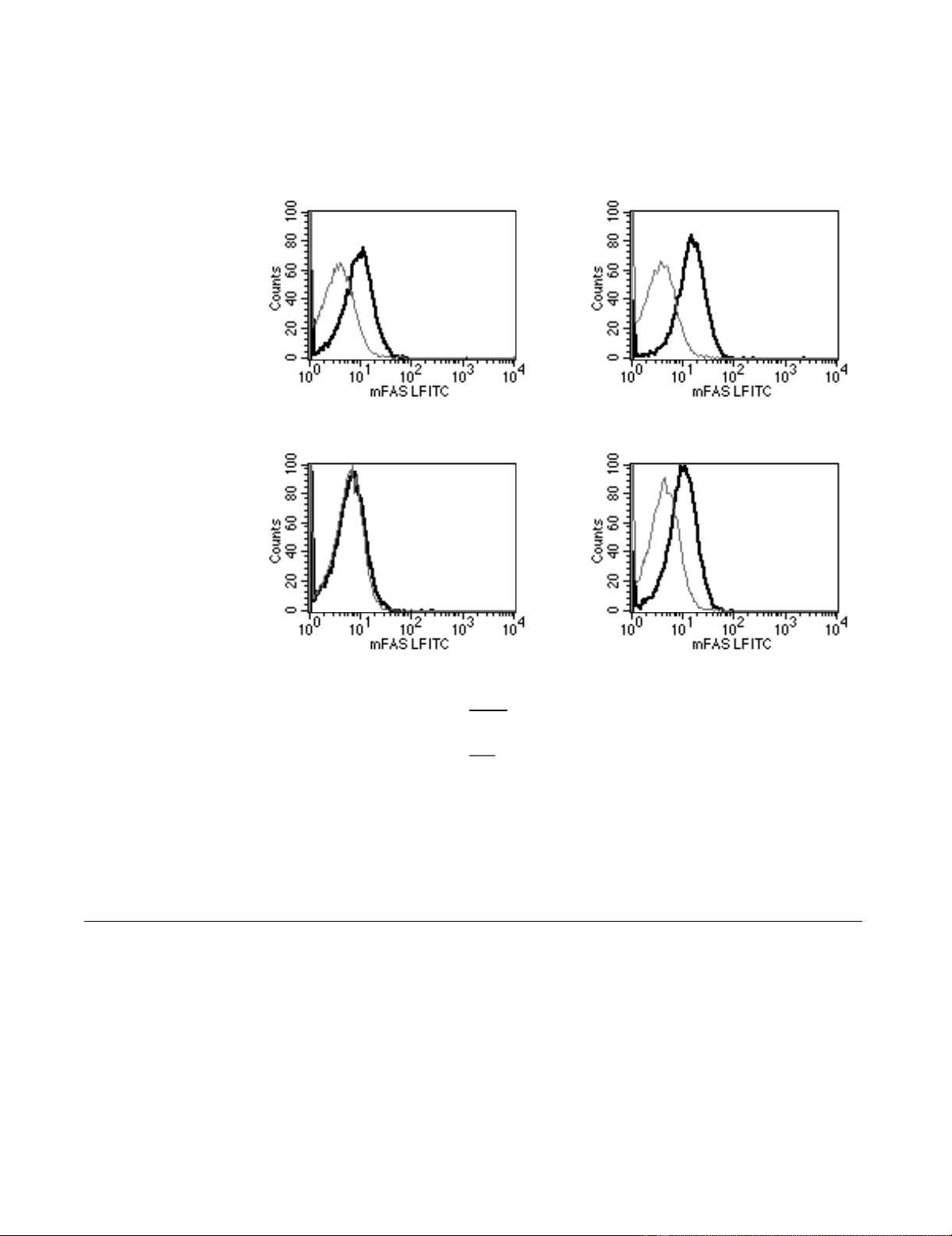
Figure 1. In these experiments, we directly measured the
rates of glucose utilization and oxidation of L1210 and
L1210DDP [34].
L1210 DDP cells express no cell surface Fas [1]. To address
the possibility that Fas is expressed, but has been targeted
to a subcellular organelle, we permeabilized and stained
L1210 and L1210DDP cells with fluorochrome conju-
gated anti-Fas antibody (J02.2, Pharmingen). The cells
were examined by flow cytometry. Our data indicate that
Journal of Immune Based Therapies and Vaccines 2004, 2http://www.jibtherapies.com/content/2/1/3
Page 3 of 6
(page number not for citation purposes)
L1210 DDP cells express no cell surface Fas; however, the
cells do express intracellular Fas. Fluorochrome-conju-
gated isotype matched antibody was used as control, and
specific antibody stains were confirmed as specific. These
data demonstrate that the Fas negative, apoptosis resistant
cells, express intracellular Fas, Figure 1 below. The rele-
vance of internal Fas in drug-selected, drug resistant
tumor cells is that the cell is rendered Fas-insensitive to
cell death unless the intracellular pool can be redistrib-
uted to the cell surface and potentially re-wired to "death-
inducing" machinery.
It is known that T cells require two signals for activation
[3]. One of these signals involves the binding of the pro-
teins CD28 or CTLA-4, which are constitutively expressed
on most resting T cells, with the proteins B7.1 (CD80) or
Distribution and Level of Fas in L1210/0 and L1210/DDP CellsFigure 1
Distribution and Level of Fas in L1210/0 and L1210/DDP Cells. Expression of cell-surface Fas, leftmost panels, and
intracellular Fas, right most panels in L1210/0, upper two panels, and L1210/DDP cells, lower two panels. The levels of cell sur-
face Fas (dark lines) were determined using fluorochrome conjugated anti-Fas antibodies (Pharmingen Inc.) and flow cytometry.
The levels of intracellular Fas were determined subsequent to cellular permeabilization and fixation. The Fas levels are meas-
ured relative to staining for fluorochrome-conjugated isotype control (grey lines).
Key
L1210/DDP
L1210/0
Intracellular FAS
FAS Expression
Isotype Control
Surface FAS
Journal of Immune Based Therapies and Vaccines 2004, 2http://www.jibtherapies.com/content/2/1/3
Page 4 of 6
(page number not for citation purposes)
B7.2 (CD86). T cell activation through CD28 binding,
results in a proliferative T cell response, enhanced T cell
survival and cytokine release [35]. Conversely, CTLA-4
engagement induces powerful inhibitory signals in T cell
activation resulting in the negative regulation of T cell
responses [36]. Collins et al. recently showed that B7.1
favors CTLA-4 over CD28 engagement [37]. This is still
controversial, nonetheless is raises the possibility that co-
stimulatory receptor/ligand pairs are multifunctional.
We propose that co-stimulatory interactions between B7
family members and CD28 or CTLA4-bearing T cells and
the resulting cytokines directly impact the subcellular dis-
tribution of Fas and the ultimate outcome of Fas engage-
ment on tumor cells.
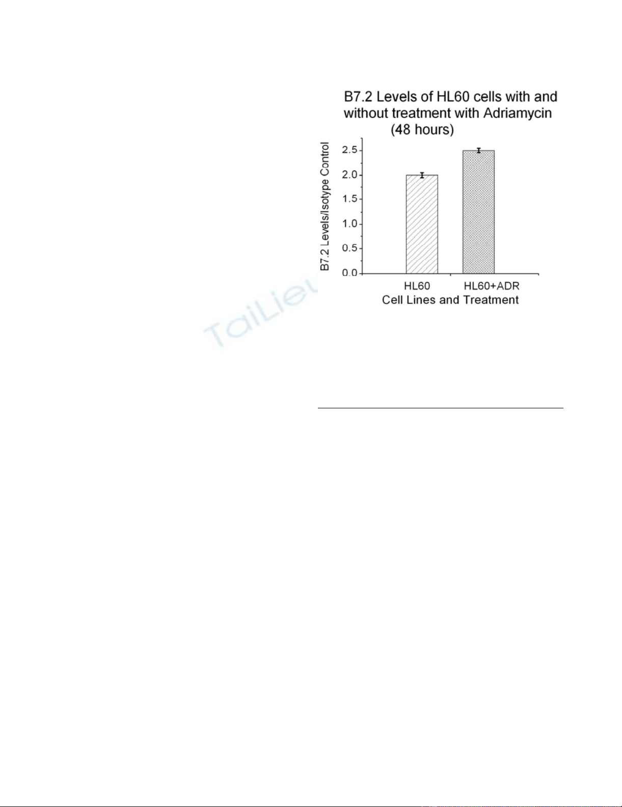
In Figure 2 we show that B7.2 levels in HL60 (human
leukemic cells) also increase after treatment with 10-8 M of
Adriamycin. We note that HL60 is a human cell line and
that the drug is different than that in previous figures. This
figure is representative of many experiments with other
cell lines and additional drugs that include methotrexate,
adriamycin, and 5-fluorouracil. While we have not tested
the ability of all drugs to promote immunogenicity, these
resuts may imply that the increase in the co-stimulatory sig-
nal as a result of drug treatment is a general phenomenon.
Which immune cell can kill the tumor cell?
The first attempts at cancer immunotherapy were made
over 100 years ago on the assumption that tumor antigens
might be recognized as foreign [38]. These studies gave
rise to animal tumor models using syngeneic tumors,
spontaneously arising tumors, and xenografts into immu-
nodeficient hosts. The collective of these studies resulted
in a variety of immunotherapeutic protocols including
adjuvant therapy, cytokines, NK cell activation, macro-
phages, and attempts to stimulate tumor antigen specific
B and/or T cell responses against tumor antigens. Some
approaches have had partial success, but what has become
clear is that tumor cells are, by definition, "immunologi-
cally privileged" and successfully evade effective tumori-
cidal immune recognition [38]. An alternate possibility is
suggested by the premise which Prehn has postulated that
effective chemotherapies may result from suppressing a
particular type of immune response that supports tumor
cell growth [39]. An example of this notion would be T
cell-produced cytokines which have been reported to sup-
port neural regeneration [40].
MHC encoded molecules were defined by Peter Gorer and
George Snell as surface molecules responsible for the
rejection of tumor cells between genetically distinct mem-
bers of the same species [41]. These molecules are also
responsible for graft rejection and T cell activation. The
mechanism for both phenomenons has been attributed to
T cell receptor recognition and effector functions that
occur only when MHC molecules and antigen are recog-
nized by the T cell receptor for antigen. Cells implicated in
tumor cell death include CD4+ T cells, CD8+ T cells, natu-
ral killer (NK) cells, or more recently, gamma delta (γδ) T
cells [38]. Immune recognition and destruction of alloge-
neic tumor cells likely results from increased expression of
MHC antigens on the tumor cell surface, processing and
presentation of tumor antigens, and expression of costim-
ulatory molecules on the tumor cell. Rejection of tumor
cells following drug treatment, therefore, may be directly
related to "recognition" of a cell which has changed in cell
surface expression of immunologically important cell sur-
face receptors and that has been metabolically "rewired"
by chemotherapeutic agents.
Conclusion
Thus, we suggest that a drug-treated tumor cell is made
susceptible by drugs or radiation to "death-inducing"
receptor/ligand pairs, including, but not limited to, Fas
and FasL expressed on candidate immune cells, such as
CD4+ T cells, CD8+ T cells, gamma delta T cells, and NK
cells. We propose that selective identification of the
Adriamycin Induced Increase in B7.2 ExpressionFigure 2
Adriamycin Induced Increase in B7.2 Expression.
Expression of the cell-surface co-stimulatory molecule B7.2
as a function of treatment with adriamycin. The level of cell-
surface B7.2 was determined using fluorochrome conjugated
anti-B7.2 antibodies and flow cytometry. The B7.2 levels are
measured relative to staining for fluorochrome-conjugated
isotype control.

Journal of Immune Based Therapies and Vaccines 2004, 2http://www.jibtherapies.com/content/2/1/3
Page 5 of 6
(page number not for citation purposes)
immunocytes proliferating in the tumor-bearing lymph
node as a key element in personalizing and selectively
sensitizing an individual's tumor cells to chemo-, radio-,
and immunotherapy.
While the potential of immune-directed cytotoxicity of
drug-treated tumor cells may provide an important new
perspective, the question arises as to how to reconcile this
idea with the accepted notion that chemotherapy can be
immunosuppressive. The key factor in resolving this
seeming paradox may be the dose of the agent or the
nature of a given chemotherapeutic agent. Clearly, there
are cases where drugs at high doses have immunosuppres-
sive effects (perhaps by direct cytotoxicity of the immune
cells). In contrast, decreased doses have recently been
shown to be more effective in the clinic. Taken together,
both views suggest that "less may be more" effective for
chemotherapy [45,46]. We propose that an in depth eval-
uation of the effects of popular chemotherapeutic agents
on induction of immunologically relevant molecules on
the tumor be rigorously evaluated.
Considering the potential importance of cells of the
immune system in controlling cancer growth, with or
without chemotherapy, an important question is raised.
Should lymph nodes, the local "home" too many
immune cells, be removed as therapy? Although axillary
node removal is still a standard regime for treatment of
invasive breast cancer, it is clear that regional lymph
nodes have biological significance for being more than
just anatomical filters. The regional lymph node is the
heart of our immunologic defense system and the present
routine practice of partial resection of the regional nodes
where they are easiest to remove undoubtedly has an
effect on immunological and physiological function.
Macroscopically involved lymph nodes should possibly
be removed for prognosis [42] and for the identification
of the immune cells involved in tumor recognition, but
the routine removal of lymph nodes is questioned as
noted above by our group and others [43]. It is becoming
clear that many patients can be spared axillary node dis-
section without adversely affecting outcome [44]. As we
begin to better understand the inter-relationships of sur-
gery, tumor cell kinetics, chemotherapy, and the host
immune response, new paradigms are developing. These
include the notion that routine surgical removal of axil-
lary nodes provides no additional benefit and could be
omitted to spare the patient unnecessary axillary node
removal [43].
In summary, we suggest a novel perspective be applied to
the clinical diagnosis and treatment of tumors. Maxi-
mally, we suggest that each tumor be screened for the
effects of potential chemotherapeutics on immunogenic-
ity. We suggest identifying cells responding to the tumor
in the node (unsuccessfully or not) so that drug-sensitized
tumor cells can be killed rather than supported by the
identified immune cells. Minimally we suggest that a re-
evaluation of the mechanism of tumor cell death and
therapeutic approaches be experimentally and clinically
considered.
Declaration of Competing Interests
None declared.
Authors' Contributions
This paper is distinct because it is an opinion paper. How-
ever, each author contributed uniquely to the manuscript.
Author 1, MKN, provided the conceptual framework for
the model presented in this paper. Author 2, RM, partici-
pated in discussions and drafts of the manuscript. Author
3, EVM, performed the flow cytometric data provided in
this manuscript. Author 4, DS, provided discussion about
a supportive role for T-Cells in the growth of a tumor.
Author 5, RT, participated in discussions and drafts of the
manuscript. Author 6, WC, contributed the effects of sur-
gery on tumor growth. Author 7, RC, participated in dis-
cussions and drafts of the manuscript.
Acknowledgements
We greatly acknowledge Jaimi Kupperman and Jeff Rogers for their assist-
ance with this work.
References
1. Bhushan A, Kupperman JL, Stone JE, Kimberly PJ, Calman NS, Hacker
MP, Birge RB, Tritton TR, Newell MK: Drug resistance results in
alterations in expression of immune recognition molecules
and failure to express Fas (CD95). Immunology and Cell Biology
1998, 76:350-356.
2. Yague J, White J: The T-cell receptor: The α and β chains define
idiotype, and antigen and MHC specificity. Cell 1985, 42:81-87.
3. Bretscher P: The two-signal model of lymphocyte activation
twenty-one years later. Immunology Today 1992, 13:74-76.
4. Delabie J, Ceuppens JL, Vandenberghe P, Coorevits L, De Wolf-
Peeters C: The B7/BB1 antigen is expressed by Reed-Stern-
berg cells of Hodgkin's disease and contributes to the stimu-
lation capacity of Hodgkin's disease derived cell lines. Blood
1993, 82:2845-2852.
5. Guinan EC, Gribben JG, Boussiotis VA, Freeman GJ, Nadle L: Pivotal
role of the B7:CD28 pathway in transplantation tolerance
and tumor immunity. Blood 1994, 84(10):3261-3282.
6. Baskar S, Clements VK, Glimcher LH, Nabavi N, Ostrand-Rosenberg
S: Rejection of MHC class II transfected tumor cells requires
induction of tumor encoded B7-1 and/or B7-2 costimulatory
molecules. Journal of Immunology 1996, 156:3821-3827.
7. Cai Z, Stancou R, Korner M, Chouaib S: Impairment of Fas-anti-
gen expression in adriamycin-resistant but not TNF resist-
ant MCF7 tumor cells. International Journal of Cancer 1996,
68(4):535-546.
8. Fulda S, Los M, Friesen C, Debatin KM: Chemosensitivity of solid
tumor cells in vitro is related to activation of the CD95
system. Int J Cancer 1998, 76(1):105-114.
9. Landowski TH, Gleason-Guzman MC, Dalton MS: Selection for
drug resistance results in resistance to Fas-mediated
apoptosis. Blood 1997, 89:1854-1861.
10. Los M, Herr I, Friesen C, Fulda S, Schulze-Osthoff K, Debatin KM:
Cross-resistance of CD95- and drug-induced apoptosis as a
consequence of deficient activation of caspases (ICE/Ced-3
proteases). Blood 1997, 90(8):3118-3129.


![Vaccine và ứng dụng: Bài tiểu luận [chuẩn SEO]](https://cdn.tailieu.vn/images/document/thumbnail/2016/20160519/3008140018/135x160/652005293.jpg)


%20--%3e%3cdefs%3e%3cstyle%3e%20.st0%20{%20fill:%20%23fff;%20}%20.st1%20{%20fill:%20%237800fa;%20}%20%3c/style%3e%3c/defs%3e%3cpath%20class='st1'%20d='M117.78,12.18H43.11c2.9,3.47,4.65,7.94,4.65,12.82,0,5.6-2.3,10.66-6.01,14.29h76.02l7.22-13.56-7.22-13.56Z'/%3e%3cg%3e%3cpath%20class='st0'%20d='M53.58,26.17h-.59v-1.46h.59v-4.96h2.83c1.78,0,2.67.94,2.67,2.82v5.76c0,1.87-.89,2.81-2.67,2.81h-2.83v-4.96ZM55.36,21.37v3.34h1.1v1.46h-1.1v3.34h1.01c.61,0,.91-.37.91-1.1v-5.93c0-.74-.3-1.1-.91-1.1h-1.01Z'/%3e%3cpath%20class='st0'%20d='M65.99,31.14h-1.8l-.31-2.07h-2.19l-.31,2.07h-1.64l1.82-11.39h2.62l1.82,11.39ZM65.28,18.04c-.25.46-.51.77-.75.94-.21.15-.47.22-.79.22-.26,0-.57-.07-.92-.22l-.38-.15c-.14-.05-.26-.07-.37-.07-.3,0-.53.18-.71.54l-.91-.68c.25-.46.51-.77.75-.94.21-.14.48-.21.79-.21.26,0,.57.07.92.21l.38.15c.14.05.26.07.37.07.3,0,.53-.18.71-.54l.91.68ZM61.91,27.52h1.73l-.87-5.76-.87,5.76Z'/%3e%3cpath%20class='st0'%20d='M74.53,26.89v1.52c0,1.91-.89,2.86-2.67,2.86s-2.67-.95-2.67-2.86v-5.93c0-1.91.89-2.86,2.67-2.86s2.67.95,2.67,2.86v1.11h-1.69v-1.22c0-.75-.31-1.12-.93-1.12s-.93.37-.93,1.12v6.15c0,.74.31,1.11.93,1.11s.93-.37.93-1.11v-1.63h1.69Z'/%3e%3cpath%20class='st0'%20d='M81.4,31.14h-1.8l-.31-2.07h-2.19l-.31,2.07h-1.64l1.82-11.39h2.62l1.82,11.39ZM75.9,19.2l1.52-1.91h1.71l1.51,1.91h-1.61l-.76-.95-.75.95h-1.61ZM77.32,27.52h1.73l-.87-5.76-.87,5.76ZM83.1,15.99l-1.76,1.91h-1.26l1.17-1.91h1.86Z'/%3e%3cpath%20class='st0'%20d='M84.86,19.75c1.78,0,2.67.94,2.67,2.82v1.48c0,1.87-.89,2.81-2.67,2.81h-.85v4.28h-1.79v-11.39h2.64ZM84.01,21.37v3.86h.85c.58,0,.87-.36.87-1.08v-1.71c0-.71-.29-1.07-.87-1.07h-.85Z'/%3e%3cpath%20class='st0'%20d='M93.51,19.75c1.78,0,2.67.94,2.67,2.82v1.48c0,1.87-.89,2.81-2.67,2.81h-.85v4.28h-1.79v-11.39h2.64ZM92.66,21.37v3.86h.85c.58,0,.87-.36.87-1.08v-1.71c0-.71-.29-1.07-.87-1.07h-.85Z'/%3e%3cpath%20class='st0'%20d='M98.8,31.14h-1.79v-11.39h1.79v4.88h2.03v-4.88h1.83v11.39h-1.83v-4.88h-2.03v4.88Z'/%3e%3cpath%20class='st0'%20d='M105.36,24.55h2.46v1.62h-2.46v3.34h3.09v1.63h-4.88v-11.39h4.88v1.63h-3.09v3.18ZM108.17,17.29l-1.76,1.91h-1.26l1.17-1.91h1.86Z'/%3e%3cpath%20class='st0'%20d='M112.2,19.75c1.78,0,2.67.94,2.67,2.82v1.48c0,1.87-.89,2.81-2.67,2.81h-.85v4.28h-1.79v-11.39h2.64ZM111.35,21.37v3.86h.85c.58,0,.87-.36.87-1.08v-1.71c0-.71-.29-1.07-.87-1.07h-.85Z'/%3e%3c/g%3e%3ccircle%20class='st1'%20cx='25'%20cy='25'%20r='20'/%3e%3cpath%20class='st0'%20d='M32.78,19.27c2.92,0,4.43,2.55,5.28,5.33l.71,2.17c.14.38-.33.75-.71.75h-5.61c.19-.33.24-.71.09-1.08l-.75-2.45c-.43-1.32-.99-2.64-1.79-3.77.75-.57,1.65-.94,2.78-.94h0ZM25,18.38c3.25,0,4.9,2.78,5.89,5.89l.76,2.45c.14.42-.33.8-.8.8h-11.69c-.42,0-.94-.38-.8-.8l.75-2.45c.99-3.11,2.64-5.89,5.89-5.89h0ZM25,11.35c1.74,0,3.11,1.37,3.11,3.11s-1.37,3.11-3.11,3.11-3.11-1.41-3.11-3.11,1.41-3.11,3.11-3.11h0ZM17.27,19.27c1.08,0,1.98.38,2.73.94-.8,1.13-1.37,2.45-1.74,3.77l-.8,2.45c-.14.38-.05.75.09,1.08h-5.56c-.42,0-.9-.38-.75-.75l.71-2.17c.9-2.78,2.41-5.33,5.33-5.33h0ZM17.27,12.91c1.51,0,2.78,1.27,2.78,2.83s-1.27,2.83-2.78,2.83-2.83-1.27-2.83-2.83,1.27-2.83,2.83-2.83h0ZM32.78,12.91c1.56,0,2.78,1.27,2.78,2.83s-1.23,2.83-2.78,2.83-2.83-1.27-2.83-2.83,1.27-2.83,2.83-2.83h0ZM27.07,28.56v.09c0,.57-.24,1.08-.61,1.46h0v.05c-.38.33-.9.57-1.46.57s-1.08-.24-1.46-.61h0c-.38-.38-.61-.9-.61-1.46v-.09h1.41v.09c0,.19.05.38.19.47v.05c.09.09.28.19.47.19s.38-.09.47-.19v-.05c.14-.09.24-.28.24-.47t-.05-.09h1.41ZM30.99,28.56v.09c0,1.65-.66,3.16-1.74,4.24-1.08,1.08-2.59,1.79-4.24,1.79s-3.16-.71-4.24-1.79l-.05-.05c-1.04-1.08-1.7-2.55-1.7-4.2v-.09h1.41v.09c0,1.27.47,2.4,1.27,3.25h.05c.85.85,1.98,1.37,3.25,1.37s2.4-.52,3.25-1.37c.85-.8,1.37-1.98,1.37-3.25v-.09h1.37ZM34.99,28.56v.09c0,2.78-1.13,5.28-2.92,7.07-1.79,1.79-4.29,2.92-7.07,2.92s-5.23-1.13-7.07-2.92c-1.79-1.79-2.92-4.29-2.92-7.07v-.09h1.41v.09c0,2.4.94,4.53,2.5,6.08,1.56,1.56,3.72,2.5,6.08,2.5s4.52-.94,6.08-2.5c1.56-1.56,2.5-3.68,2.5-6.08v-.09h1.41Z'/%3e%3c/svg%3e)