
Van Steendam et al. Arthritis Research & Therapy 2010, 12:R132
http://arthritis-research.com/content/12/4/R132
Open Access
RESEARCH ARTICLE
© 2010 Van Steendam et al.; licensee BioMed Central Ltd. This is an open access article distributed under the terms of the Creative Com-
mons Attribution License (http://creativecommons.org/licenses/by/2.0), which permits unrestricted use, distribution, and reproduc-
tion in any medium, provided the original work is properly cited.
Research article
Citrullinated vimentin as an important antigen in
immune complexes from synovial fluid of
rheumatoid arthritis patients with antibodies
against citrullinated proteins
Katleen Van Steendam
1
, Kelly Tilleman
1
, Marlies De Ceuleneer
1
, Filip De Keyser
2
, Dirk Elewaut
2
and Dieter Deforce*
1
Abstract
Introduction: Rheumatoid arthritis (RA) is an inflammatory disease, which results in destruction of the joint. The
presence of immune complexes (IC) in serum and synovial fluid of RA patients might contribute to this articular
damage through different mechanisms, such as complement activation. Therefore, identification of the antigens from
these IC is important to gain more insight into the pathogenesis of RA. Since RA patients have antibodies against
citrullinated proteins (ACPA) in their serum and synovial fluid (SF) and since elevated levels of citrullinated proteins are
detected in the joints of RA patients, citrullinated antigens are possibly present in IC from RA patients.
Methods: IC from serum of healthy persons, serum of RA patients and IC from synovial fluid of RA patients and
Spondyloarthropathy (SpA) patients were isolated by immunoprecipitation. Identification of the antigens was
performed by SDS-PAGE, mass spectrometry and immunodetection. The presence of citrullinated proteins was
evaluated by anti-modified citrulline (AMC) staining.
Results: Circulating IC in the serum of RA patients and healthy controls contain fibrinogenβ and fibronectin, both in a
non-citrullinated form. Additionally, in IC isolated from RA SF, fibrinogenγ and vimentin were identified as well. More
importantly, vimentin and a minor portion of fibrinogenβ were found to be citrullinated in the isolated complexes.
Moreover these citrullinated antigens were only found in ACPA+ patients. No citrullinated antigens were found in IC
from SF of SpA patients.
Conclusions: Citrullinated fibrinogenβ and citrullinated vimentin were found in IC from SF of ACPA+ RA patients, while
no citrullinated antigens were found in IC from SF of ACPA- RA patients or SpA patients or in IC from serum of RA
patients or healthy volunteers. The identification of citrullinated vimentin as a prominent citrullinated antigen in IC
from SF of ACPA+ RA patients strengthens the hypothesis that citrullinated vimentin plays an important role in the
pathogenesis of RA.
Introduction
Rheumatoid arthritis (RA) is a progressive autoimmune
disease characterized by chronic inflammation of the
peripheral joints. It is a complex multifactorial pathology,
in which genetic and environmental factors, like smok-
ing, can play an important role in the onset of disease and
the progression of the joint damage [1,2]. The presence of
immune complexes (IC) in serum and synovial fluid (SF)
of RA patients is likely to contribute to the pathogenesis
of the disease and to articular damage, since they are
responsible for the activation of complement, the stimu-
lation of phagocytes through their Fc receptor and the
release of chemotactic factors, cytokines, metalloprotei-
nases and reactive oxygen intermediates [3-6]. The for-
mation of IC as such is not specifically related to
autoimmune pathologies as it is a natural process, com-
pleting an immune response in the body. The antigen-
antibody complexes are usually effectively removed by
phagocytosis. However, it is known that an impaired
* Correspondence: dieter.deforce@ugent.be
1 Laboratory for Pharmaceutical Biotechnology, Ghent University,
Harelbekestraat 72, B-9000 Ghent, Belgium
Full list of author information is available at the end of the article

Van Steendam et al. Arthritis Research & Therapy 2010, 12:R132
http://arthritis-research.com/content/12/4/R132
Page 2 of 10
clearance of these complexes can elicit or sustain an
inflammatory response [7,8].
The pathological nature of IC has been suggested by
several groups based on in vitro studies. The effect of the
SF IC from juvenile RA patients on healthy PBMCs was
studied by Jarvis et al. They found that especially the high
molecular weight IC, separated by size exclusion chroma-
tography from the other immunoglobulins and low
molecular weight IC, were responsible for inducing a
spectrum of pro-inflammatory cytokines, such as TNFα,
IL-1β, IL6, IL8 and granulocyte-macrophage colony-
stimulating factor (GM-CSF) [9]. A comparison between
IC from SF of RA patients, serum of RA patients and
serum of healthy persons was made by Schuerwegh et al.
They demonstrated that IC isolated from RA serum and
RA SF, in contrast to IC from healthy persons, had an
effect on chondrocyte growth, NO production and apop-
tosis, thereby contributing directly to cartilage destruc-
tion in RA [10]. Mathsson et al. showed that polyethylene
glycol (PEG) precipitated IC from RA SF induced the
production of the pro-inflammatory cytokine TNFα in
peripheral blood mononuclear cell (PBMC) cultures from
healthy donors. When IC from RA serum or healthy
serum were used, no elevated levels in TNFα could be
seen [11]. These reports show the relevance of IC in the
joint destruction and the pathogenesis of RA.
The best known IC in RA is the rheumatoid factor (RF)
bound to its antigen, the Fc domain of IgG. The RF, which
is mainly IgM [12], is used in diagnostic tests for RA and
has a sensitivity of 78.6% and a specificity of 80.8% [13].
The RF factor is also found in other diseases such as sys-
temic sclerosis (20 to 30%) [14] and occasionally in
healthy persons (1.3 to 4%) [5]. Besides the RF, immuno-
globulins and complement factors, other components can
also be present in IC from serum of RA patients. Indeed,
recently, it has been shown that fibrinogen and citrulline-
containing fibrinogen are present in the IC of RA patients
[15]. Because of the pathogenic nature of IC in RA, it is
important to identify the antigens in these complexes.
After identification of these antigens, a better under-
standing of the immunological process in the affected
joints can be achieved.
Since anti-citrullinated protein/peptide antibodies
(ACPA) are very specific for RA (specificity of 98%, sensi-
tivity 68%) [16] and high amounts of citrullinated pro-
teins, like fibrinogen, have been detected in the joint of
RA patients, it is likely that some antigens in IC of RA
patients are citrullinated.
The isolation of IC and subsequent identification of the
antigens is therefore of great importance in the under-
standing of RA. The isolation of IC from biological matri-
ces has been tackled by many different techniques such as
PEG precipitation [10,11,17], C1q ELISA [15] and immu-
noprecipitation [18]. PEG precipitation is broadly used
for the isolation of IC but the IC-fraction also contains a
considerable amount of non immune complex (IC)-
related proteins, such as albumine, haptoglobin and α1-
antitrypsin [17]. C1q ELISA will isolate IC that are bound
to the C1q component of the complement and this
method is gaining interest because of the high through-
put possibilities. However, to capture IC by C1q ELISA,
C1q must be present and accessible in the IC. IC from
serum and SF can be isolated with a high purity by means
of immunoprecipitation with proteinG, but it has the dis-
advantage of isolating the free immunoglobulins as well.
For the identification of the antigens in IC, a sensitive
method like mass spectrometry and immunodetection is
necessary because of their low abundance.
In this report a broad range proteome approach, by
means of mass spectrometry, is used in order to find new
antigens in IC. Because of the low abundance of the anti-
gens and the excess of immunoglobulins, it is possible
that not all antigens will be detected by this approach,
especially antigens that have a molecular weight that cor-
respond to those of immunoglobulins. Therefore, a sec-
ond, very sensitive method such as immunodetection on
2D-PAGE, was chosen to confirm the results of the broad
range proteome approach and to investigate whether
known antigens in RA (e.g. fibrinogenβ (Fibβ), fibrino-
genγ (Fibγ), fibronectin and vimentin) are present in
these complexes. Besides the high sensitivity of immuno-
detection, Western blot makes it also possible to visualize
different isoforms of a certain protein.
Since not only the identification of the antigens, but
also their citrullination status was of interest, the choice
of antibodies for immunodetection was based on previ-
ous reports on citrullinated proteins either in serum, SF
or synovial tissue of RA patients. The comparison
between citrullinated proteins in serum and SF was
already reported by Takizawa et al. [19]. In their study,
soluble antigens were studied in RA serum, RA SF and
osteo-arthritis (OA) SF. They could only identify citrulli-
nated fibrinogen in RA SF. However, two years later, also
citrullinated fibronectin and citrullinated vimentin were
found as soluble antigens in RA SF and synovial tissue
[20-22]. Citrullinated fibronectin was also detected in RA
SF and synovial exosomes [23,24]. Additionally, the pres-
ence of citrullinated Fibβ and Fibγ in RA synovium has
been reported by Matsuo et al. [25]. Based on these find-
ings, immunodetection was performed with anti-Fibβ,
anti-Fibγ, anti-fibronectin and anti-vimentin antibodies
on 2D-PAGE with IC, followed by anti-modified citrul-
line (AMC) detection.
The citrullination of the antigens perfectly fits the
model for the development and chronic nature of RA
proposed by van Venrooij and Pruijn. They divided the
process of autoimmunity in RA into five steps: an inno-
cent inflammation in combination with massive apopto-

Van Steendam et al. Arthritis Research & Therapy 2010, 12:R132
http://arthritis-research.com/content/12/4/R132
Page 3 of 10
sis or impaired clearance can lead to the elevation of
cytosolic Ca2+ concentrations (1) followed by the activa-
tion of peptidylarginine deiminase (PAD) and the citrulli-
nation of proteins (2). When citrullinated antigens are
presented to T cells, the production of ACPAs is trig-
gered (3). Immune complexes can be formed if the anti-
gens react with the auto-antibodies (4). These IC
stimulate inflammatory processes (5) and cause a vicious
circle of inflammation resulting in joint destruction for
years [26].
This study describes the isolation and characterization
of antigens residing in IC of RA patients. We found that
circulating IC in the serum of RA patients and healthy
controls contain Fibβ and fibronectin, both in a non-cit-
rullinated form. In IC, isolated from RA SF, on the other
hand, Fibrinogenβ, Fibrinogenγ, fibronectin and vimen-
tin were identified. More importantly, vimentin and a
minor portion of Fibβ were found to be citrullinated in
the isolated complexes from RA SF. However, these cit-
rullinated antigens were only found in IC from SF of
ACPA+ RA patients, while no citrullinated antigens were
found in IC from SF of ACPA- RA patients or SpA
patients.
Materials and methods
Patients and controls
Serum and synovial fluid were collected from patients
fulfilling the American College of Rheumatology criteria
for RA [27] and European Spondyloarthropathy Study
Group criteria for SpA [28] (for patient information see
Table 1). Sera from healthy donors were used as controls.
Informed consent was obtained from patients and
healthy controls and the study was approved by the local
ethics committee. Detailed information on the identity of
the samples used in each experiment is provided in Table
1. RF titers were determined with the Waaler Rose test
and ACPA titers were measured with anti-CCP-EliA
(Phadia, Freiburg, Germany).
Immunoprecipitation with Immobilized Protein G
IC were further purified by affinity immunoprecipitation
with Immobilized Protein G (Pierce, Rockford, IL, USA).
400 μL beads were washed twice with 500 μL phosphate
buffered saline (PBS). A total of 50 μL serum or SF was
mixed with 450 μL PBS and added to the beads. Sample
and beads were placed on a rocker for four hours at 4°C.
The beads with the bound IC were washed five times in
500 μL PBS. The pellet of protein G beads was resus-
pended in reducing Laemmli sample buffer for five min-
utes at 95°C. After centrifugation (5 minutes, 460 g) the
supernatant was stored at -20°C.
Protein concentrations were determined by Coomassie
(Bradford) Protein Assay (Pierce, Rockford, IL, USA) and
2 D Quant (GE Healthcare, Uppsala, Sweden).
One-dimensional gel electrophoresis (1D-PAGE)
Protein samples were dissolved in Laemmli buffer (50
mM TrisHCl, pH 6.8, 2% SDS, 10% glycerol, bromophe-
nol blue) with 5% β-mercapto-ethanol and incubated at
95°C for five minutes. The samples were loaded on a 10%
TrisHCl polyacrylamidegel (Biorad, Hercules, CA, USA)
and electrophoresis was performed by applying 150 V for
30 minutes, followed by 200 V for one hour.
Two-dimensional gel electrophoresis (2D-PAGE)
For 2D-PAGE, protein samples were first precipitated
overnight in acetone at -20°C. After centrifugation at
18,000 g for 10 minutes the samples were air dried. A
total of 100 μg was resuspended in 200 μL rehydration
buffer (7 M Urea, 2 M Thiourea, 2% CHAPS, 0.2% carrier
ampholytes, 100 mM DTT, bromophenol blue). The sam-
ple was introduced passively in an IPG strip (11 cm, pH 4
to 7) (Biorad) as previously described [29]. Iso-electric
focussing was performed in a Protean IEF Chamber (Bio-
rad) according to the following program: 100 V, 30 min-
utes, linear voltage slope - 250 V, 30 minutes, linear - 500
V, one hour, linear - 1,000 V, one hour, linear - 8,000 V,
four hours, rapid - 8,000 V, 35,000 V hours, rapid - 500 V,
20 h, rapid. Subsequently the strips were equilibrated in
equilibration buffer (50 mM TrisHCl, pH 8.8, 6 M Urea,
20% glycerol, 2% SDS) containing 1.5% DTT for 15 min-
utes, followed by 4% IAA in equilibration buffer for 15
minutes.
Gel electrophoresis was carried out on a 10% TrisHCl
PAGE using 150 V for 30 minutes, followed by 200 V for
one hour.
Western blot
After a 15-minute equilibration of the gels and the nitro-
cellulose membranes (Biorad) in CAPS (pH 11), electro-
phoretic transfer of proteins was performed by tank
blotting in a Trans Blot Cell (Biorad), with CAPS (pH =
11), at 50 V for three hours. Successful transfer of pro-
teins was checked by means of Ponceau S staining.
Detection of citrullinated proteins
The presence of citrullinated proteins on the nitrocellu-
lose blots was detected using the anti-modified citrulline
(AMC) detection kit (Upstate, Charlottesville, VA, USA)
according to the manufacturer's protocol. Each AMC
detection was accompanied with a positive control, as
indicated in the manufacturer's protocol.
Protein identification
Visualization of proteins in the gels was performed using
Sypro Ruby Protein Gel staining (Invitrogen, Carlsbad,
CA, USA) for at least three hours after a 30-minute fixa-
tion in a 10% MeOH, 7% acetic acid solution. After stain-
ing, the gel was washed twice with a 10% MeOH, 7%
acetic acid solution. Proteins of interest were excised

Van Steendam et al. Arthritis Research & Therapy 2010, 12:R132
http://arthritis-research.com/content/12/4/R132
Page 4 of 10
Table 1: Rheumatoid factor and CCP values of RA patients
Used in experiment diagnosis RF (U/ml) CCP (U/ml)
RA1 results 1&2 RA 1,280 2,839
RA2 results 1&2 RA 320 265
RA3 results 1&2 RA 0 > 1,600
SF1 results 3 RA 6 0
SF2 results 1&2&3 RA 80 1
SF3 results 3 RA 0 1
SF4 results 3 RA 0 1,6
SF5 results 3 RA 0 2
SF6 results 3 RA 5 2
SF7 results 3 RA 0 3
SF8 results 3 RA 40 4
SF9 results 3 RA 0 5
SF10 results 3 RA 0 5
SF11 results 3 RA 80 7
SF12 results 3 RA 160 10
SF13 results 1&2&3 RA 351 107
SF14 results 3 RA 183 340
SF15 results 1&2&3 RA 1,280 533
SF16 results 3 RA 1,280 608
SF17 results 3 RA 1,280 710
SF18 results 3 RA 2,560 740
SF19 results 3 RA 10,240 767
SF20 results 1 RA 1,280 1,141
SF21 results 3 RA 640 1,294
SF22 results 3 RA 0 > 1,600
SF23 results 3 RA 160 > 1,600
SF24 results 3 RA 0 > 1,600
SF25 results 3 RA 640 1,775
SF26 results 2 RA 227 ND
SF27 results 3 SpA 0 0
SF28 results 3 SpA ND 1,6
SF29 results 3 SpA 0 2
SF30 results 3 SpA 0 2
SF31 results 3 SpA 0 3
SF32 results 3 SpA 0 4
SF33 results 3 SpA 0 11
SF34 results 3 SpA 0 ND
SF35 results 3 SpA ND ND
SF36 results 3 SpA 0 ND
SF37 results 3 SpA 0 ND
SF38 results 3 SpA 0 ND
The rheumatoid factor was determined with Waaler Rose (U/ml) and CCP (U/ml) values were determined with anti-CCP-EliA. Serum (RA1-RA3)
as well as SF was used. The section of the article in which the sera and SF are used is mentioned in the second column. ND, not determined
Van Steendam et al. Arthritis Research & Therapy 2010, 12:R132
http://arthritis-research.com/content/12/4/R132
Page 5 of 10
from the gel and digested with modified sequence grade
porcine trypsin (Promega, Madison, WI, USA) as
described earlier [30]. Proteins were analyzed and identi-
fied by LC-MSMS, using a Q-TOF Ultima Mass spec-
trometer (Waters, Milford, MA, USA) combined with ESI
source. The data were processed using Mascot Distiller
and searched against the Swissprot human database,
using the in-house mascot daemon searching algorithm.
Identification was considered positive with a P-value <
0.05.
Immunodetection
Before immunodetection, each blot was blocked for one
hour in 0.3% Tween-20 in PBS. Vimentin was detected
with the mouse anti-human vimentin antibody (clone V9,
Sigma, St. Louis, MI, USA) at a concentration of 1/400 in
0.3% Tween-20 in PBS. After overnight incubation,
vimentine was detected with HRP labelled goat anti-
mouse IgG followed by ECL detection. Detection of Fibβ,
Fibγ and fibronectin was performed using respectively
rabbit anti-human Fibβ, rabbit anti-human Fibγ and rab-
bit anti-human fibronectin. Anti-rabbit HRP labelled
antibody was used as a secondary antibody. ECL detec-
tion was carried out by means of Supersignal West Dura
Extended Duration Substrate (Pierce).
Following each immunodetection, the blot was stripped
for 30 minutes at 50°C with stripping buffer (2% SDS, 0.1
M β-mercapto-ethanol, 0.05 M Tris pH 6.8) and washed
three times with 0.3% Tween20 in PBS. To check the
stripping efficiency, the blot was re-incubated with sec-
ondary antibody and detected with ECL. Afterwards the
blot was stripped for another 15 minutes, before incuba-
tion with a new primary antibody. Additionally, the
sequence of antibodies used for immunodetection varied
throughout the different experiments in order to exclude
false positive results. Protein patterns were scanned and
digitized using the VersaDoc Imaging System (Biorad).
Results
Broad range proteome approach to identify potential
antigens in RA serum and RA SF
Immunoprecipitation (IP) was used in order to isolate the
IC from serum and synovial fluid. Because of the high vis-
cosity of SF, a hyaluronidase treatment was necessary.
Both, the flow-through and the eluted IC fraction from a
pool of RA SF (SF2; SF13; SF15; SF26) were subjected to 1
D gel electrophoresis (20 μg/lane). In order to identify
potential autoantigens in the eluted IC fractions from SF,
each lane (20 μg) from the gel was divided in 30 different
plugs and analysed separately by mass spectrometry after
in gel digestion. Mass spectrometric analysis revealed
that the eluted IC fraction from RA SF contained mainly
immunoglobulins, while almost none were detected in
the flow-through fraction (data not shown). Additionally,
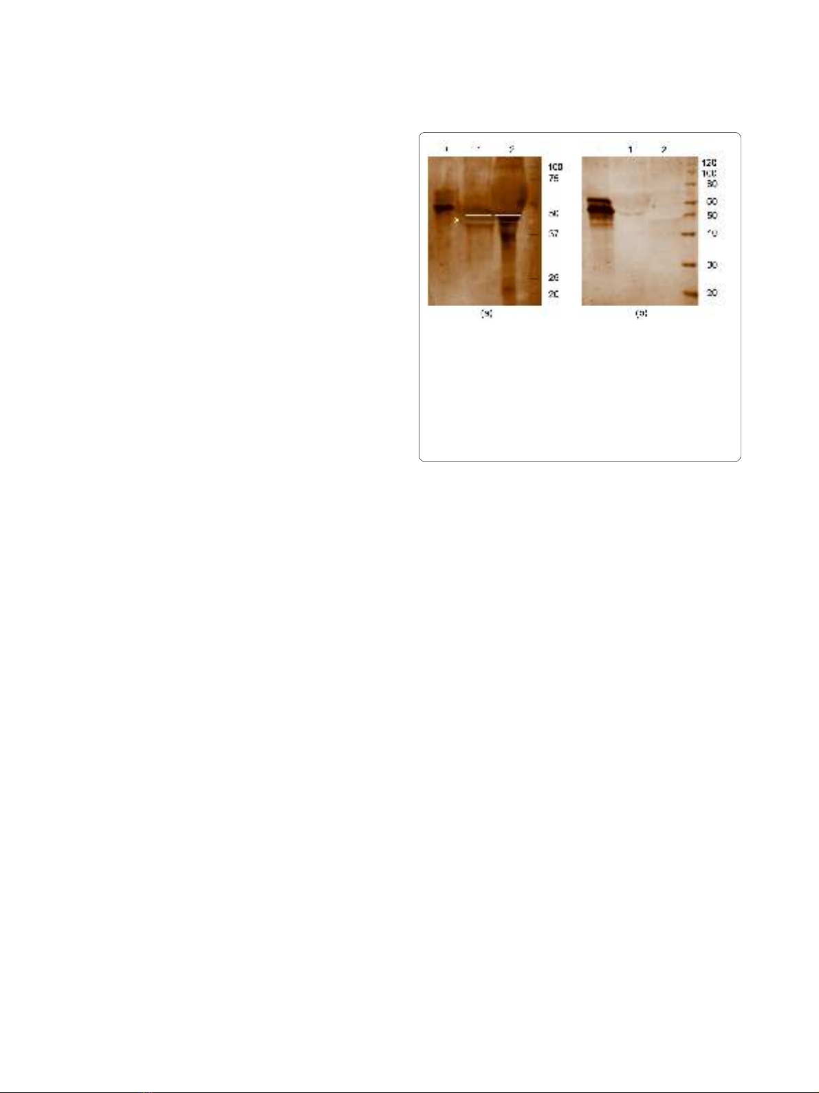
Fibβ (at MW 50 kDa, Figure 1a box (x)) could be identi-
fied in the SF IC fraction as well as in the flow-through.
At this MW, a clear positive AMC staining was detected
in the isolated IC fraction from RA SF (Figure 1a lane 2),
while no citrullinated proteins could be detected in the
flow through of RA SF (Figure 1a lane 1). On the contrary,
when the same setup was repeated with a pool of RA sera
no citrullinated proteins were detected in the IC from RA
sera (RA1 to RA3) (Figure 1b lane 2), while the positive
control for AMC staining was explicit. In order to con-
firm these findings, a set of immunoblotting experiments
was performed.
Immunodetection of potential IC antigens in RA serum and
SF
First, a pool of serum obtained from healthy persons (n =
4), a pool of serum from RA patients (RA1 to RA3) and a
pool of SF from RA patients (SF2; SF13; SF14; SF15) were
used to isolate IC by IP. Subsequent identification of
potential antigens in the isolated IC was performed by
sequential immunodetection with anti-vimentin, anti-
Fibβ, anti-Fibγ and anti-fibronectin on a 2D-Western
blot. Between the different immunodetections, the blot
was carefully stripped and adequate stripping was
checked each time before subsequent primary antibody
addition. The results of these experiments are summa-
rized in Figure 2.
Fibβ was detected in IC from healthy serum and RA
serum and from RA SF at a molecular weight of 50-60
kDa and pI 5-6. However in IC from RA SF, and not in IC
from RA or healthy serum, some extra spots that reacted
with anti-Fibβ could be detected at MW 37-50 kDa and
pI 6-7. These are probably processed isoforms of Fibβ
Figure 1 Detection of citrullinated proteins in IC of RA SF (a) and
RA serum (b) after immunoprecipitation. 1D-PAGE and AMC stain-
ing were performed on the isolated IC and the flow through after IP,
from synovial fluid and serum of RA patients. Where (+) is the positive
control for AMC staining; the flow-through is shown in lane 1 and the
IC-fraction in lane 2. Citrullinated proteins could be detected in the IC
isolated by IP in the SF of RA patients (a) and were absent in the sera of
RA patients (b). In the fraction indicated by "x";, immunoglobulins, se-
rum albumin and Fibβ were identified by mass spectrometry.


