
REVIE W Open Access
Down-regulation of UHRF1, associated with
re-expression of tumor suppressor genes, is a
common feature of natural compounds
exhibiting anti-cancer properties
Mahmoud Alhosin, Tanveer Sharif, Marc Mousli, Nelly Etienne-Selloum, Guy Fuhrmann, Valérie B Schini-Kerth and
Christian Bronner
*
Abstract
Over-expressed in numerous cancers, Ubiquitin-like containing PHD Ring Finger 1 (UHRF1, also known as ICBP90 or
Np95)is characterized by a SRA domain (Set and Ring Associated) which is found only in the UHRF family. UHRF1
constitutes a complex with histone deacetylase 1 (HDAC1) and DNA methyltransferase 1 (DNMT1) via its SRA
domain and represses the expression of several tumour suppressor genes (TSGs) including p16
INK4A
,hMLH1, BRCA1
and RB1. Conversely, UHRF1 is regulated by other TSGs such as p53 and p73. UHRF1 is hypothetically involved in a
macro-molecular protein complex called “ECREM”for “Epigenetic Code Replication Machinery”. This complex would
be able to duplicate the epigenetic code by acting at the DNA replication fork and by activating the right
enzymatic activity at the right moment. There are increasing evidence that UHRF1 is the conductor of this
replication process by ensuring the crosstalk between DNA methylation and histone modifications via the SRA and
Tandem Tudor Domains, respectively. This cross-talk allows cancer cells to maintain the repression of TSGs during
cell proliferation. Several studies showed that down-regulation of UHRF1 expression in cancer cells by natural
pharmacological active compounds, favors enhanced expression or re-expression of TSGs, suppresses cell growth
and induces apoptosis. This suggests that hindering UHRF1 to exert its role in the duplication of the methylation
patterns (DNA + histones) is responsible for inducing apoptosis. In this review, we present UHRF1 expression as a
target of several natural products and we discuss their underlying molecular mechanisms and benefits for
chemoprevention and chemotherapy.
1. Introduction
Cancer is one of the main causes of death among Wes-
ternized countries and is principally due to environmen-
tal risk factors, including diet [1]. It is caused by a series
of genetic and epigenetic abnormalities that induce the
activation of oncogenes and/or the inactivation of
tumour suppressor genes (TSGs) [2,3]. For instance, col-
orectal cancer is known to be a consequence of succes-
sive genetic and epigenetic changes [4,5]. Indeed, an
aberrant promoter hypermethylation of the hMLH1
gene (Human Mutant L homologue 1) is a potential
major cause of colon carcinogenesis suggesting that an
epigenetic mechanism is underlying tumorogenesis [6].
The term epigenetic is defined as heritable modification
in gene expression without any variation in the DNA
sequence [2,3,7,8]. DNA methylation and histone post-
translational changes are the two main hallmarks of the
epigenetic process. Unlike the genetic abnormalities
which are irreversible, epigenetic alterations could be
reversible making them as interesting therapeutic tar-
gets. Epigenetic regulation of gene expression is particu-
larly sensitive to environmental conditions, including
diet [9]. A few examples clearly demonstrate that dietary
behaviours can affect the future health of subsequent
generations, by increasing the risk of cardio-metabolic
diseases such as diabetes mellitus, hypertension and
obesity [9].
* Correspondence: christian.bronner@unistra.fr
CNRS UMR 7213 Laboratoire de Biophotonique et Pharmacologie, Université
de Strasbourg, Faculté de Pharmacie, 74 route du Rhin, 67401 Illkirch, France
Alhosin et al.Journal of Experimental & Clinical Cancer Research 2011, 30:41
http://www.jeccr.com/content/30/1/41
© 2011 Alhosin et al; licensee BioMed Central Ltd. This is an Open Access article distributed under the terms of the Creative Commons
Attribution License (http://creativecommons.org/licenses/by/2.0), which permits unrestricted use, distribution, and reproduction in
any medium, provided the original work is properly cited.

Concerning cancer and transgenerational epigenetic
effect of diets, in terms of increased risk, no evidence
has so far yet been reported. However, cancerogenesis is
now recognised as being the result of profound dietary-
influenced epigenetic modifications, among which
hypermethylation of the promoters of several TSGs
occupies a main place [3,10]. Reversing promoter
methylation of silenced tumor suppressor genes repre-
sents a current challenge for anti-cancer therapy.
2. DNA methylation and histone modifications in cancer
In mammalians, DNA methylation is the most widely
studied epigenetic modification. It is mediated by a
family of DNA methyltransferases (DNMTs) that trans-
fer a methyl group (CH3) from the methyl donor S-ade-
nosylmethionine at the carbon in the fifth position of
cytosine in CpG dinucleotides [11,12]. This family
includes several members, i.e.DNMT1,DNMT3Aand
DNMT3B [13]. DNMT2 and DNMT3L have very little
methyltransferase activity and will not be discussed here
[13]. While about 80% of isolated CpG sites in the gen-
ome are methylated, the « CpG islands » (CpG-rich
short regions of DNA) are usually unmethylated [14].
Exceptions are some CpG island promoters which
remain methylated during development. X-chromosome
inactivation and imprinted genes are the two known
examples of these exceptions [15]. In cancer cells, in
contrast to genome-wide hypomethylation which
increases genomic instability and activates growth-pro-
moting genes (proto-oncogenes), promoters of tumour
suppressor genes are frequently hypermethylated and
this contributes to carcinogenesis [16]. Various TSGs
are silenced in cancer cells by promoter hypermethyla-
tion such as RB1,H1C1 (Hypermethylated In Cancer 1),
p16
INK4A
,MLH1 (Human Mutant L homologue 1),
BRCA1 (BReast CAncer 1) and p73 [17-23]. While the
capacity of CpG island hypermethylation to induce
TSGs silencing is well studied, the mechanism by which
these TSGs are specifically targeted is still unclear. One
hypothesis is that CpG island hypermethylation of TSGs
is driven by a mechanism involving unknown DNA
binding factors that selectively recruit DNMT1 to the
promoters of TSGs which will lead to pathological
hypermethylation and subsequently to unpaired
apoptosis.
Many evidences of the crosstalk between DNA methy-
lation and histone modifications have been reported
[24,25]. The most important histones modifications,
having effects on gene expression, are located on histone
H3 and histone H4 [26]. One of them, that is known to
have a gene silencing role and to have a strong relation-
ship with DNA methylation, is the di- or tri-methylation
oflysine9ofhistone3(H3K9me2orH3K9me3).But
methylation on the same histone on lysine 4 (H3K4me)
is related to gene activation. All these modifications are
catalysed by a broad variety of specific enzymes, some
of which can catalyse the same reaction but at different
location in the nucleus, i.e., heterochromatin or euchro-
matin [26].
Histones undergo specific changes in their acetylation
and methylation degrees during cancerogenesis [27].
Both deacetylation of H4K16 and accumulation of
H3K9me2 are found on many repressed genes, including
TSGs [27,28]. These modifications are mediated by
HDACs (histone deacetylases) and G9a (histone 3
methyltransferase) respectively. HDACs are often over-
expressed in various types of cancer such as renal can-
cer [29] or gastric cancer [30] and have become essen-
tial targets for anticancer therapy. G9a is co-localized
near the methylated promoters of numerous genes in
cancer cells [31]. Interestingly, it has been found that
the inhibition of G9a is sufficient to induce a reactiva-
tion of TSGs [32]. Therefore, over-expression of
enzymes catalysing histone modifications (epigenetic
writers), might be one explanation for the occurrence of
altered epigenetic marks found in cancer.
There is increasing evidence that Ubiquitin-like con-
taining PHD Ring Finger 1 (UHRF1, also known as
ICBP90 or Np95) plays a fundamental role in these pro-
cesses by being involved in DNA methylation, histone
methylation, histone acetylation, cell proliferation and
apoptosis. This is due to the fact that UHRF1 possesses
several domains (Figure 1) able to read both DNA
methylation and histone methylation, thus, physically
linking these two epigenetic marks [26,33,34].
3. UHRF1 and DNA methylation and histone modifications
patterns
UHRF1, a putative oncogenic factor, is over-expressed in
numerous cancers [35,36] and has been suggested to be
an important biomarker to discriminate between cervical
high-grade and low-grade cancer lesions [37]. Another
study has highlighted the efficiency of UHRF1 as a mar-
ker to differentially diagnose pancreatic adenocarcinoma,
chronic pancreatitis and normal pancreas [38]. UHRF1
over-expression was also found in bladder cancer and
the intensity of its over-expression appears to be related
to the stage of the cancer [39], suggesting that the pre-
sence of UHRF1 in urine sediment or surgical speci-
mens could be a useful diagnostic marker and may
improve the diagnosis of the bladder cancer. Recently,
UHRF1’s overpression has also been described in lung
cancer cells, particularly in non-adenocarcinomas [40].
This alteration in UHRF1 expression could be linked to
the degree of the lung cancer aggressiveness and was
detectable in half of the patients in an early pathological
stage. This suggests therefore that UHRF1 could be a
novel diagnostic tool for lung cancer [40]. Altogether,
Alhosin et al.Journal of Experimental & Clinical Cancer Research 2011, 30:41
http://www.jeccr.com/content/30/1/41
Page 2 of 10
these clinical studies show that immuno-histochemical
staining of UHRF1 may improve the specificity and sen-
sitivity of current tests for cancer diagnosis. These stu-
dies also emphasize that over-expression of UHRF1
might be involved in the establishment of aberrant his-
tone code and altered DNA methylation patterns. The
consequences of UHRF1 over-expression are cell contact
inhibition loss [41] and inhibition of TSGs expression,
such as CDKN2A and RASSF1 [42]. Furthermore, very
recently, it was shown that UHRF1 down-regulation in
p53 containing and deficient cancer cells induced cell
cycle arrest in G2/M and caspase-8-dependent apoptosis
[43]. This is consistent with previous studies showing
that down-regulation of UHRF1 leads to cell growth
inhibition [44-46].
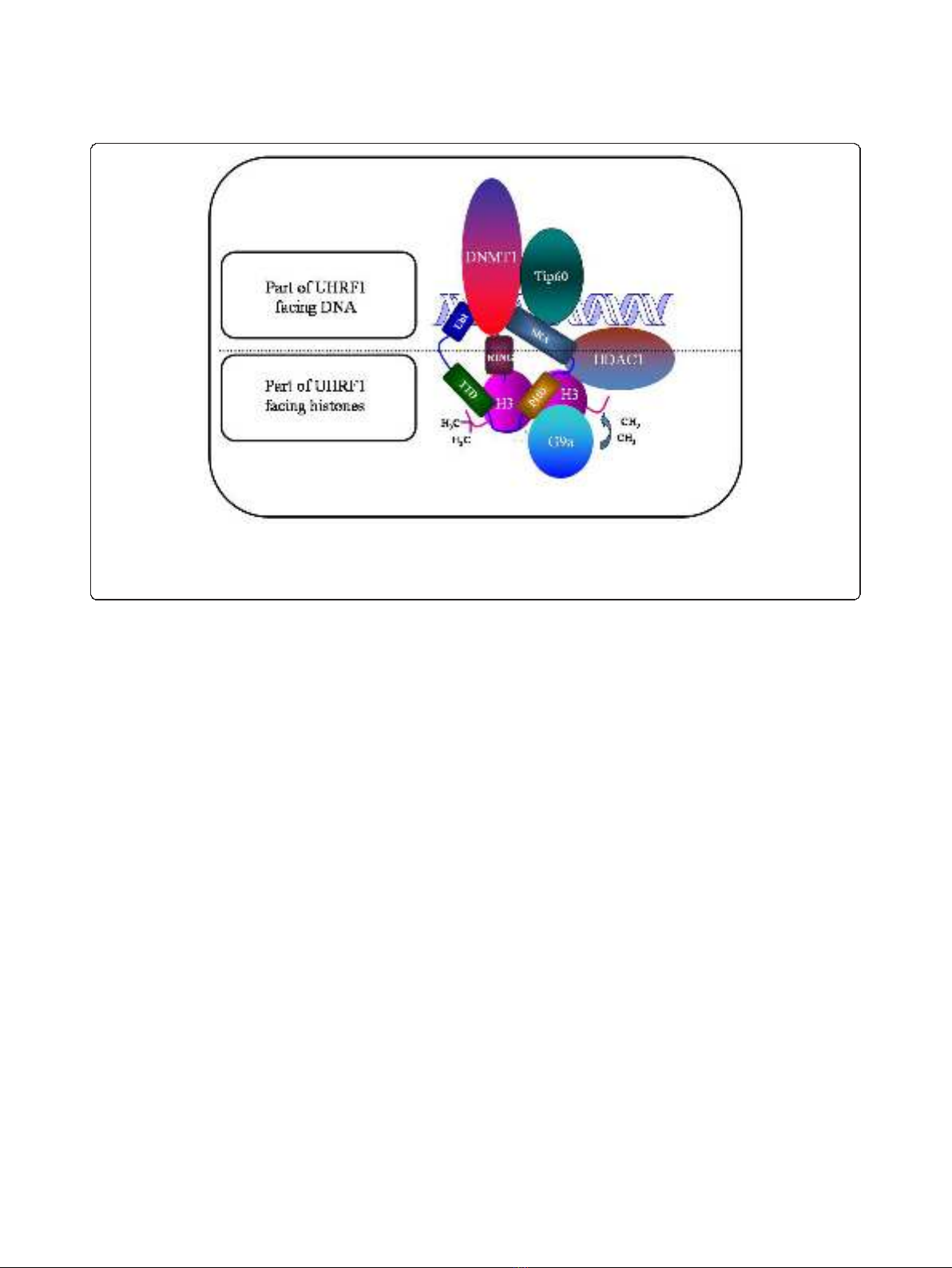
UHRF1 is characterized by the presence of several
structural domains, some facing DNA and others facing
histones (Figure 1). Among them, one of the most
amazing domain is undoubtedly the SRA domain (Set
and Ring Associated) which, in vertebrates, is found
only in the UHRF family [35]. Thanks to this domain,
UHRF1 interacts with histone deacetylase 1 (HDAC1)
and can bind to methylated promoter regions of various
TSGs, including p16
INK4A
and p14
ARF
[44]. Moreover,
we have shown that UHRF1, via the SRA domain,
associates with DNA methyltransferase 1 (DNMT1) to
formacouplecooperatingintheduplicationofthe
DNA methylation patterns but other domains of UHRF1
could also be involved [26,47-49]. The mechanism of
DNA methylation pattern duplication, involves the SRA
domain which is able to detect the hemi-methylated
state of the DNA that occurs after the synthesis of the
new DNA strand [50-52]. This domain behaves as a
“hand”with a palm which holds the methylated cyto-
sine, after that two “fingers”have flipped the methylated
cytosine out from the DNA helix into the major DNA
groove. The flipped methylated cytosine allows UHRF1
to be anchored at the hemi-methylated site to give the
time necessary for DNMT1 to methylate the newly
synthesized DNA strand [26,53], thus ensuring the
maintenance of the DNA methylation patterns through
successive cell divisions. Altogether, these observations
show that immediately after DNA replication which
generates hemi-methylated strands, UHRF1 is recruited
withDNMT1and/orlikelyDNMT3aandDNMT3b,in
order to perpetuate gene repression, and particularly
that of TSGs in cancer cells.
Recently, two novel and interesting partners of
UHRF1, namely Tip60 (Tat-Interactive Protein) and
HAUSP (Herpes virus-Associated Ubiquitin Specific
Protease) have been identified [54,55]. Indeed, we
showed that Tip60 is present in the same macromolecu-
lar complex as UHRF1, DNMT1, and HDAC1. Tip60 is
a histone acetyltransferase with specificity toward lysine
5 of histone H2A (H2AK5) [54]. Interestingly, we
observed that UHRF1 down-regulation correlated with
Figure 1 Schematic representation of UHRF1 with the structural domains facing either DNA or histones. Abbreviation: UBL, Ubiquitin-like
domain; TTD, cryptic Tandem Tudor Domain; PHD, Plant Homeo Domain; SRA, Set and Ring Associated; RING, Really Interesting New Gene. The
major partners of UHRF1, namely Tat-Interactive Protein of 60 kDA (Tip60), DNA methyltransferase 1 (DNMT1), histone methyltransferase G9a
(G9a) and Histone DeAcetylase (HDAC1) are also depicted.
Alhosin et al.Journal of Experimental & Clinical Cancer Research 2011, 30:41
http://www.jeccr.com/content/30/1/41
Page 3 of 10

an increase in Tip60 expression, which was associated
with a decrease of acetylated H2AK5, suggesting that
Tip60 requires UHRF1 for H2AK5 acetylation [54]. This
mark could be involved in the epigenetic silencing of
TSGs, but this possibility requires further investigations.
The other studies reported that through an acetylation-
dependent process UHRF1/Tip60 acts as destroyers of
DNMT1 whereas HDAC1/HAUSP act as protectors for
DNMT1 [55-57]. The paradigm resulting from this
study additionally supports the idea of the existence of a
macromolecular complex involved in the duplication of
theepigeneticcodethatiscapableofselfregulation
through external signals [57]. This complex is able to
duplicate the epigenetic code after DNA replication and
thus, allows cancer cells to maintain the repression of
TSGs, including for instance BRCA1 and p16
INK4A
[49,58]. Indeed, it has been reported that UHRF1 is
responsible for the repression of BRCA1 gene in spora-
dic breast cancer through DNA methylation, by recruit-
ing DNMT1, and histone deacetylation or methylation,
by recruiting HDAC1, or G9a, respectively [58]. As a
platform protein, UHRF1 is expected to be the major
conductor of the epigenetic orchestra by using various
executors to facilitate the conservation of the silencing
marks, especially those concerning TSGs repression in
the cancer cells. Thus, targeting this epigenetic conduc-
tor may be a new promising approach for anticancer
therapy.
Until today, only the two key partners of UHRF1
(DNMT1 and HDAC1) are targeted therapeutically.
Indeed, two large families of specific inhibitors of
DNMT1 (DNMTi) and HDAC1 (HDACi) are commer-
cially available but which efficiency in solid tumors is
often questioned [59,60]. The current challenge is there-
fore to find new targets which will enable to treat more
efficiently cancer, with lower toxicity and more specifi-
city to reduce the side effects of these chemical com-
pounds. Considering that DNMT1 and probably
HDAC1 require UHRF1 to fully exert their effects, inhi-
biting the UHRF1 activity or expression would theoreti-
cally mimic the cumulative effects of HDAC1 and
DNMT1 inhibitors and thus would be highly efficient,
especially in solid tumors in which DNMTs are particu-
larly less active.
4. Targeting UHRF1 abundance by natural compounds
Targeting UHRF1 abundance and/or UHRF1’senzy-
matic activity would have application in several types of
cancer. UHRF1 is essential for cell proliferation and
therefore, to our opinion it would be more rational to
target cancer types in which UHRF1 is actually found in
high abundance, i.e., over-expressed. UHRF1 has been
reported to be over-expressed in various cancers such as
breast, bladder, kidney, lung, prostate, cervical, and
pancreatic cancers, as well as in astrocytomas and glio-
blastoma [35,40,61]. The anticancer strategic idea would
be not to completely inhibit UHRF1 expression consid-
ering that UHRF1 is also necessary for non cancerous to
proliferate [44,62,63], hence, for instance, for physiologic
tissue regeneration. Thus, to consolidate the anti-
UHRF1 therapeutic interest, it would be interesting to
show that diminishing but not abolishing UHRF1’s
expression by chronic treatment of natural compound is
sufficient for re-expression of silenced tumor suppressor
genes. An ideal property for future natural compounds
as anti-cancer drugs, would be that cancer cells but not
normal cells are affected by them in order to undergo
apoptosis via an UHRF1 down-regulation. Targeting
UHRF1 is particularly interesting because this protein
regulates the G1/S transition [47-49,62,63]. The arrest at
G1/S checkpoint is mediated by the action of the tumor
suppressor gene p53 or its functional homologue p73
[64,65]. Recent years have seen a dramatic progress in
understanding mechanisms that regulate the cell divi-
sion.Inthiscontext,weandothergroupshaveshown
that UHRF1 is essential for G1/S transition [63]. Loss of
p53 activity, as a result of genetic mutations or epige-
netic alterations in cancer, prevents G1/S checkpoints.
DNA damage induces a p53 or p73 up-regulation (in
p53-deficient cells) that activates the expression of
p21
cip/waf
or p16
INK4A
, resulting in cell cycle arrest at
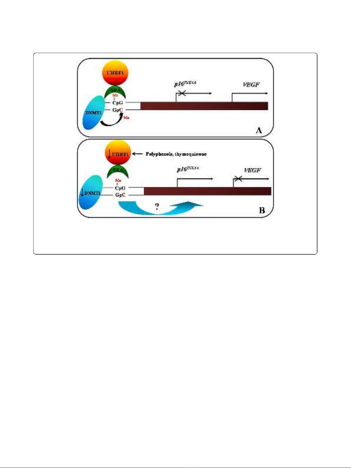
G1/S transition [65,66]. We have shown that UHRF1
represses the expression of tumour suppressor genes
such as p16
INK4A
&RB1 leading to a down-regulation of
the Vascular Endothelial Growth Factor (VEGF, Figure
2A) [49] and by a feedback mechanism, UHRF1 may be
regulated by other tumour suppressor genes such as p53
and p73 products [46,67]. This suggests that the appear-
ance of genetic and/or epigenetic abnormalities of TSGs
including p53 and p73 genes, in various human cancers
would be an explanation for the observed UHRF1 over-
expression. Since UHRF1 controls the duplication of the
epigenetic code after DNA replication, the inability of
p53 and P73 to down-regulate UHRF1, allows the
daughter cancer cells to maintain the repression of
tumour suppressor genes observed in the mother cancer
cell [26,68].
Over the last millenium, herbal products have been
commonly used for prevention and treatment of various
diseases including cancer [69-71]. One of these natural
products is curcumin which has potent anti-cancer
properties in experimental systems. Curcumin is con-
sumed in high quantities in Asian countries and epide-
miological studies have attributed the lower rate of
colon cancer in these countries to its consumption [72].
Green tea is also widely consumed in Asia countries.
This natural product, which is rich in polyphenols, has
been shown to significantly decrease the risk of breast
Alhosin et al.Journal of Experimental & Clinical Cancer Research 2011, 30:41
http://www.jeccr.com/content/30/1/41
Page 4 of 10
and ovarian cancers in women in Asian countries [73].
Black seed (nigella sativia)belongstotheRanuncula-
ceae family which grows in the Mediterranean sea and
Western Asia countries, including Pakistan, India and
China [74]. This plant is used in traditional folk medi-
cine for the prevention and the treatment of numerous
diseases such as eczema, cough, bacterial and viral infec-
tions, hypertension and diabetes [75]. The chemothera-
peutic and chemopreventive activities of black cumin oil
are attributed to thymoquinone (TQ). Several in vitro
and in vivo studies have shown that TQ has potent
cytotoxic and genotoxic activities on a wide range of
cancer cells [76-80]. TQ exerts its anti-cancer effects by
inhibiting cell proliferation, arresting cell cycle progres-
sion and inducing subsequently apoptosis by p53-
dependent or -independent pathways. By using the
acute lymphoblastic leukemia jurkat cell model (p53
mutated cell line), we have demonstrated that TQ trig-
gers apoptosis through the production of reactive oxy-
gen species (ROS) and the activation of the p73 gene
[67]. This tumor suppressor gene seems to act as a cel-
lular gatekeeper by preventing the proliferation of TQ-
exposed Jurkat cells [67]. Obviously, the observed p73
activation triggers G1 cell cycle arrest and apoptosis.
Interestingly, a transient TQ concentration-dependent
up-regulation of caspase 3 cleaved subunits was also
observed, suggesting that TQ exerts its apoptotic activity
through a p73-dependent caspase-dependent cell death
pathway. Consistently with our study, it was recently
reported that catechin, a natural polyphenolic com-
pound, induces apoptosis, in a similar way as does TQ,
by its ability to increase the expression of pro-apoptotic
genes such as caspase-3, -8, and -9 and p53 [81]. Inter-
estingly, our study also showed that TQ down-regulated
UHRF1, DNMT1 and HDAC1 expressions [67]. We
determined that p73 was responsible for UHRF1 down-
regulation through a caspase-3 dependent process. A
subsequent study allowed us to propose that down-regu-
lation of phosphodiesterase 1A (PDE1A), a modulator of
cAMP and cGMP cyclic nucleotides, could be the key
event to explain the TQ-induced down-regulation of
UHRF1 and the occurrence of apoptosis [82]. All these
findings showed for the first time that a natural com-
pound induces apoptosis by acting on the epigenetic
integrator UHRF1 through a p73-dependent mitochon-
drial pathway.
Figure 2 Schematic model of the role of UHRF1/DNMT1 complex in the regulation of p16
INK4A
and VEGF gene expressions.A.When
the SRA domain of UHRF1 meets hemi-methylated DNA present in the p16
INK4A
promoter, UHRF1 acts as a guide for DNMT1 to methylate the
complementary DNA strand. Subsequently a p16
INK4A
gene repression and VEGF gene activation are maintained on the DNA daughter strands,
i.e., in the daughter cancer cells. B. The UHRF1 down-regulation, by natural compounds such as TQ or polyphenols, induces the DNMT1
abundance decrease, that is accompanied by a p16
INK4A
gene re-expression and a down-regulation of VEGF gene expression.
Alhosin et al.Journal of Experimental & Clinical Cancer Research 2011, 30:41
http://www.jeccr.com/content/30/1/41
Page 5 of 10


![Vaccine và ứng dụng: Bài tiểu luận [chuẩn SEO]](https://cdn.tailieu.vn/images/document/thumbnail/2016/20160519/3008140018/135x160/652005293.jpg)


%20--%3e%3cdefs%3e%3cstyle%3e%20.st0%20{%20fill:%20%23fff;%20}%20.st1%20{%20fill:%20%237800fa;%20}%20%3c/style%3e%3c/defs%3e%3cpath%20class='st1'%20d='M117.78,12.18H43.11c2.9,3.47,4.65,7.94,4.65,12.82,0,5.6-2.3,10.66-6.01,14.29h76.02l7.22-13.56-7.22-13.56Z'/%3e%3cg%3e%3cpath%20class='st0'%20d='M53.58,26.17h-.59v-1.46h.59v-4.96h2.83c1.78,0,2.67.94,2.67,2.82v5.76c0,1.87-.89,2.81-2.67,2.81h-2.83v-4.96ZM55.36,21.37v3.34h1.1v1.46h-1.1v3.34h1.01c.61,0,.91-.37.91-1.1v-5.93c0-.74-.3-1.1-.91-1.1h-1.01Z'/%3e%3cpath%20class='st0'%20d='M65.99,31.14h-1.8l-.31-2.07h-2.19l-.31,2.07h-1.64l1.82-11.39h2.62l1.82,11.39ZM65.28,18.04c-.25.46-.51.77-.75.94-.21.15-.47.22-.79.22-.26,0-.57-.07-.92-.22l-.38-.15c-.14-.05-.26-.07-.37-.07-.3,0-.53.18-.71.54l-.91-.68c.25-.46.51-.77.75-.94.21-.14.48-.21.79-.21.26,0,.57.07.92.21l.38.15c.14.05.26.07.37.07.3,0,.53-.18.71-.54l.91.68ZM61.91,27.52h1.73l-.87-5.76-.87,5.76Z'/%3e%3cpath%20class='st0'%20d='M74.53,26.89v1.52c0,1.91-.89,2.86-2.67,2.86s-2.67-.95-2.67-2.86v-5.93c0-1.91.89-2.86,2.67-2.86s2.67.95,2.67,2.86v1.11h-1.69v-1.22c0-.75-.31-1.12-.93-1.12s-.93.37-.93,1.12v6.15c0,.74.31,1.11.93,1.11s.93-.37.93-1.11v-1.63h1.69Z'/%3e%3cpath%20class='st0'%20d='M81.4,31.14h-1.8l-.31-2.07h-2.19l-.31,2.07h-1.64l1.82-11.39h2.62l1.82,11.39ZM75.9,19.2l1.52-1.91h1.71l1.51,1.91h-1.61l-.76-.95-.75.95h-1.61ZM77.32,27.52h1.73l-.87-5.76-.87,5.76ZM83.1,15.99l-1.76,1.91h-1.26l1.17-1.91h1.86Z'/%3e%3cpath%20class='st0'%20d='M84.86,19.75c1.78,0,2.67.94,2.67,2.82v1.48c0,1.87-.89,2.81-2.67,2.81h-.85v4.28h-1.79v-11.39h2.64ZM84.01,21.37v3.86h.85c.58,0,.87-.36.87-1.08v-1.71c0-.71-.29-1.07-.87-1.07h-.85Z'/%3e%3cpath%20class='st0'%20d='M93.51,19.75c1.78,0,2.67.94,2.67,2.82v1.48c0,1.87-.89,2.81-2.67,2.81h-.85v4.28h-1.79v-11.39h2.64ZM92.66,21.37v3.86h.85c.58,0,.87-.36.87-1.08v-1.71c0-.71-.29-1.07-.87-1.07h-.85Z'/%3e%3cpath%20class='st0'%20d='M98.8,31.14h-1.79v-11.39h1.79v4.88h2.03v-4.88h1.83v11.39h-1.83v-4.88h-2.03v4.88Z'/%3e%3cpath%20class='st0'%20d='M105.36,24.55h2.46v1.62h-2.46v3.34h3.09v1.63h-4.88v-11.39h4.88v1.63h-3.09v3.18ZM108.17,17.29l-1.76,1.91h-1.26l1.17-1.91h1.86Z'/%3e%3cpath%20class='st0'%20d='M112.2,19.75c1.78,0,2.67.94,2.67,2.82v1.48c0,1.87-.89,2.81-2.67,2.81h-.85v4.28h-1.79v-11.39h2.64ZM111.35,21.37v3.86h.85c.58,0,.87-.36.87-1.08v-1.71c0-.71-.29-1.07-.87-1.07h-.85Z'/%3e%3c/g%3e%3ccircle%20class='st1'%20cx='25'%20cy='25'%20r='20'/%3e%3cpath%20class='st0'%20d='M32.78,19.27c2.92,0,4.43,2.55,5.28,5.33l.71,2.17c.14.38-.33.75-.71.75h-5.61c.19-.33.24-.71.09-1.08l-.75-2.45c-.43-1.32-.99-2.64-1.79-3.77.75-.57,1.65-.94,2.78-.94h0ZM25,18.38c3.25,0,4.9,2.78,5.89,5.89l.76,2.45c.14.42-.33.8-.8.8h-11.69c-.42,0-.94-.38-.8-.8l.75-2.45c.99-3.11,2.64-5.89,5.89-5.89h0ZM25,11.35c1.74,0,3.11,1.37,3.11,3.11s-1.37,3.11-3.11,3.11-3.11-1.41-3.11-3.11,1.41-3.11,3.11-3.11h0ZM17.27,19.27c1.08,0,1.98.38,2.73.94-.8,1.13-1.37,2.45-1.74,3.77l-.8,2.45c-.14.38-.05.75.09,1.08h-5.56c-.42,0-.9-.38-.75-.75l.71-2.17c.9-2.78,2.41-5.33,5.33-5.33h0ZM17.27,12.91c1.51,0,2.78,1.27,2.78,2.83s-1.27,2.83-2.78,2.83-2.83-1.27-2.83-2.83,1.27-2.83,2.83-2.83h0ZM32.78,12.91c1.56,0,2.78,1.27,2.78,2.83s-1.23,2.83-2.78,2.83-2.83-1.27-2.83-2.83,1.27-2.83,2.83-2.83h0ZM27.07,28.56v.09c0,.57-.24,1.08-.61,1.46h0v.05c-.38.33-.9.57-1.46.57s-1.08-.24-1.46-.61h0c-.38-.38-.61-.9-.61-1.46v-.09h1.41v.09c0,.19.05.38.19.47v.05c.09.09.28.19.47.19s.38-.09.47-.19v-.05c.14-.09.24-.28.24-.47t-.05-.09h1.41ZM30.99,28.56v.09c0,1.65-.66,3.16-1.74,4.24-1.08,1.08-2.59,1.79-4.24,1.79s-3.16-.71-4.24-1.79l-.05-.05c-1.04-1.08-1.7-2.55-1.7-4.2v-.09h1.41v.09c0,1.27.47,2.4,1.27,3.25h.05c.85.85,1.98,1.37,3.25,1.37s2.4-.52,3.25-1.37c.85-.8,1.37-1.98,1.37-3.25v-.09h1.37ZM34.99,28.56v.09c0,2.78-1.13,5.28-2.92,7.07-1.79,1.79-4.29,2.92-7.07,2.92s-5.23-1.13-7.07-2.92c-1.79-1.79-2.92-4.29-2.92-7.07v-.09h1.41v.09c0,2.4.94,4.53,2.5,6.08,1.56,1.56,3.72,2.5,6.08,2.5s4.52-.94,6.08-2.5c1.56-1.56,2.5-3.68,2.5-6.08v-.09h1.41Z'/%3e%3c/svg%3e)